Esophageal Cancer Prevention (PDQ®)–Health Professional Version

Esophageal Cancer Prevention (PDQ®)–Health Professional Version

Overview

Note: The Overview section summarizes the published evidence on this topic. The rest of the summary describes the evidence in more detail.

Other PDQ summaries on Esophageal Cancer Screening; Esophageal Cancer Treatment; and Levels of Evidence for Cancer Screening and Prevention Studies are also available.

Who Is at Risk?

Smoking and drinking alcohol may account for roughly 90% of esophageal squamous cell carcinoma cases in Western countries like the United States.[1] Gastroesophageal reflux/Barrett esophagus is associated with an increased risk of esophageal adenocarcinoma. Other factors that may explain the increased risk of adenocarcinoma of the esophagus include obesity [2] and the use of medications such as anticholinergics that can predispose to gastroesophageal reflux disease (GERD) by relaxing the lower esophageal sphincter.[3]

Squamous Cell Carcinoma of the Esophagus

Factors with adequate evidence of increased risk of squamous cell carcinoma of the esophagus

Cigarette smoking and drinking alcohol

Based on solid evidence, smoking cigarettes and drinking alcohol increases the risk of esophageal squamous cell carcinoma. Smoking and drinking alcohol may account for roughly 90% of esophageal squamous cell carcinomas in Western countries like the United States.[1]

Magnitude of Effect: Increased risk, moderate magnitude.

  • Study Design: Evidence from population-based case-control and cohort studies.
  • Internal Validity: Fair.
  • Consistency: Good.
  • External Validity: Fair.

Factors with adequate evidence of decreased risk of squamous cell carcinoma of the esophagus

Avoidance of tobacco and alcohol

Based on solid evidence, avoidance of tobacco and alcohol would decrease the risk of squamous cell carcinoma.[1,4]

Magnitude of Effect: Large positive benefit.

  • Study Design: Evidence obtained from cohort or case-control studies.
  • Internal Validity: Fair.
  • Consistency: Multiple studies.
  • External Validity: Fair.
Chemoprevention
Aspirin and nonsteroidal anti-inflammatory drug (NSAID) use: Benefits

Based on fair evidence, epidemiological studies have found that aspirin or NSAID use is associated with decreased risk of developing or dying of esophageal cancer (odds ratio [OR], 0.57; 95% confidence interval [CI], 0.47–0.71).[5]

Magnitude of Effect: Small positive.

  • Study Design: Evidence obtained from cohort or case-control studies.
  • Internal Validity: Fair.
  • Consistency: Good.
  • External Validity: Fair.
Aspirin and NSAID use: Harms

Based on solid evidence, harms of NSAID use include upper gastrointestinal bleeding and serious cardiovascular events, such as myocardial infarction, heart failure, hemorrhagic stroke, and renal impairment.

Magnitude of Effect: Increased risk, small magnitude.

  • Study Design: Evidence obtained from randomized controlled trials.
  • Internal Validity: Fair.
  • Consistency: Good.
  • External Validity: Fair.

Adenocarcinoma of the Esophagus

Factors with adequate evidence of increased risk of adenocarcinoma of the esophagus

Gastroesophageal reflux

Based on fair evidence, an association exists between GERD and adenocarcinoma, particularly if the GERD is long-standing and symptoms are severe.[6,7] In a case-control study from Sweden, the OR for patients with recurrent reflux symptoms was 7.7, while the OR for patients with long-standing and severe symptoms was 43.5 (95% CI, 18.3–103.5).[8] A meta-analysis of 1,128 individuals with esophageal adenocarcinoma from five case-control studies reported statistically significant increases in risk with recurrent heartburn (OR, 4.6; 95% CI, 3.3–6.6), regurgitation (OR, 4.6; 95% CI, 3.4–6.1), or both (OR, 4.8; 95% CI, 3.4–6.8). Daily heartburn and regurgitation was associated with an eightfold increase in risk (OR, 8.0; 95% CI, 4.5–14.0).[7]

It is unknown whether elimination of gastroesophageal reflux by surgical or medical means will reduce the risk of adenocarcinoma of the esophagus.[8,9]

Magnitude of Effect: Unknown.

  • Study Design: Case-control studies.
  • Internal Validity: Fair.
  • Consistency: Good; multiple studies.
  • External Validity: Fair.

Interventions with adequate evidence of decreased risk of adenocarcinoma of the esophagus

Aspirin and NSAID use: Benefits

Based on fair evidence, epidemiological studies have found that aspirin or NSAID use is associated with decreased risk of developing or dying from esophageal cancer (OR, 0.57; 95% CI, 0.47–0.71).[5,10]

Magnitude of Effect: Unknown magnitude.

  • Study Design: Evidence obtained from cohort or case-control studies.
  • Internal Validity: Fair.
  • Consistency: Good.
  • External Validity: Fair.
Aspirin and NSAID use: Harms

Based on solid evidence, harms of NSAID use include upper gastrointestinal bleeding and serious cardiovascular events, such as myocardial infarction, heart failure, hemorrhagic stroke, and renal impairment.

Magnitude of Effect: Increased risk; small magnitude.

  • Study Design: Evidence obtained from randomized controlled trials.
  • Internal Validity: Good.
  • Consistency: Good.
  • External Validity: Good.
Ablation of Barrett esophagus with dysplasia: Benefits

A randomized controlled trial has found that radiofrequency ablation of Barrett esophagus with severe dysplasia may lead to eradication of both dysplasia and intestinal metaplasia and a reduced risk of disease progression.[11]

Magnitude of Effect: Impact on cancer mortality not known.

  • Study Design: Evidence obtained from a randomized controlled trial.
  • Internal Validity: Good.
  • Consistency: Single study.
  • External Validity: Good.
Ablation of Barrett esophagus with dysplasia: Harms

Based on solid evidence, harms of radiofrequency ablation include esophageal stricture and requirement for dilatation and upper gastrointestinal hemorrhage but at low rates. It is possible that overdiagnosis and overtreatment of Barrett esophagus, particularly without severe dysplasia, could lead to a substantial number of harms.

Magnitude of Effect: The low rates of esophageal stricture and requirement for dilatation and upper gastrointestinal hemorrhage may be an understatement of the risks if this practice is widely adopted by less-experienced physicians.

  • Study Design: Evidence obtained from a randomized controlled trial.
  • Internal Validity: Good.
  • Consistency: Single study.
  • External Validity: Patients representative of a subset of people with dysplasia, particularly severe dysplasia; physicians may not be representative of practicing physicians because this is a new technology and requires specialized knowledge.
References
  1. Engel LS, Chow WH, Vaughan TL, et al.: Population attributable risks of esophageal and gastric cancers. J Natl Cancer Inst 95 (18): 1404-13, 2003. [PUBMED Abstract]
  2. Lagergren J: Controversies surrounding body mass, reflux, and risk of oesophageal adenocarcinoma. Lancet Oncol 7 (4): 347-9, 2006. [PUBMED Abstract]
  3. Lagergren J, Bergström R, Adami HO, et al.: Association between medications that relax the lower esophageal sphincter and risk for esophageal adenocarcinoma. Ann Intern Med 133 (3): 165-75, 2000. [PUBMED Abstract]
  4. Siemiatycki J, Krewski D, Franco E, et al.: Associations between cigarette smoking and each of 21 types of cancer: a multi-site case-control study. Int J Epidemiol 24 (3): 504-14, 1995. [PUBMED Abstract]
  5. Corley DA, Kerlikowske K, Verma R, et al.: Protective association of aspirin/NSAIDs and esophageal cancer: a systematic review and meta-analysis. Gastroenterology 124 (1): 47-56, 2003. [PUBMED Abstract]
  6. Lagergren J, Bergström R, Lindgren A, et al.: Symptomatic gastroesophageal reflux as a risk factor for esophageal adenocarcinoma. N Engl J Med 340 (11): 825-31, 1999. [PUBMED Abstract]
  7. Cook MB, Corley DA, Murray LJ, et al.: Gastroesophageal reflux in relation to adenocarcinomas of the esophagus: a pooled analysis from the Barrett’s and Esophageal Adenocarcinoma Consortium (BEACON). PLoS One 9 (7): e103508, 2014. [PUBMED Abstract]
  8. Lagergren J, Ye W, Lagergren P, et al.: The risk of esophageal adenocarcinoma after antireflux surgery. Gastroenterology 138 (4): 1297-301, 2010. [PUBMED Abstract]
  9. Spechler SJ, Goyal RK: The columnar-lined esophagus, intestinal metaplasia, and Norman Barrett. Gastroenterology 110 (2): 614-21, 1996. [PUBMED Abstract]
  10. Liao LM, Vaughan TL, Corley DA, et al.: Nonsteroidal anti-inflammatory drug use reduces risk of adenocarcinomas of the esophagus and esophagogastric junction in a pooled analysis. Gastroenterology 142 (3): 442-452.e5; quiz e22-3, 2012. [PUBMED Abstract]
  11. Shaheen NJ, Sharma P, Overholt BF, et al.: Radiofrequency ablation in Barrett’s esophagus with dysplasia. N Engl J Med 360 (22): 2277-88, 2009. [PUBMED Abstract]

Background

Two histological types account for most malignant esophageal neoplasms: adenocarcinoma and squamous cell carcinoma. The epidemiology of these types varies markedly. In the 1960s, squamous cell carcinomas comprised over 90% of all esophageal tumors. The incidence of esophageal adenocarcinomas has risen markedly for the past two decades. Adenocarcinoma is now more prevalent than squamous cell carcinomas in the United States and Western Europe, with most tumors located in the distal esophagus.[1]

References
  1. Holmes RS, Vaughan TL: Epidemiology and pathogenesis of esophageal cancer. Semin Radiat Oncol 17 (1): 2-9, 2007. [PUBMED Abstract]

Incidence and Mortality

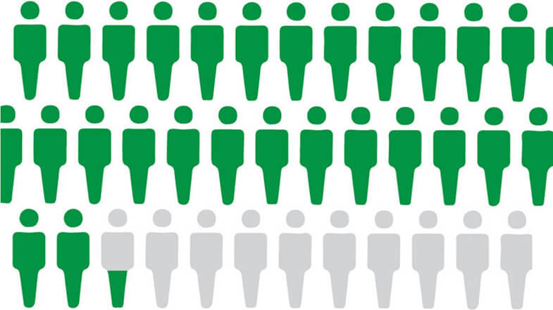
In 2025, an estimated 22,070 Americans will be diagnosed with esophageal cancer, and 16,250 will die of this disease. Of the new cases, an estimated 17,430 will occur in men, and 4,640 will occur in women.[1] Incidence rates for esophageal cancer have been falling on average 0.4% each year from 2012 to 2021. Death rates have been falling on average 1.1% each year from 2013 to 2022. Incidence rates generally increase with age in all racial and ethnic groups. Incidence rates are higher in White men compared with Black men in all age groups. In women, incidence rates are higher in Black women through age 74 years, at which point the rates become higher in White women.[2] Death rates are higher for White men compared with Black men at all ages. In women, death rates are higher in Black women through age 69 years, at which point the rates become higher in White women.

Although the overall incidence of squamous cell carcinoma of the esophagus is declining, this histological type remains six times more likely to occur in Black men than in White men.[3] In contrast, the incidence of adenocarcinoma of the esophagus rapidly increased from the 1970s to the mid-1990s.[4]

Male sex is an important predictor of adenocarcinoma of the esophagus. The attributable risk is low enough in women that, although the risk from sex is not modifiable, other risk factors necessarily have limited impact.[4]

References
  1. American Cancer Society: Cancer Facts and Figures 2025. American Cancer Society, 2025. Available online. Last accessed January 16, 2025.
  2. Surveillance Research Program, National Cancer Institute: SEER*Explorer: An interactive website for SEER cancer statistics. Bethesda, MD: National Cancer Institute. Available online. Last accessed December 30, 2024.
  3. Devesa SS, Blot WJ, Fraumeni JF: Changing patterns in the incidence of esophageal and gastric carcinoma in the United States. Cancer 83 (10): 2049-53, 1998. [PUBMED Abstract]
  4. Hur C, Miller M, Kong CY, et al.: Trends in esophageal adenocarcinoma incidence and mortality. Cancer 119 (6): 1149-58, 2013. [PUBMED Abstract]

Squamous Cell Carcinoma of the Esophagus

Factors With Adequate Evidence of Increased Risk of Squamous Cell Carcinoma of the Esophagus

Smoking cigarettes and drinking alcohol

In the United States, squamous cell carcinoma of the esophagus is strongly associated with tobacco and alcohol abuse. The relative risk associated with tobacco use is 2.4, and the population attributable risk is 54.2% (95% confidence interval [CI], 3.0%–76.2%).[1,2] Retrospective cohort studies adjusted for tobacco use have shown a twofold to sevenfold increase in the risk of esophageal cancer in individuals with alcohol addiction, compared with rates for the general population.[1] Case-control studies have also suggested a significantly increased risk of cancer of the esophagus associated with alcohol abuse.

A multicenter, population-based, case-control study included 221 patients with esophageal squamous cell carcinoma and 695 controls. In this study, ever-smoking, alcohol consumption, and low fruit and vegetable consumption accounted for 56.9% (95% CI, 36.6%–75.1%), 72.4% (95% CI, 53.3%–85.8%), and 28.7% (95% CI, 11.1%–56.5%) of esophageal squamous cell carcinomas, respectively, with a combined population attributable risk of 89.4% (95% CI, 79.1%–95.0%).[3]

In China, where the overall prevalence of esophageal carcinoma is much higher than in the United States, esophageal cancer is associated with deficiencies of nutrients, such as retinol, riboflavin, alpha-carotene, beta-carotene, alpha-tocopherol, ascorbate and zinc, and with exposure to specific carcinogens (e.g., N-nitroso compounds).[1]

Factors With Adequate Evidence of Decreased Risk of Squamous Cell Carcinoma of the Esophagus

Chemoprevention

A prospective, placebo-controlled, esophagus chemoprevention study randomly assigned 610 high-risk Chinese patients.[4] Patients were aged 35 to 64 years and received either placebo or combined low-dose retinol (15 mg or 50,000 IU) plus riboflavin (200 mg) and zinc gluconate (50 mg) for 13.5 months. Standard histological evaluations (including two endoscopic biopsies) were conducted for 93% of all participants. Micronuclei from esophageal cells were obtained before therapy began and after the 13.5 months of treatment. Serum levels of vitamin A, beta-carotene, riboflavin, and zinc were obtained at 0, 2, and 13.5 months.

The second report of this study presented micronuclei frequency results.[5] A statistically significant reduction in the mean percentage of micronucleated esophageal cells occurred in the active-treatment group compared with the placebo group. The pattern of cell proliferation, another potential intermediate end point marker, also improved.[6]

Aspirin and nonsteroidal anti-inflammatory drug (NSAID) use

A systematic review and meta-analysis of the association between aspirin and NSAID use and esophageal cancer identified two cohort and seven case-control studies published between 1980 and 2001.[7] Pooled results showed a protective association between aspirin/NSAID use and esophageal cancer (odds ratio [OR], 0.57; 95% CI, 0.47–0.71). The association with aspirin use was statistically significant (OR, 0.50; 95% CI, 0.38–0.66), and the association with NSAIDs was borderline significant (OR, 0.75; 95% CI, 0.54–1.0). Aspirin/NSAID use was associated with lower risk of both adenocarcinoma (OR, 0.67; 95% CI, 0.51–0.87) and squamous cell carcinoma (OR, 0.58; 95% CI, 0.43–0.78).[7]

References
  1. Oesophagus. In: World Cancer Research Fund, American Institute for Cancer Research: Food, Nutrition and the Prevention of Cancer: A Global Perspective. The Institute, 1997, pp 118-129.
  2. Siemiatycki J, Krewski D, Franco E, et al.: Associations between cigarette smoking and each of 21 types of cancer: a multi-site case-control study. Int J Epidemiol 24 (3): 504-14, 1995. [PUBMED Abstract]
  3. Engel LS, Chow WH, Vaughan TL, et al.: Population attributable risks of esophageal and gastric cancers. J Natl Cancer Inst 95 (18): 1404-13, 2003. [PUBMED Abstract]
  4. Muñoz N, Wahrendorf J, Bang LJ, et al.: No effect of riboflavine, retinol, and zinc on prevalence of precancerous lesions of oesophagus. Randomised double-blind intervention study in high-risk population of China. Lancet 2 (8447): 111-4, 1985. [PUBMED Abstract]
  5. Muñoz N, Hayashi M, Bang LJ, et al.: Effect of riboflavin, retinol, and zinc on micronuclei of buccal mucosa and of esophagus: a randomized double-blind intervention study in China. J Natl Cancer Inst 79 (4): 687-91, 1987. [PUBMED Abstract]
  6. Yang GC, Lipkin M, Yang K, et al.: Proliferation of esophageal epithelial cells among residents of Linxian, People’s Republic of China. J Natl Cancer Inst 79 (6): 1241-6, 1987. [PUBMED Abstract]
  7. Corley DA, Kerlikowske K, Verma R, et al.: Protective association of aspirin/NSAIDs and esophageal cancer: a systematic review and meta-analysis. Gastroenterology 124 (1): 47-56, 2003. [PUBMED Abstract]

Adenocarcinoma of the Esophagus

Factors Associated With Increased Risk of Adenocarcinoma of the Esophagus

Gastroesophageal reflux disease (GERD)

The most important epidemiological difference between squamous cell carcinoma and adenocarcinoma is the strong association between GERD and adenocarcinoma. The results of a population-based case-controlled study suggest that symptomatic gastroesophageal reflux is a risk factor for adenocarcinoma of the esophagus. The frequency, severity, and duration of reflux symptoms were positively associated with an increased risk of adenocarcinoma of the esophagus.[1] In a case-control study from Sweden, the odds ratio (OR) was 7.7 for patients with recurrent reflux symptoms, while the OR for patients with long-standing and severe symptoms was 43.5 (95% confidence interval [CI], 18.3–103.5).[1] A meta-analysis of 1,128 individuals with esophageal adenocarcinoma from five case-control studies reported statistically significant increases in risk with recurrent heartburn (OR, 4.6; 95% CI, 3.3–6.6), regurgitation (OR, 4.6; 95% CI, 3.4–6.1), or both (OR, 4.8; 95% CI, 3.4–6.8). Daily heartburn and regurgitation was associated with an eightfold increase in risk (OR, 8.0; 95% CI, 4.5–14.0).[2] The probable mechanism is that long-standing GERD is associated with the development of Barrett esophagus, a condition in which an abnormal intestinal-type epithelium replaces the stratified squamous epithelium that normally lines the distal esophagus. Barrett esophagus is considered a precursor of esophageal adenocarcinoma.[3] The intestinal-type epithelium of Barrett esophagus has a characteristic endoscopic appearance that differs from squamous epithelium.[4] Dysplasia in Barrett epithelium represents a neoplastic alteration of the columnar epithelium that may progress to invasive adenocarcinoma.[5]

A population-based cohort study in Sweden showed that patients with Barrett esophagus developed adenocarcinoma of the esophagus at about 1.2 cases per 1,000 person-years of follow-up monitoring, which is about 11.3 times higher than in the general population. Thus, while the relative risk may be elevated, the absolute risk is still not high. Furthermore, over half of the cases of adenocarcinoma of the esophagus are not associated with GERD symptoms.

Interventions With Adequate Evidence of Decreased Risk of Adenocarcinoma of the Esophagus

Aspirin and NSAID use

A systematic review and meta-analysis of the association between aspirin and nonsteroidal anti-inflammatory drug (NSAID) use and esophageal cancer identified two cohort and seven case-control studies published between 1980 and 2001.[6] Pooled results showed a protective association between aspirin/NSAID use and esophageal cancer (OR, 0.57; 95% CI, 0.47–0.71). The association with aspirin use was statistically significant (OR, 0.50; 95% CI, 0.38–0.66), and the association with NSAIDs was borderline significant (OR, 0.75; 95% CI, 0.54–1.0). Aspirin/NSAID use was associated with lower risk of both adenocarcinoma (OR, 0.67; 95% CI, 0.51–0.87) and squamous cell carcinoma (OR, 0.58; 95% CI, 0.43–0.78).[6]

Radiofrequency ablation in dysplastic Barrett esophagus

A randomized controlled trial [7] assessed whether radiofrequency ablation (vs. sham ablation) could eradicate dysplastic Barrett esophagus and decrease the rate of neoplastic progression in patients with Barrett esophagus and dysplasia. Among patients with low-grade dysplasia, eradication of dysplasia occurred in 90.5% of the treatment group, compared with 22.7% in the control group. In the high-grade dysplasia group, rates were 81.0% in the treatment group, compared with 19.0% in the control group. Additionally, 77.4% of patients in the ablation group had complete eradication of intestinal metaplasia, compared with 2.3% in the control group. Patients in the ablation group had less disease progression, and although cancer was not a primary outcome because expected numbers were small, there were fewer cancers in the ablation group (1.2% vs. 9.3%; P = .045). The complication rate was relatively low. Among 84 treated patients, there was one upper gastrointestinal hemorrhage and five strictures that were easily treated.[7]

This study suggests that the treatment of patients with Barrett esophagus and dysplasia may ablate Barrett esophagus and prevent disease progression. However, the study provides only weak evidence about whether treatment reduces the outcome of esophageal cancer (because it was not designed to answer that question). Evidence from the study suggests that ablation does not simply coagulate and hide dangerous cells under the surface of the esophagus (those cells could later evolve to cancer). A question entirely separate from this study is whether patients should be screened for Barrett esophagus (this study focused on the treatment of patients with Barrett esophagus who had been identified as having dysplasia). Furthermore, the study does not discuss the net benefits and harms of an overall program of screening (e.g., screening of patients with GERD or certain GERD symptoms) and the surveillance of patients with Barrett esophagus. The potential for overdiagnosis and overtreatment may be considerable if physicians used results of this study to treat patients with Barrett esophagus and no dysplasia.

References
  1. Lagergren J, Bergström R, Lindgren A, et al.: Symptomatic gastroesophageal reflux as a risk factor for esophageal adenocarcinoma. N Engl J Med 340 (11): 825-31, 1999. [PUBMED Abstract]
  2. Cook MB, Corley DA, Murray LJ, et al.: Gastroesophageal reflux in relation to adenocarcinomas of the esophagus: a pooled analysis from the Barrett’s and Esophageal Adenocarcinoma Consortium (BEACON). PLoS One 9 (7): e103508, 2014. [PUBMED Abstract]
  3. Spechler SJ, Goyal RK: The columnar-lined esophagus, intestinal metaplasia, and Norman Barrett. Gastroenterology 110 (2): 614-21, 1996. [PUBMED Abstract]
  4. Van Dam J, Brugge WR: Endoscopy of the upper gastrointestinal tract. N Engl J Med 341 (23): 1738-48, 1999. [PUBMED Abstract]
  5. Reid BJ, Blount PL, Rabinovitch PS: Biomarkers in Barrett’s esophagus. Gastrointest Endosc Clin N Am 13 (2): 369-97, 2003. [PUBMED Abstract]
  6. Corley DA, Kerlikowske K, Verma R, et al.: Protective association of aspirin/NSAIDs and esophageal cancer: a systematic review and meta-analysis. Gastroenterology 124 (1): 47-56, 2003. [PUBMED Abstract]
  7. Shaheen NJ, Sharma P, Overholt BF, et al.: Radiofrequency ablation in Barrett’s esophagus with dysplasia. N Engl J Med 360 (22): 2277-88, 2009. [PUBMED Abstract]

Latest Updates to This Summary (04/10/2025)

The PDQ cancer information summaries are reviewed regularly and updated as new information becomes available. This section describes the latest changes made to this summary as of the date above.

Incidence and Mortality

Updated statistics with estimated new cases and deaths for 2025 (cited American Cancer Society as reference 1).

This summary is written and maintained by the PDQ Screening and Prevention Editorial Board, which is editorially independent of NCI. The summary reflects an independent review of the literature and does not represent a policy statement of NCI or NIH. More information about summary policies and the role of the PDQ Editorial Boards in maintaining the PDQ summaries can be found on the About This PDQ Summary and PDQ® Cancer Information for Health Professionals pages.

About This PDQ Summary

Purpose of This Summary

This PDQ cancer information summary for health professionals provides comprehensive, peer-reviewed, evidence-based information about esophageal cancer prevention. It is intended as a resource to inform and assist clinicians in the care of their patients. It does not provide formal guidelines or recommendations for making health care decisions.

Reviewers and Updates

This summary is reviewed regularly and updated as necessary by the PDQ Screening and Prevention Editorial Board, which is editorially independent of the National Cancer Institute (NCI). The summary reflects an independent review of the literature and does not represent a policy statement of NCI or the National Institutes of Health (NIH).

Board members review recently published articles each month to determine whether an article should:

  • be discussed at a meeting,
  • be cited with text, or
  • replace or update an existing article that is already cited.

Changes to the summaries are made through a consensus process in which Board members evaluate the strength of the evidence in the published articles and determine how the article should be included in the summary.

Any comments or questions about the summary content should be submitted to Cancer.gov through the NCI website’s Email Us. Do not contact the individual Board Members with questions or comments about the summaries. Board members will not respond to individual inquiries.

Levels of Evidence

Some of the reference citations in this summary are accompanied by a level-of-evidence designation. These designations are intended to help readers assess the strength of the evidence supporting the use of specific interventions or approaches. The PDQ Screening and Prevention Editorial Board uses a formal evidence ranking system in developing its level-of-evidence designations.

Permission to Use This Summary

PDQ is a registered trademark. Although the content of PDQ documents can be used freely as text, it cannot be identified as an NCI PDQ cancer information summary unless it is presented in its entirety and is regularly updated. However, an author would be permitted to write a sentence such as “NCI’s PDQ cancer information summary about breast cancer prevention states the risks succinctly: [include excerpt from the summary].”

The preferred citation for this PDQ summary is:

PDQ® Screening and Prevention Editorial Board. PDQ Esophageal Cancer Prevention. Bethesda, MD: National Cancer Institute. Updated <MM/DD/YYYY>. Available at: /types/esophageal/hp/esophageal-prevention-pdq. Accessed <MM/DD/YYYY>. [PMID: 26389392]

Images in this summary are used with permission of the author(s), artist, and/or publisher for use within the PDQ summaries only. Permission to use images outside the context of PDQ information must be obtained from the owner(s) and cannot be granted by the National Cancer Institute. Information about using the illustrations in this summary, along with many other cancer-related images, is available in Visuals Online, a collection of over 2,000 scientific images.

Disclaimer

The information in these summaries should not be used as a basis for insurance reimbursement determinations. More information on insurance coverage is available on Cancer.gov on the Managing Cancer Care page.

Contact Us

More information about contacting us or receiving help with the Cancer.gov website can be found on our Contact Us for Help page. Questions can also be submitted to Cancer.gov through the website’s Email Us.

Esophageal Cancer Prevention (PDQ®)–Patient Version

Esophageal Cancer Prevention (PDQ®)–Patient Version

What Is Prevention?

Cancer prevention is action taken to lower the chance of getting cancer. By preventing cancer, the number of new cases of cancer in a group or population is lowered. Hopefully, this will lower the number of deaths caused by cancer.

To prevent new cancers from starting, scientists look at risk factors and protective factors. Anything that increases your chance of developing cancer is called a cancer risk factor; anything that decreases your chance of developing cancer is called a cancer protective factor.

Some risk factors for cancer can be avoided, but many cannot. For example, both smoking and inheriting certain genes are risk factors for some types of cancer, but only smoking can be avoided. Regular exercise and a healthy diet may be protective factors for some types of cancer. Avoiding risk factors and increasing protective factors may lower your risk, but it does not mean that you will not get cancer.

Different ways to prevent cancer are being studied.

General Information About Esophageal Cancer

Key Points

  • Esophageal cancer is a disease in which malignant (cancer) cells form in the tissues of the esophagus.
  • Esophageal cancer is found more often in men.

Esophageal cancer is a disease in which malignant (cancer) cells form in the tissues of the esophagus.

The esophagus is the hollow, muscular tube that moves food and liquid from the throat to the stomach. The wall of the esophagus is made up of several tissue layers, including mucous membrane, muscle, and connective tissue. Esophageal cancer starts in the inner lining of the esophagus and spreads outward through the other layers as it grows.

EnlargeGastrointestinal (digestive) system anatomy; drawing shows the esophagus, liver, stomach, small intestine, and large intestine.
The esophagus and stomach are part of the upper gastrointestinal (digestive) system.

The two most common types of esophageal cancer are named for the type of cells that become cancerous:

  • Squamous cell carcinoma: Cancer forms in the thin, flat cells lining the inside of the esophagus. This cancer is most often found in the upper and middle part of the esophagus but can occur anywhere along the esophagus. This is also called epidermoid carcinoma.
  • Adenocarcinoma: Cancer begins in glandular cells. Glandular cells in the lining of the esophagus produce and release fluids such as mucus. Adenocarcinoma usually forms in the lower part of the esophagus, near the stomach.

Other PDQ summaries containing information related to esophageal cancer include:

Esophageal cancer is found more often in men.

Rates of esophageal cancer cases and deaths have decreased slightly over recent years. Men are about four times more likely than women to develop esophageal cancer. The chance of developing this disease increases with age in all racial and ethnic groups. White men are more likely to develop esophageal cancer and have higher death rates from it than Black men across all age groups. Black women are more likely to develop esophageal cancer until age 74 years, after which White women have a higher risk. Black women have higher death rates from this disease until age 69 years, after which White women have higher death rates.

In the United States, rates of adenocarcinoma of the esophagus rose rapidly through the 1990s, overtaking rates of squamous cell carcinoma. Although the rates of squamous cell carcinoma are declining overall, they remain much higher among Black men than White men, while adenocarcinoma rates are higher among White men.

Esophageal Cancer Prevention

Key Points

  • Avoiding risk factors and increasing protective factors may help prevent cancer.
  • The following risk factors increase the risk of squamous cell carcinoma of the esophagus:
    • Smoking and alcohol use
  • The following protective factors may decrease the risk of squamous cell carcinoma of the esophagus:
    • Avoiding tobacco and alcohol use
    • Chemoprevention with nonsteroidal anti-inflammatory drugs
  • The following risk factors increase the risk of adenocarcinoma of the esophagus:
    • Gastric reflux
  • The following protective factors may decrease the risk of adenocarcinoma of the esophagus:
    • Chemoprevention with nonsteroidal anti-inflammatory drugs
    • Radiofrequency ablation of the esophagus
  • Cancer prevention clinical trials are used to study ways to prevent cancer.
  • New ways to prevent esophageal cancer are being studied in clinical trials.

Avoiding risk factors and increasing protective factors may help prevent cancer.

Avoiding cancer risk factors may help prevent certain cancers. Risk factors include smoking, having overweight, and not getting enough exercise. Increasing protective factors such as quitting smoking and exercising may also help prevent some cancers. Talk to your doctor or other health care professional about how you might lower your risk of cancer.

The risk factors and protective factors for squamous cell carcinoma of the esophagus and adenocarcinoma of the esophagus are not the same.

The following risk factors increase the risk of squamous cell carcinoma of the esophagus:

Smoking and alcohol use

Studies have shown that the risk of squamous cell carcinoma of the esophagus is increased in people who smoke or drink a lot.

The following protective factors may decrease the risk of squamous cell carcinoma of the esophagus:

Avoiding tobacco and alcohol use

Studies have shown that the risk of squamous cell carcinoma of the esophagus is lower in people who do not use tobacco and alcohol.

Chemoprevention with nonsteroidal anti-inflammatory drugs

Chemoprevention is the use of drugs, vitamins, or other substances to help lower a person’s risk of developing cancer. Nonsteroidal anti-inflammatory drugs (NSAIDs) include aspirin and other drugs that reduce swelling and pain.

Some studies have shown that the use of NSAIDs may lower the risk of squamous cell carcinoma of the esophagus. However, the use of NSAIDs increases the risk of heart attack, heart failure, stroke, bleeding in the stomach and intestines, and kidney damage.

The following risk factors increase the risk of adenocarcinoma of the esophagus:

Gastric reflux

Adenocarcinoma of the esophagus is strongly linked to gastroesophageal reflux disease (GERD), especially when the GERD lasts a long time and severe symptoms occur daily. GERD is a condition in which the contents of the stomach, including stomach acid, flow up into the lower part of the esophagus. This irritates the inside of the esophagus, and over time, may affect the cells lining the lower part of the esophagus. This condition is called Barrett esophagus. Over time, the affected cells are replaced with abnormal cells, which may later become adenocarcinoma of the esophagus. Obesity in combination with GERD may further increase the risk of adenocarcinoma of the esophagus.

The use of medicines that relax the lower sphincter muscle of the esophagus may increase the likelihood of developing GERD. When the lower sphincter muscle is relaxed, stomach acid may flow up into the lower part of the esophagus.

It is not known if surgery or other medical treatment to stop gastric reflux lowers the risk of adenocarcinoma of the esophagus. Clinical trials are being done to see if surgery or medical treatments can prevent Barrett esophagus.

The following protective factors may decrease the risk of adenocarcinoma of the esophagus:

Chemoprevention with nonsteroidal anti-inflammatory drugs

Chemoprevention is the use of drugs, vitamins, or other substances to help lower a person’s risk of developing cancer. Nonsteroidal anti-inflammatory drugs (NSAIDs) include aspirin and other drugs that reduce swelling and pain.

Some studies have shown that the use of NSAIDs may lower the risk of adenocarcinoma of the esophagus. However, the use of NSAIDs increases the risk of heart attack, heart failure, stroke, bleeding in the stomach and intestines, and kidney damage.

Radiofrequency ablation of the esophagus

Patients with Barrett esophagus who have abnormal cells in the lower esophagus may be treated with radiofrequency ablation. This procedure uses radio waves to heat and destroy abnormal cells, which may become cancer. Risks of using radiofrequency ablation include narrowing of the esophagus and bleeding in the esophagus, stomach, or intestines.

One study of patients who have Barrett esophagus and abnormal cells in the esophagus compared patients who received radiofrequency ablation with patients who did not. Patients who received radiofrequency ablation were less likely to be diagnosed with esophageal cancer. More studies are needed to know whether radiofrequency ablation decreases the risk of adenocarcinoma of the esophagus in patients with these conditions.

Cancer prevention clinical trials are used to study ways to prevent cancer.

Cancer prevention clinical trials are used to study ways to lower the risk of developing certain types of cancer. Some cancer prevention trials include healthy people who may or may not have an increased risk of cancer. Other prevention trials include people who have had cancer and are trying to prevent recurrence or a second cancer.

The purpose of some cancer prevention clinical trials is to find out whether actions people take can prevent cancer. These may include eating fruits and vegetables, exercising, quitting smoking, or taking certain medicines, vitamins, minerals, or food supplements.

New ways to prevent esophageal cancer are being studied in clinical trials.

Information about clinical trials supported by NCI can be found on NCI’s clinical trials search webpage. Clinical trials supported by other organizations can be found on the ClinicalTrials.gov website.

About This PDQ Summary

About PDQ

Physician Data Query (PDQ) is the National Cancer Institute’s (NCI’s) comprehensive cancer information database. The PDQ database contains summaries of the latest published information on cancer prevention, detection, genetics, treatment, supportive care, and complementary and alternative medicine. Most summaries come in two versions. The health professional versions have detailed information written in technical language. The patient versions are written in easy-to-understand, nontechnical language. Both versions have cancer information that is accurate and up to date and most versions are also available in Spanish.

PDQ is a service of the NCI. The NCI is part of the National Institutes of Health (NIH). NIH is the federal government’s center of biomedical research. The PDQ summaries are based on an independent review of the medical literature. They are not policy statements of the NCI or the NIH.

Purpose of This Summary

This PDQ cancer information summary has current information about esophageal cancer prevention. It is meant to inform and help patients, families, and caregivers. It does not give formal guidelines or recommendations for making decisions about health care.

Reviewers and Updates

Editorial Boards write the PDQ cancer information summaries and keep them up to date. These Boards are made up of experts in cancer treatment and other specialties related to cancer. The summaries are reviewed regularly and changes are made when there is new information. The date on each summary (“Updated”) is the date of the most recent change.

The information in this patient summary was taken from the health professional version, which is reviewed regularly and updated as needed, by the PDQ Screening and Prevention Editorial Board.

Clinical Trial Information

A clinical trial is a study to answer a scientific question, such as whether one treatment is better than another. Trials are based on past studies and what has been learned in the laboratory. Each trial answers certain scientific questions in order to find new and better ways to help cancer patients. During treatment clinical trials, information is collected about the effects of a new treatment and how well it works. If a clinical trial shows that a new treatment is better than one currently being used, the new treatment may become “standard.” Patients may want to think about taking part in a clinical trial. Some clinical trials are open only to patients who have not started treatment.

Clinical trials can be found online at NCI’s website. For more information, call the Cancer Information Service (CIS), NCI’s contact center, at 1-800-4-CANCER (1-800-422-6237).

Permission to Use This Summary

PDQ is a registered trademark. The content of PDQ documents can be used freely as text. It cannot be identified as an NCI PDQ cancer information summary unless the whole summary is shown and it is updated regularly. However, a user would be allowed to write a sentence such as “NCI’s PDQ cancer information summary about breast cancer prevention states the risks in the following way: [include excerpt from the summary].”

The best way to cite this PDQ summary is:

PDQ® Screening and Prevention Editorial Board. PDQ Esophageal Cancer Prevention. Bethesda, MD: National Cancer Institute. Updated <MM/DD/YYYY>. Available at: /types/esophageal/patient/esophageal-prevention-pdq. Accessed <MM/DD/YYYY>. [PMID: 26389280]

Images in this summary are used with permission of the author(s), artist, and/or publisher for use in the PDQ summaries only. If you want to use an image from a PDQ summary and you are not using the whole summary, you must get permission from the owner. It cannot be given by the National Cancer Institute. Information about using the images in this summary, along with many other images related to cancer can be found in Visuals Online. Visuals Online is a collection of more than 3,000 scientific images.

Disclaimer

The information in these summaries should not be used to make decisions about insurance reimbursement. More information on insurance coverage is available on Cancer.gov on the Managing Cancer Care page.

Contact Us

More information about contacting us or receiving help with the Cancer.gov website can be found on our Contact Us for Help page. Questions can also be submitted to Cancer.gov through the website’s E-mail Us.

Esophageal Cancer Treatment (PDQ®)–Patient Version

Esophageal Cancer Treatment (PDQ®)–Patient Version

General Information About Esophageal Cancer

Key Points

  • Esophageal cancer is a disease in which malignant (cancer) cells form in the tissues of the esophagus.
  • Smoking, heavy alcohol use, and Barrett esophagus can increase the risk of esophageal cancer.
  • Signs and symptoms of esophageal cancer are weight loss and painful or difficult swallowing.
  • Tests that examine the esophagus are used to diagnose esophageal cancer.
  • Certain factors affect prognosis (chance of recovery) and treatment options.

Esophageal cancer is a disease in which malignant (cancer) cells form in the tissues of the esophagus.

The esophagus is the hollow, muscular tube that moves food and liquid from the throat to the stomach. The wall of the esophagus is made up of several layers of tissue, including mucous membrane, muscle, and connective tissue. Esophageal cancer starts on the inside lining of the esophagus and spreads outward through the other layers as it grows.

EnlargeGastrointestinal (digestive) system anatomy; drawing shows the esophagus, liver, stomach, small intestine, and large intestine.
The esophagus and stomach are part of the upper gastrointestinal (digestive) system.

The two most common forms of esophageal cancer are named for the type of cells that become malignant (cancerous):

  • Squamous cell carcinoma: Cancer that forms in the thin, flat cells lining the inside of the esophagus. This cancer is most often found in the upper and middle part of the esophagus, but can occur anywhere along the esophagus. This is also called epidermoid carcinoma.
  • Adenocarcinoma: Cancer that begins in glandular cells. Glandular cells in the lining of the esophagus produce and release fluids such as mucus. Adenocarcinomas usually form in the lower part of the esophagus, near the stomach.

Smoking, heavy alcohol use, and Barrett esophagus can increase the risk of esophageal cancer.

Anything that increases your risk of getting a disease is called a risk factor. Having a risk factor does not mean that you will get cancer; not having risk factors doesn’t mean that you will not get cancer. Talk with your doctor if you think you may be at risk. Risk factors include:

  • Tobacco use.
  • Heavy alcohol use.
  • Barrett esophagus: A condition in which the cells lining the lower part of the esophagus have changed or been replaced with abnormal cells that could lead to cancer of the esophagus. Gastric reflux (heartburn) is the most common cause of Barrett esophagus.
  • Older age.

Signs and symptoms of esophageal cancer are weight loss and painful or difficult swallowing.

These and other signs and symptoms may be caused by esophageal cancer or by other conditions. Check with your doctor if you have:

  • Painful or difficult swallowing.
  • Weight loss.
  • Pain behind the breastbone.
  • Hoarseness and cough.
  • Indigestion and heartburn.
  • A lump under the skin.

Tests that examine the esophagus are used to diagnose esophageal cancer.

The following tests and procedures may be used:

  • Physical exam and health history: An exam of the body to check general signs of health, including checking for signs of disease, such as lumps or anything else that seems unusual. A history of the patient’s health habits and past illnesses and treatments will also be taken.
  • Chest x-ray: An x-ray of the organs and bones inside the chest. An x-ray is a type of energy beam that can go through the body and onto film, making a picture of areas inside the body.
  • Esophagoscopy: A procedure to look inside the esophagus to check for abnormal areas. An esophagoscope is inserted through the mouth or nose and down the throat into the esophagus. An esophagoscope is a thin, tube-like instrument with a light and a lens for viewing. It may also have a tool to remove tissue samples, which are checked under a microscope for signs of cancer. When the esophagus and stomach are looked at, it is called an upper endoscopy.
    EnlargeEsophagoscopy; shows endoscope inserted through the mouth and into the esophagus. Inset shows patient on table having an esophagoscopy.
    Esophagoscopy. A thin, lighted tube is inserted through the mouth and into the esophagus to look for abnormal areas.
  • Biopsy: The removal of cells or tissues so they can be viewed under a microscope by a pathologist to check for signs of cancer. The biopsy is usually done during an esophagoscopy. Sometimes a biopsy shows changes in the esophagus that are not cancer but may lead to cancer.

Certain factors affect prognosis (chance of recovery) and treatment options.

The prognosis and treatment options depend on:

  • The stage of the cancer (whether it affects part of the esophagus, involves the whole esophagus, or has spread to other places in the body).
  • Whether the tumor can be completely removed by surgery.
  • The patient’s general health.

When esophageal cancer is found very early, there is a better chance of recovery. Esophageal cancer is often in an advanced stage when it is diagnosed. At later stages, esophageal cancer can be treated but rarely can be cured. Taking part in one of the clinical trials being done to improve treatment should be considered. Information about ongoing clinical trials is available from the NCI website.

Stages of Esophageal Cancer

Key Points

  • After esophageal cancer has been diagnosed, tests are done to find out if cancer cells have spread within the esophagus or to other parts of the body.
  • There are three ways that cancer spreads in the body.
  • Cancer may spread from where it began to other parts of the body.
  • The grade of the tumor is also used to describe the cancer and plan treatment.
  • The following stages are used for squamous cell carcinoma of the esophagus:
    • Stage 0 (High-grade Dysplasia)
    • Stage I squamous cell carcinoma of the esophagus
    • Stage II squamous cell carcinoma of the esophagus
    • Stage III squamous cell carcinoma of the esophagus
    • Stage IV squamous cell carcinoma of the esophagus
  • The following stages are used for adenocarcinoma of the esophagus:
    • Stage 0 (High-grade Dysplasia)
    • Stage I adenocarcinoma of the esophagus
    • Stage II adenocarcinoma of the esophagus
    • Stage III adenocarcinoma of the esophagus
    • Stage IV adenocarcinoma of the esophagus
  • Esophageal cancer can recur (come back) after it has been treated.

After esophageal cancer has been diagnosed, tests are done to find out if cancer cells have spread within the esophagus or to other parts of the body.

The process used to find out if cancer cells have spread within the esophagus or to other parts of the body is called staging. The information gathered from the staging process determines the stage of the disease. It is important to know the stage in order to plan treatment. The following tests and procedures may be used in the staging process:

  • Endoscopic ultrasound (EUS): A procedure in which an endoscope is inserted into the body, usually through the mouth or rectum. For esophageal cancer, the endoscope is inserted through the mouth. An endoscope is a thin, tube-like instrument with a light and a lens for viewing. A probe at the end of the endoscope is used to bounce high-energy sound waves (ultrasound) off internal tissues or organs and make echoes. The echoes form a picture of body tissues called a sonogram. A biopsy may also be done. This procedure is also called endosonography.
  • CT scan (CAT scan): A procedure that makes a series of detailed pictures of areas inside the body, such as the chest, abdomen, and pelvis, taken from different angles. The pictures are made by a computer linked to an x-ray machine. A dye may be injected into a vein or swallowed to help the organs or tissues show up more clearly. This procedure is also called computed tomography, computerized tomography, or computerized axial tomography.
  • PET scan (positron emission tomography scan): A procedure to find malignant tumor cells in the body. A small amount of radioactive glucose (sugar) is injected into a vein. The PET scanner rotates around the body and makes a picture of where glucose is being used in the body. Malignant tumor cells show up brighter in the picture because they are more active and take up more glucose than normal cells do. A PET scan and CT scan may be done at the same time. This is called a PET-CT.
  • MRI (magnetic resonance imaging): A procedure that uses a magnet, radio waves, and a computer to make a series of detailed pictures of areas inside the body. This procedure is also called nuclear magnetic resonance imaging (NMRI).
  • Thoracoscopy: A surgical procedure to look at the organs inside the chest to check for abnormal areas. An incision (cut) is made between two ribs and a thoracoscope is inserted into the chest. A thoracoscope is a thin, tube-like instrument with a light and a lens for viewing. It may also have a tool to remove tissue or lymph node samples, which are checked under a microscope for signs of cancer. In some cases, this procedure may be used to remove part of the esophagus or lung.
  • Laparoscopy: A surgical procedure to look at the organs inside the abdomen to check for signs of disease. Small incisions (cuts) are made in the wall of the abdomen and a laparoscope (a thin, lighted tube) is inserted into one of the incisions. Other instruments may be inserted through the same or other incisions to perform procedures such as removing organs or taking tissue samples to be checked under a microscope for signs of disease.
  • Ultrasound exam: A procedure in which high-energy sound waves (ultrasound) are bounced off internal tissues or organs, such as those in the neck, and make echoes. The echoes form a picture of body tissues called a sonogram. The picture can be printed to be looked at later.

There are three ways that cancer spreads in the body.

Cancer can spread through tissue, the lymph system, and the blood:

  • Tissue. The cancer spreads from where it began by growing into nearby areas.
  • Lymph system. The cancer spreads from where it began by getting into the lymph system. The cancer travels through the lymph vessels to other parts of the body.
  • Blood. The cancer spreads from where it began by getting into the blood. The cancer travels through the blood vessels to other parts of the body.

Cancer may spread from where it began to other parts of the body.

When cancer spreads to another part of the body, it is called metastasis. Cancer cells break away from where they began (the primary tumor) and travel through the lymph system or blood.

  • Lymph system. The cancer gets into the lymph system, travels through the lymph vessels, and forms a tumor (metastatic tumor) in another part of the body.
  • Blood. The cancer gets into the blood, travels through the blood vessels, and forms a tumor (metastatic tumor) in another part of the body.

The metastatic tumor is the same type of cancer as the primary tumor. For example, if esophageal cancer spreads to the lung, the cancer cells in the lung are actually esophageal cancer cells. The disease is metastatic esophageal cancer, not lung cancer.

Many cancer deaths are caused when cancer moves from the original tumor and spreads to other tissues and organs. This is called metastatic cancer. This animation shows how cancer cells travel from the place in the body where they first formed to other parts of the body.

The grade of the tumor is also used to describe the cancer and plan treatment.

The grade of the tumor describes how abnormal the cancer cells look under a microscope and how quickly the tumor is likely to grow and spread. Grades 1 to 3 are used to describe esophageal cancer:

  • In grade 1, the cancer cells look more like normal cells under a microscope and grow and spread more slowly than grade 2 and 3 cancer cells.
  • In grade 2, the cancer cells look more abnormal under a microscope and grow and spread more quickly than grade 1 cancer cells.
  • In grade 3, the cancer cells look more abnormal under a microscope and grow and spread more quickly than grade 1 and 2 cancer cells.

The following stages are used for squamous cell carcinoma of the esophagus:

Stage 0 (High-grade Dysplasia)

In stage 0, cancer has formed in the inner lining of the esophagus wall. Stage 0 is also called high-grade dysplasia.

EnlargeStage 0 squamous cell carcinoma of the esophagus; drawing shows the esophagus and stomach. An inset shows cancer cells in the inner lining of the esophagus wall. Also shown are the mucosa layer, thin muscle layer, submucosa layer, thick muscle layer, and connective tissue layer of the esophagus wall. The lymph nodes are also shown.
Stage 0 squamous cell carcinoma of the esophagus. Cancer has formed in the inner lining of the esophagus wall.

Stage I squamous cell carcinoma of the esophagus

Stage I is divided into stages IA and IB, depending on where the cancer has spread.

  • Stage IA: Cancer has spread into the mucosa layer or thin muscle layer of the esophagus wall. The cancer cells are grade 1 or the grade is not known.
    EnlargeStage IA squamous cell carcinoma of the esophagus; drawing shows the esophagus and stomach. An inset shows grade 1 cancer cells or cancer cells of an unknown grade in the mucosa layer and thin muscle layer of the esophagus wall. Also shown are the submucosa layer, thick muscle layer, and connective tissue layer of the esophagus wall. The lymph nodes are also shown.
    Stage IA squamous cell carcinoma of the esophagus. Cancer has spread into the mucosa layer or thin muscle layer of the esophagus wall. The cancer cells are grade 1 or the grade is not known. Grade 1 cancer cells look more like normal cells under a microscope and grow and spread more slowly than grade 2 and 3 cancer cells.
  • Stage IB: Cancer has spread:
    • into the mucosa layer, thin muscle layer, or submucosa layer of the esophagus wall. The cancer cells are any grade or the grade is not known; or
    • into the thick muscle layer of the esophagus wall. The cancer cells are grade 1.
    EnlargeStage IB squamous cell carcinoma of the esophagus; drawing shows the esophagus and stomach. A two-panel inset shows the layers of the esophagus wall: the mucosa layer, thin muscle layer, submucosa layer, thick muscle layer, and connective tissue layer. The lymph nodes are also shown. The left panel shows cancer cells that are any grade or of an unknown grade in the mucosa layer, thin muscle layer, and submucosa layer. The right panel shows grade 1 cancer cells in the mucosa layer, thin muscle layer, submucosa layer, and thick muscle layer.
    Stage IB squamous cell carcinoma of the esophagus. Cancer has spread into the mucosa layer, thin muscle layer, or submucosa layer of the esophagus wall. The cancer cells are any grade or the grade is not known; OR cancer has spread into the thick muscle layer of the esophagus wall. The cancer cells are grade 1. Grade 1 cancer cells look more like normal cells under a microscope and grow and spread more slowly than grade 2 and 3 cancer cells.

Stage II squamous cell carcinoma of the esophagus

Stage II is divided into stages IIA and IIB, depending on where the cancer has spread.

  • Stage IIA: Cancer has spread:
    • into the thick muscle layer of the esophagus wall. The cancer cells are grade 2 or 3 or the grade is not known; or
      EnlargeStage IIA squamous cell carcinoma of the esophagus (1); drawing shows the esophagus and stomach. An inset shows grade 2 or 3 cancer cells or cancer cells of an unknown grade in the mucosa layer, thin muscle layer, submucosa layer, and thick muscle layer of the esophagus wall. Also shown are the connective tissue layer of the esophagus wall and the lymph nodes.
      Stage IIA squamous cell carcinoma of the esophagus (1). Cancer has spread into the thick muscle layer of the esophagus wall. The cancer cells are grade 2 or 3 or the grade is not known. Grade 2 and 3 cancer cells look more abnormal under a microscope and grow and spread more quickly than grade 1 cancer cells.
    • into the connective tissue layer of the esophagus wall. The tumor is in the lower esophagus; or
      EnlargeStage IIA squamous cell carcinoma of the esophagus (2); drawing shows the esophagus, including the lower part of the esophagus, and the stomach. An inset shows cancer cells of any grade in the mucosa layer, thin muscle layer, submucosa layer, thick muscle layer, and connective tissue layer of the lower esophagus wall. The lymph nodes are also shown.
      Stage IIA squamous cell carcinoma of the esophagus (2). Cancer has spread into the connective tissue layer of the esophagus wall. The tumor is in the lower esophagus.
    • into the connective tissue layer of the esophagus wall. The cancer cells are grade 1. The tumor is in either the upper or middle esophagus.
      EnlargeStage IIA squamous cell carcinoma of the esophagus (3); drawing shows the upper and middle parts of the esophagus and the stomach. An inset shows grade 1 cancer cells in the mucosa layer, thin muscle layer, submucosa layer, thick muscle layer, and connective tissue layer of the upper and middle esophagus wall. The lymph nodes are also shown.
      Stage IIA squamous cell carcinoma of the esophagus (3). Cancer has spread into the connective tissue layer of the esophagus wall. The cancer cells are grade 1. Grade 1 cancer cells look more like normal cells under a microscope and grow and spread more slowly than grade 2 and 3 cancer cells. The tumor is in either the upper or middle esophagus.
  • Stage IIB: Cancer has spread:
    • into the connective tissue layer of the esophagus wall. The cancer cells are grade 2 or 3. The tumor is in either the upper or middle esophagus; or
      EnlargeStage IIB squamous cell carcinoma of the esophagus (1); drawing shows the upper and middle parts of the esophagus and the stomach. An inset shows grade 2 or 3 cancer cells in the mucosa layer, thin muscle layer, submucosa layer, thick muscle layer, and connective tissue layer of the upper and middle esophagus wall.
      Stage IIB squamous cell carcinoma of the esophagus (1). Cancer has spread into the connective tissue layer of the esophagus wall. The cancer cells are grade 2 or 3. Grade 2 and 3 cancer cells look more abnormal under a microscope and grow and spread more quickly than grade 1 cancer cells. The tumor is in either the upper or middle esophagus.
    • into the connective tissue layer of the esophagus wall. The grade of the cancer cells is not known, or it is not known where the tumor has formed in the esophagus; or
      EnlargeStage IIB squamous cell carcinoma of the esophagus (2); drawing shows the esophagus and stomach. An inset shows (a) cancer cells of an unknown grade in the mucosa layer, thin muscle layer, submucosa layer, thick muscle layer, and connective tissue layer of the esophagus wall. Also shown is (b) the location of the tumor in the esophagus is unknown.
      Stage IIB squamous cell carcinoma of the esophagus (2). Cancer has spread into the connective tissue layer of the esophagus wall. The grade of the cancer cells is not known, or it is not known where the tumor has formed in the esophagus.
    • into the mucosa layer, thin muscle layer, or submucosa layer of the esophagus wall. Cancer is found in 1 or 2 lymph nodes near the tumor.
      EnlargeStage IIB squamous cell carcinoma of the esophagus (3); drawing shows the esophagus and stomach. An inset shows cancer cells of any grade in the mucosa layer, thin muscle layer, and submucosa layer of the esophagus wall. Also shown are the thick muscle layer and connective tissue layer of the esophagus wall and cancer in 1 lymph node near the tumor.
      Stage IIB squamous cell carcinoma of the esophagus (3). Cancer has spread into the mucosa layer, thin muscle layer, or submucosa layer of the esophagus wall. Cancer is found in 1 to 2 lymph nodes near the tumor.

Stage III squamous cell carcinoma of the esophagus

Stage III is divided into stages IIIA and IIIB, depending on where the cancer has spread.

  • Stage IIIA: Cancer has spread:
    • into the mucosa layer, thin muscle layer, or submucosa layer of the esophagus wall. Cancer is found in 3 to 6 lymph nodes near the tumor; or
    • into the thick muscle layer of the esophagus wall. Cancer is found in 1 or 2 lymph nodes near the tumor.
    EnlargeStage IIIA squamous cell carcinoma of the esophagus; drawing shows the esophagus and stomach. A two-panel inset shows the layers of the esophagus wall: the mucosa layer, thin muscle layer, submucosa layer, thick muscle layer, and connective tissue layer. The left panel shows cancer in the mucosa layer, thin muscle layer, and submucosa layer and in 3 lymph nodes near the tumor. The right panel shows cancer in the mucosa layer, thin muscle layer, submucosa layer, and thick muscle layer and in 1 lymph node near the tumor.
    Stage IIIA squamous cell carcinoma of the esophagus. Cancer has spread into the mucosa layer, thin muscle layer, or submucosa layer of the esophagus wall. Cancer is found in 3 to 6 lymph nodes near the tumor; OR cancer has spread into the thick muscle layer of the esophagus wall. Cancer is found in 1 to 2 lymph nodes near the tumor.
  • Stage IIIB: Cancer has spread:
    • into the thick muscle layer or the connective tissue layer of the esophagus wall. Cancer is found in 1 to 6 lymph nodes near the tumor; or
      EnlargeStage IIIB squamous cell carcinoma of the esophagus (1); drawing shows the esophagus and stomach. An inset shows cancer cells in the mucosa layer, thin muscle layer, submucosa layer, thick muscle layer, and connective tissue layer of the esophagus wall and in 4 lymph nodes near the tumor.
      Stage IIIB squamous cell carcinoma of the esophagus (1). Cancer has spread into the thick muscle layer or the connective tissue layer of the esophagus wall. Cancer is found in 1 to 6 lymph nodes near the tumor.
    • into the diaphragm, azygos vein, pleura, sac around the heart, or peritoneum. Cancer may be found in 0 to 2 lymph nodes near the tumor.
      EnlargeStage IIIB squamous cell carcinoma of the esophagus (2); drawing shows cancer in the esophagus and in the (a) diaphragm, (b) azygos vein, (c) pleura, and (d) membrane (sac) around the heart. Also shown are the airway, lung, aorta, chest wall, heart, and rib.
      Stage IIIB squamous cell carcinoma of the esophagus (2). Cancer has spread into the (a) diaphragm, (b) azygos vein, (c) pleura, (d) sac around the heart, or peritoneum (not shown). Cancer may be found in 0 to 2 lymph nodes near the tumor.

Stage IV squamous cell carcinoma of the esophagus

Stage IV is divided into stages IVA and IVB, depending on where the cancer has spread.

  • Stage IVA: Cancer has spread:
    • into the diaphragm, azygos vein, pleura, sac around the heart, or peritoneum. Cancer is found in 3 to 6 lymph nodes near the tumor; or
      EnlargeStage IVA squamous cell carcinoma of the esophagus (1); drawing shows cancer in the esophagus and in the (a) diaphragm, (b) azygos vein, (c) pleura, and (d) membrane (sac) around the heart. Also shown is cancer in 3 lymph nodes near the tumor. The airway, lung, aorta, chest wall, heart, and rib are also shown.
      Stage IVA squamous cell carcinoma of the esophagus (1). Cancer has spread into the (a) diaphragm, (b) azygos vein, (c) pleura, (d) sac around the heart, or peritoneum (not shown). Cancer is found in 3 to 6 lymph nodes near the tumor.
    • into nearby structures, such as the aorta, airway, or spine. Cancer may be found in 0 to 6 lymph nodes near the tumor; or
      EnlargeStage IVA squamous cell carcinoma of the esophagus (2); drawing shows cancer in the esophagus, airway, aorta, and spine.
      Stage IVA squamous cell carcinoma of the esophagus (2). Cancer has spread into nearby structures, such as the airway, aorta, or spine. Cancer may be found in 0 to 6 lymph nodes near the tumor.
    • to 7 or more lymph nodes near the tumor.
      EnlargeStage IVA squamous cell carcinoma of the esophagus (3); drawing shows cancer in the esophagus and in 9 lymph nodes near the tumor.
      Stage IVA squamous cell carcinoma of the esophagus (3). Cancer has spread to 7 or more lymph nodes near the tumor.
  • Stage IVB: Cancer has spread to other parts of the body, such as the liver or lung.
    EnlargeStage IVB squamous cell carcinoma of the esophagus; drawing shows other parts of the body where esophagus cancer may spread, including the lung and liver. An inset shows cancer cells spreading from the esophagus, through the blood and lymph system, to another part of the body where metastatic cancer has formed.
    Stage IVB squamous cell carcinoma of the esophagus. Cancer has spread to other parts of the body, such as the liver or lung.

The following stages are used for adenocarcinoma of the esophagus:

Stage 0 (High-grade Dysplasia)

In stage 0, cancer has formed in the inner lining of the esophagus wall. Stage 0 is also called high-grade dysplasia.

EnlargeStage 0 adenocarcinoma of the esophagus; drawing shows the esophagus and stomach. An inset shows cancer cells in the inner lining of the esophagus wall. Also shown are the mucosa layer, thin muscle layer, submucosa layer, thick muscle layer, and connective tissue layer of the esophagus wall. The lymph nodes are also shown.
Stage 0 adenocarcinoma of the esophagus. Cancer has formed in the inner lining of the esophagus wall.

Stage I adenocarcinoma of the esophagus

Stage I is divided into stages IA, IB, and IC, depending on where the cancer has spread.

  • Stage IA: Cancer has spread into the mucosa layer or thin muscle layer of the esophagus wall. The cancer cells are grade 1 or the grade is not known.
    EnlargeStage IA adenocarcinoma of the esophagus; drawing shows the esophagus and stomach. An inset shows cancer cells in the mucosa layer and thin muscle layer of the esophagus wall. The cancer cells are grade 1 or the grade is not known. Also shown are the submucosa layer, thick muscle layer, and connective tissue layer of the esophagus wall. The lymph nodes are also shown.
    Stage IA adenocarcinoma of the esophagus. Cancer has spread into the mucosa layer or thin muscle layer of the esophagus wall. The cancer cells are grade 1 or the grade is not known. Grade 1 cancer cells look more like normal cells under a microscope and grow and spread more slowly than grade 2 and 3 cancer cells.
  • Stage IB: Cancer has spread:
    • into the mucosa layer or thin muscle layer of the esophagus wall. The cancer cells are grade 2; or
    • into the submucosa layer of the esophagus wall. The cancer cells are grade 1 or 2 or the grade is not known.
    EnlargeStage IB adenocarcinoma of the esophagus; drawing shows the esophagus and stomach. A two-panel inset shows the layers of the esophagus wall: the mucosa layer, thin muscle layer, submucosa layer, thick muscle layer, and connective tissue layer. The lymph nodes are also shown. The left panel shows grade 2 cancer cells in the mucosa layer and thin muscle layer. The right panel shows cancer cells in the mucosa layer, thin muscle layer, and submucosa layer. The cancer cells are grade 1 or 2 or the grade is not known.
    Stage IB adenocarcinoma of the esophagus. Cancer has spread into the mucosa layer or thin muscle layer of the esophagus wall. The cancer cells are grade 2. Grade 2 cancer cells look more abnormal under a microscope and grow and spread more quickly than grade 1 cancer cells; OR cancer has spread into the submucosa layer of the esophagus wall. The cancer cells are grade 1 or 2 or the grade is not known.
  • Stage IC: Cancer has spread:
    • into the mucosa layer, thin muscle layer, or submucosa layer of the esophagus wall. The cancer cells are grade 3; or
    • into the thick muscle layer of the esophagus wall. The cancer cells are grade 1 or 2.
    EnlargeStage IC adenocarcinoma of the esophagus; drawing shows the esophagus and stomach. A two-panel inset shows the layers of the esophagus wall: the mucosa layer, thin muscle layer, submucosa layer, thick muscle layer, and connective tissue layer. The lymph nodes are also shown. The left panel shows grade 3 cancer cells in the mucosa layer, thin muscle layer, and submucosa layer. The right panel shows grade 1 or 2 cancer cells in the mucosa layer, thin muscle layer, submucosa layer, and thick muscle layer.
    Stage IC adenocarcinoma of the esophagus. Cancer has spread into the mucosa layer, thin muscle layer, or submucosa layer of the esophagus wall. The cancer cells are grade 3. Grade 3 cancer cells look more abnormal under a microscope and grow and spread more quickly than grade 1 and 2 cancer cells; OR cancer has spread into the thick muscle layer of the esophagus wall. The cancer cells are grade 1 or 2.

Stage II adenocarcinoma of the esophagus

Stage II is divided into stages IIA and IIB, depending on where the cancer has spread.

  • Stage IIA: Cancer has spread into the thick muscle layer of the esophagus wall. The cancer cells are grade 3 or the grade is not known.
    EnlargeStage IIA adenocarcinoma of the esophagus; drawing shows the esophagus and stomach. An inset shows cancer cells in the mucosa layer, thin muscle layer, submucosa layer, and thick muscle layer of the esophagus wall. The cancer cells are grade 3 or the grade is not known. Also shown is the connective tissue layer of the esophagus wall and the lymph nodes.
    Stage IIA adenocarcinoma of the esophagus. Cancer has spread into the thick muscle layer of the esophagus wall. The cancer cells are grade 3 or the grade is not known. Grade 3 cancer cells look more abnormal under a microscope and grow and spread more quickly than grade 1 and 2 cancer cells.
  • Stage IIB: Cancer has spread:
    • into the layer of the esophagus wall; or
    • into the mucosa layer, thin muscle layer, or submucosa layer of the esophagus wall. Cancer is found in 1 or 2 lymph nodes near the tumor.
    EnlargeStage IIB adenocarcinoma of the esophagus; drawing shows the esophagus and stomach. A two-panel inset shows the layers of the esophagus wall: the mucosa layer, thin muscle layer, submucosa layer, thick muscle layer, and connective tissue layer. The left panel shows cancer in the mucosa layer, thin muscle layer, submucosa layer, thick muscle layer, and connective tissue layer. The right panel shows cancer in the mucosa layer, thin muscle layer, and submucosa layer and in 1 lymph node near the tumor.
    Stage IIB adenocarcinoma of the esophagus. Cancer has spread into the connective tissue layer of the esophagus wall; OR cancer has spread into the mucosa layer, thin muscle layer, or submucosa layer of the esophagus wall. Cancer is found in 1 to 2 lymph nodes near the tumor.

Stage III adenocarcinoma of the esophagus

Stage III is divided into stages IIIA and IIIB, depending on where the cancer has spread.

  • Stage IIIA: Cancer has spread:
    • into the mucosa layer, thin muscle layer, or submucosa layer of the esophagus wall. Cancer is found in 3 to 6 lymph nodes near the tumor; or
    • into the thick muscle layer of the esophagus wall. Cancer is found in 1 or 2 lymph nodes near the tumor.
    EnlargeStage IIIA adenocarcinoma of the esophagus; drawing shows the esophagus and stomach. A two-panel inset shows the layers of the esophagus wall: the mucosa layer, thin muscle layer, submucosa layer, thick muscle layer, and connective tissue layer. The left panel shows cancer in the mucosa layer, thin muscle layer, and submucosa layer and in 3 lymph nodes near the tumor. The right panel shows cancer in the mucosa layer, thin muscle layer, submucosa layer, and thick muscle layer and in 1 lymph node near the tumor.
    Stage IIIA adenocarcinoma of the esophagus. Cancer has spread into the mucosa layer, thin muscle layer, or submucosa layer of the esophagus wall. Cancer is found in 3 to 6 lymph nodes near the tumor; OR cancer has spread into the thick muscle layer of the esophagus wall. Cancer is found in 1 to 2 lymph nodes near the tumor.
  • Stage IIIB: Cancer has spread:
    • into the thick muscle layer of the esophagus wall. Cancer is found in 3 to 6 lymph nodes near the tumor; or
    • into the connective tissue layer of the esophagus wall. Cancer is found in 1 to 6 lymph nodes near the tumor; or
      EnlargeStage IIIB adenocarcinoma of the esophagus (1); drawing shows the esophagus and stomach. An inset shows the layers of the esophagus wall: the mucosa layer, thin muscle layer, submucosa layer, thick muscle layer, and connective tissue layer. The left panel shows cancer in the mucosa layer, thin muscle layer, submucosa layer, and thick muscle layer and in 3 lymph nodes near the tumor. The right panel shows cancer in the mucosa layer, thin muscle layer, submucosa layer, thick muscle layer, and connective tissue layer and in 4 lymph nodes near the tumor.
      Stage IIIB adenocarcinoma of the esophagus (1). Cancer has spread into the thick muscle layer of the esophagus wall. Cancer is found in 3 to 6 lymph nodes near the tumor; OR cancer has spread into the connective tissue layer of the esophagus wall. Cancer is found in 1 to 6 lymph nodes near the tumor.
    • into the diaphragm, azygos vein, pleura, sac around the heart, or peritoneum. Cancer may be found in 0 to 2 lymph nodes near the tumor.
      EnlargeStage IIIB adenocarcinoma of the esophagus (2); drawing shows cancer in the esophagus and in the (a) diaphragm, (b) azygos vein, (c) pleura, and (d) membrane (sac) around the heart. Also shown are the airway, lung, aorta, chest wall, heart, and rib.
      Stage IIIB adenocarcinoma of the esophagus (2). Cancer has spread into the (a) diaphragm, (b) azygos vein, (c) pleura, (d) sac around the heart, or peritoneum (not shown). Cancer may be found in 0 to 2 lymph nodes near the tumor.

Stage IV adenocarcinoma of the esophagus

Stage IV is divided into stages IVA and IVB, depending on where the cancer has spread.

  • Stage IVA: Cancer has spread:
    • into the diaphragm, azygos vein, pleura, sac around the heart, or peritoneum. Cancer is found in 3 to 6 lymph nodes near the tumor; or
      EnlargeStage IVA adenocarcinoma of the esophagus (1); drawing shows cancer in the esophagus and in the (a) diaphragm, (b) azygos vein, (c) pleura, and (d) membrane (sac) around the heart. Also shown is cancer in 3 lymph nodes near the tumor. The airway, lung, aorta, chest wall, heart, and rib are also shown.
      Stage IVA adenocarcinoma of the esophagus (1). Cancer has spread into the (a) diaphragm, (b) azygos vein, (c) pleura, (d) sac around the heart, or peritoneum (not shown). Cancer is found in 3 to 6 lymph nodes near the tumor.
    • into nearby structures, such as the aorta, airway, or spine. Cancer may be found in 0 to 6 lymph nodes near the tumor; or
      EnlargeStage IVA adenocarcinoma of the esophagus (2); drawing shows cancer in the esophagus, airway, aorta, and spine.
      Stage IVA adenocarcinoma of the esophagus (2). Cancer has spread into nearby structures, such as the airway, aorta, or spine. Cancer may be found in 0 to 6 lymph nodes near the tumor.
    • to 7 or more lymph nodes near the tumor.
      EnlargeStage IVA adenocarcinoma of the esophagus (3); drawing shows cancer in the esophagus and in 9 lymph nodes near the tumor.
      Stage IVA adenocarcinoma of the esophagus (3). Cancer has spread to 7 or more lymph nodes near the tumor.
  • Stage IVB: Cancer has spread to other parts of the body, such as the liver or lung.
    EnlargeStage IVB adenocarcinoma of the esophagus; drawing showing other parts of the body where esophagus cancer may spread, including the lung and liver. An inset shows cancer cells spreading from the esophagus, through the blood and lymph system, to another part of the body where metastatic cancer has formed.
    Stage IVB adenocarcinoma of the esophagus. Cancer has spread to other parts of the body, such as the liver or lung.

Esophageal cancer can recur (come back) after it has been treated.

The cancer may come back in the esophagus or in other parts of the body.

Treatment Option Overview

Key Points

  • There are different types of treatment for patients with esophageal cancer.
  • Patients have special nutritional needs during treatment for esophageal cancer.
  • The following types of treatment are used:
    • Surgery
    • Radiation therapy
    • Chemotherapy
    • Chemoradiation therapy
    • Laser therapy
    • Electrocoagulation
    • Immunotherapy
  • New types of treatment are being tested in clinical trials.
    • Targeted therapy
  • Treatment for esophageal cancer may cause side effects.
  • Patients may want to think about taking part in a clinical trial.
  • Patients can enter clinical trials before, during, or after starting their cancer treatment.
  • Follow-up tests may be needed.

There are different types of treatment for patients with esophageal cancer.

Different types of treatment are available for patients with esophageal cancer. Some treatments are standard (the currently used treatment), and some are being tested in clinical trials. A treatment clinical trial is a research study meant to help improve current treatments or obtain information on new treatments for patients with cancer. When clinical trials show that a new treatment is better than the standard treatment, the new treatment may become the standard treatment. Patients may want to think about taking part in a clinical trial. Some clinical trials are open only to patients who have not started treatment.

Patients have special nutritional needs during treatment for esophageal cancer.

Many people with esophageal cancer find it hard to eat because they have trouble swallowing. The esophagus may be narrowed by the tumor or as a side effect of treatment. Some patients may receive nutrients directly into a vein. Others may need a feeding tube (a flexible plastic tube that is passed through the nose or mouth into the stomach) until they are able to eat on their own.

The following types of treatment are used:

Surgery

Surgery is the most common treatment for cancer of the esophagus. Part of the esophagus may be removed in an operation called an esophagectomy.

EnlargeThree-panel drawing showing esophageal cancer surgery; first panel shows area of esophagus with cancer, middle panel shows cancer and nearby tissue removed, last panel shows the stomach pulled up and joined to the remaining esophagus.
Esophagectomy. A portion of the esophagus is removed and the stomach is pulled up and joined to the remaining esophagus.

The doctor will connect the remaining healthy part of the esophagus to the stomach so the patient can still swallow. A plastic tube or part of the intestine may be used to make the connection. Lymph nodes near the esophagus may also be removed and viewed under a microscope to see if they contain cancer. If the esophagus is partly blocked by the tumor, an expandable metal stent (tube) may be placed inside the esophagus to help keep it open.

EnlargeEsophageal stent. Shows cancer blocking esophagus. Insets show enlarged area of cancer and a stent placed in the esophagus to keep it open.
Esophageal stent. A device (stent) is placed in the esophagus to keep it open to allow food and liquids to pass through into the stomach.

Small, early-stage cancer and high-grade dysplasia of the esophagus may be removed by endoscopic resection. An endoscope (a thin, tube-like instrument with a light and a lens for viewing) is inserted through a small incision (cut) in the skin or through an opening in the body, such as the mouth. A tool attached to the endoscope is used to remove tissue.

Radiation therapy

Radiation therapy is a cancer treatment that uses high-energy x-rays or other types of radiation to kill cancer cells or keep them from growing. There are two types of radiation therapy:

The way the radiation therapy is given depends on the type and stage of the cancer being treated. External and internal radiation therapy are used to treat esophageal cancer.

A plastic tube may be inserted into the esophagus to keep it open during radiation therapy. This is called intraluminal intubation and dilation.

Chemotherapy

Chemotherapy is a cancer treatment that uses drugs to stop the growth of cancer cells, either by killing the cells or by stopping them from dividing. When chemotherapy is taken by mouth or injected into a vein or muscle, the drugs enter the bloodstream and can reach cancer cells throughout the body (systemic chemotherapy). When chemotherapy is placed directly into the cerebrospinal fluid, an organ, or a body cavity such as the abdomen, the drugs mainly affect cancer cells in those areas (regional chemotherapy). The way the chemotherapy is given depends on the type and stage of the cancer being treated.

Learn more at Drugs Approved for Esophageal Cancer.

Chemoradiation therapy

Chemoradiation therapy combines chemotherapy and radiation therapy to increase the effects of both.

Laser therapy

Laser therapy is a cancer treatment that uses a laser beam (a narrow beam of intense light) to kill cancer cells.

Electrocoagulation

Electrocoagulation is the use of an electric current to kill cancer cells.

Immunotherapy

Immunotherapy is a treatment that uses the patient’s immune system to fight cancer. Substances made by the body or made in a laboratory are used to boost, direct, or restore the body’s natural defenses against cancer.

Immune checkpoint inhibitor therapy is a type of immunotherapy being studied to treat patients with advanced esophageal cancer that cannot be removed by surgery and recurrent esophageal cancer. Some types of immune cells, such as T cells, and some cancer cells have certain proteins, called checkpoint proteins, on their surface that keep immune responses in check. When cancer cells have large amounts of these proteins, they will not be attacked and killed by T cells. Immune checkpoint inhibitors block these proteins and the ability of T cells to kill cancer cells is increased.

There are two types of immune checkpoint inhibitor therapy:

  • CTLA-4 inhibitor therapy: CTLA-4 is a protein on the surface of T cells that helps keep the body’s immune responses in check. When CTLA-4 attaches to another protein called B7 on a cancer cell, it stops the T cell from killing the cancer cell. CTLA-4 inhibitors attach to CTLA-4 and allow the T cells to kill cancer cells. Ipilimumab is a type of CTLA-4 inhibitor.
    EnlargeImmune checkpoint inhibitor; the panel on the left shows the binding of the T-cell receptor (TCR) to antigen and MHC proteins on the antigen-presenting cell (APC) and the binding of CD28 on the T cell to B7-1/B7-2 on the APC. It also shows the binding of B7-1/B7-2 to CTLA-4 on the T cell, which keeps the T cells in the inactive state. The panel on the right shows immune checkpoint inhibitor (anti-CTLA antibody) blocking the binding of B7-1/B7-2 to CTLA-4, which allows the T cells to be active and to kill tumor cells.
    Immune checkpoint inhibitor. Checkpoint proteins, such as B7-1/B7-2 on antigen-presenting cells (APC) and CTLA-4 on T cells, help keep the body’s immune responses in check. When the T-cell receptor (TCR) binds to antigen and major histocompatibility complex (MHC) proteins on the APC and CD28 binds to B7-1/B7-2 on the APC, the T cell can be activated. However, the binding of B7-1/B7-2 to CTLA-4 keeps the T cells in the inactive state so they are not able to kill tumor cells in the body (left panel). Blocking the binding of B7-1/B7-2 to CTLA-4 with an immune checkpoint inhibitor (anti-CTLA-4 antibody) allows the T cells to be active and to kill tumor cells (right panel).
  • PD-1 and PD-L1 inhibitor therapy: PD-1 is a protein on the surface of T cells that helps keep the body’s immune responses in check. PD-L1 is a protein found on some types of cancer cells. When PD-1 attaches to PD-L1, it stops the T cell from killing the cancer cell. PD-1 and PD-L1 inhibitors keep PD-1 and PD-L1 proteins from attaching to each other. This allows the T cells to kill cancer cells. Nivolumab is a type of PD-1 inhibitor.
    EnlargeImmune checkpoint inhibitor; the panel on the left shows the binding of proteins PD-L1 (on the tumor cell) to PD-1 (on the T cell), which keeps T cells from killing tumor cells in the body. Also shown are a tumor cell antigen and T cell receptor. The panel on the right shows immune checkpoint inhibitors (anti-PD-L1 and anti-PD-1) blocking the binding of PD-L1 to PD-1, which allows the T cells to kill tumor cells.
    Immune checkpoint inhibitor. Checkpoint proteins, such as PD-L1 on tumor cells and PD-1 on T cells, help keep immune responses in check. The binding of PD-L1 to PD-1 keeps T cells from killing tumor cells in the body (left panel). Blocking the binding of PD-L1 to PD-1 with an immune checkpoint inhibitor (anti-PD-L1 or anti-PD-1) allows the T cells to kill tumor cells (right panel).
    Immunotherapy uses the body’s immune system to fight cancer. This animation explains one type of immunotherapy that uses immune checkpoint inhibitors to treat cancer.

New types of treatment are being tested in clinical trials.

This summary section describes treatments that are being studied in clinical trials. It may not mention every new treatment being studied. Information about clinical trials is available from the NCI website.

Targeted therapy

Targeted therapy is a type of treatment that uses drugs or other substances to identify and attack specific cancer cells. Monoclonal antibody therapy is a type of targeted therapy used in the treatment of esophageal cancer.

Monoclonal antibodies are immune system proteins made in the laboratory to treat many diseases, including cancer. As a cancer treatment, these antibodies can attach to a specific target on cancer cells or other cells that may help cancer cells grow. The antibodies are able to then kill the cancer cells, block their growth, or keep them from spreading. Monoclonal antibodies are given by infusion. They may be used alone or to carry drugs, toxins, or radioactive material directly to cancer cells.

How do monoclonal antibodies work to treat cancer? This video shows how monoclonal antibodies, such as trastuzumab, pembrolizumab, and rituximab, block molecules cancer cells need to grow, flag cancer cells for destruction by the body’s immune system, or deliver harmful substances to cancer cells.

Treatment for esophageal cancer may cause side effects.

For information about side effects caused by treatment for cancer, visit our Side Effects page.

Patients may want to think about taking part in a clinical trial.

For some patients, taking part in a clinical trial may be the best treatment choice. Clinical trials are part of the cancer research process. Clinical trials are done to find out if new cancer treatments are safe and effective or better than the standard treatment.

Many of today’s standard treatments for cancer are based on earlier clinical trials. Patients who take part in a clinical trial may receive the standard treatment or be among the first to receive a new treatment.

Patients who take part in clinical trials also help improve the way cancer will be treated in the future. Even when clinical trials do not lead to effective new treatments, they often answer important questions and help move research forward.

Patients can enter clinical trials before, during, or after starting their cancer treatment.

Some clinical trials only include patients who have not yet received treatment. Other trials test treatments for patients whose cancer has not gotten better. There are also clinical trials that test new ways to stop cancer from recurring (coming back) or reduce the side effects of cancer treatment.

Clinical trials are taking place in many parts of the country. Information about clinical trials supported by NCI can be found on NCI’s clinical trials search webpage. Clinical trials supported by other organizations can be found on the ClinicalTrials.gov website.

Follow-up tests may be needed.

As you go through treatment, you will have follow-up tests or check-ups. Some tests that were done to diagnose or stage the cancer may be repeated to see how well the treatment is working. Decisions about whether to continue, change, or stop treatment may be based on the results of these tests.

Some of the tests will continue to be done from time to time after treatment has ended. The results of these tests can show if your condition has changed or if the cancer has recurred (come back).

Treatment of Stage 0 (High-grade Dysplasia)

Learn more about these treatments in the Treatment Option Overview.

Treatment of stage 0 may include:

Use our clinical trial search to find NCI-supported cancer clinical trials that are accepting patients. You can search for trials based on the type of cancer, the age of the patient, and where the trials are being done. General information about clinical trials is also available.

Treatment of Stage I Esophageal Cancer

Learn more about these treatments in the Treatment Option Overview.

Treatment of stage I esophageal squamous cell carcinoma or adenocarcinoma may include:

Use our clinical trial search to find NCI-supported cancer clinical trials that are accepting patients. You can search for trials based on the type of cancer, the age of the patient, and where the trials are being done. General information about clinical trials is also available.

Treatment of Stage II Esophageal Cancer

Learn more about these treatments in the Treatment Option Overview.

Treatment of stage II esophageal squamous cell carcinoma or adenocarcinoma may include:

Use our clinical trial search to find NCI-supported cancer clinical trials that are accepting patients. You can search for trials based on the type of cancer, the age of the patient, and where the trials are being done. General information about clinical trials is also available.

Treatment of Stage III Esophageal Cancer

Learn more about these treatments in the Treatment Option Overview.

Treatment of stage III esophageal squamous cell carcinoma or adenocarcinoma may include:

Use our clinical trial search to find NCI-supported cancer clinical trials that are accepting patients. You can search for trials based on the type of cancer, the age of the patient, and where the trials are being done. General information about clinical trials is also available.

Treatment of Stage IV Esophageal Cancer

Learn more about these treatments in the Treatment Option Overview.

Treatment of stage IV esophageal squamous cell carcinoma or stage IV esophageal adenocarcinoma may include:

Use our clinical trial search to find NCI-supported cancer clinical trials that are accepting patients. You can search for trials based on the type of cancer, the age of the patient, and where the trials are being done. General information about clinical trials is also available.

Treatment of Recurrent Esophageal Cancer

All patients with recurrent esophageal cancer should consider entering a clinical trial as outlined in the Treatment Option Overview.

Treatment of recurrent esophageal cancer may include:

Use our clinical trial search to find NCI-supported cancer clinical trials that are accepting patients. You can search for trials based on the type of cancer, the age of the patient, and where the trials are being done. General information about clinical trials is also available.

To Learn More About Esophageal Cancer

About This PDQ Summary

About PDQ

Physician Data Query (PDQ) is the National Cancer Institute’s (NCI’s) comprehensive cancer information database. The PDQ database contains summaries of the latest published information on cancer prevention, detection, genetics, treatment, supportive care, and complementary and alternative medicine. Most summaries come in two versions. The health professional versions have detailed information written in technical language. The patient versions are written in easy-to-understand, nontechnical language. Both versions have cancer information that is accurate and up to date and most versions are also available in Spanish.

PDQ is a service of the NCI. The NCI is part of the National Institutes of Health (NIH). NIH is the federal government’s center of biomedical research. The PDQ summaries are based on an independent review of the medical literature. They are not policy statements of the NCI or the NIH.

Purpose of This Summary

This PDQ cancer information summary has current information about the treatment of adult esophageal cancer. It is meant to inform and help patients, families, and caregivers. It does not give formal guidelines or recommendations for making decisions about health care.

Reviewers and Updates

Editorial Boards write the PDQ cancer information summaries and keep them up to date. These Boards are made up of experts in cancer treatment and other specialties related to cancer. The summaries are reviewed regularly and changes are made when there is new information. The date on each summary (“Updated”) is the date of the most recent change.

The information in this patient summary was taken from the health professional version, which is reviewed regularly and updated as needed, by the PDQ Adult Treatment Editorial Board.

Clinical Trial Information

A clinical trial is a study to answer a scientific question, such as whether one treatment is better than another. Trials are based on past studies and what has been learned in the laboratory. Each trial answers certain scientific questions in order to find new and better ways to help cancer patients. During treatment clinical trials, information is collected about the effects of a new treatment and how well it works. If a clinical trial shows that a new treatment is better than one currently being used, the new treatment may become “standard.” Patients may want to think about taking part in a clinical trial. Some clinical trials are open only to patients who have not started treatment.

Clinical trials can be found online at NCI’s website. For more information, call the Cancer Information Service (CIS), NCI’s contact center, at 1-800-4-CANCER (1-800-422-6237).

Permission to Use This Summary

PDQ is a registered trademark. The content of PDQ documents can be used freely as text. It cannot be identified as an NCI PDQ cancer information summary unless the whole summary is shown and it is updated regularly. However, a user would be allowed to write a sentence such as “NCI’s PDQ cancer information summary about breast cancer prevention states the risks in the following way: [include excerpt from the summary].”

The best way to cite this PDQ summary is:

PDQ® Adult Treatment Editorial Board. PDQ Esophageal Cancer Treatment. Bethesda, MD: National Cancer Institute. Updated <MM/DD/YYYY>. Available at: /types/esophageal/patient/esophageal-treatment-pdq. Accessed <MM/DD/YYYY>. [PMID: 26389463]

Images in this summary are used with permission of the author(s), artist, and/or publisher for use in the PDQ summaries only. If you want to use an image from a PDQ summary and you are not using the whole summary, you must get permission from the owner. It cannot be given by the National Cancer Institute. Information about using the images in this summary, along with many other images related to cancer can be found in Visuals Online. Visuals Online is a collection of more than 3,000 scientific images.

Disclaimer

The information in these summaries should not be used to make decisions about insurance reimbursement. More information on insurance coverage is available on Cancer.gov on the Managing Cancer Care page.

Contact Us

More information about contacting us or receiving help with the Cancer.gov website can be found on our Contact Us for Help page. Questions can also be submitted to Cancer.gov through the website’s E-mail Us.

Esophageal Cancer Treatment (PDQ®)–Health Professional Version

Esophageal Cancer Treatment (PDQ®)–Health Professional Version

General Information About Esophageal Cancer

Two histological types account for most malignant esophageal neoplasms: adenocarcinoma and squamous cell carcinoma. Adenocarcinomas typically start in the lower esophagus, and squamous cell carcinoma can develop throughout the esophagus. The epidemiology of these types varies markedly.

Incidence and Mortality

Estimated new cases and deaths from esophageal cancer in the United States in 2025:[1]

  • New cases: 22,070.
  • Deaths: 16,250.

The incidence of esophageal cancer has risen in recent decades, coinciding with a shift in histological type and primary tumor location. Worldwide, squamous cell carcinoma is the predominant histology, and was historically more prevalent in the United States. However, the incidence of adenocarcinoma has risen dramatically in the last few decades and is now more prevalent than squamous cell carcinoma in the United States and western Europe.[24] The incidence of adenocarcinoma has increased most notably among White men.[5] In the United States, the median age of patients who present with esophageal cancer is 68 years.[6] Most adenocarcinomas are located in the distal esophagus. The cause of the rising incidence and demographic alterations is unknown.

Anatomy

EnlargeGastrointestinal (digestive) system anatomy; drawing shows the esophagus, liver, stomach, small intestine, and large intestine.
The esophagus and stomach are part of the upper gastrointestinal (digestive) system.

The esophagus serves as a conduit to the gastrointestinal tract for food. The esophagus extends from the larynx to the stomach and lies in the posterior mediastinum within the thorax near the lung pleura, peritoneum, pericardium, and diaphragm. As it travels into the abdominal cavity, the esophagus makes an abrupt turn and enters the stomach. The most muscular segment of the gastrointestinal system, the esophagus is composed of inner circular and outer longitudinal muscle layers. The upper and lower esophagus are controlled by the sphincter function of the cricopharyngeus muscle and gastroesophageal sphincter, respectively. The esophagus has a rich network of lymphatic channels concentrated in the lamina propria and submucosa, which drains longitudinally along the submucosa.

Tumors of the esophagus are conventionally described in terms of distance of the upper border of the tumor to the incisors. When measured from the incisors via endoscopy, the esophagus extends approximately 30 cm to 40 cm. The esophagus is divided into four main segments:

  1. Cervical esophagus (~15–20 cm from the incisors).
  2. Upper thoracic esophagus (~20–25 cm from the incisors).
  3. Middle thoracic esophagus (~25–30 cm from the incisors).
  4. Lower thoracic esophagus and gastroesophageal junction (~30–40 cm from the incisors).

Risk Factors

Risk factors for squamous cell carcinoma of the esophagus include:

  • Tobacco use.
  • Alcohol use.

Risk factors associated with esophageal adenocarcinoma are less clear.[3] Barrett esophagus is an exception, and its presence is associated with an increased risk of developing adenocarcinoma of the esophagus. Chronic reflux is considered the predominant cause of Barrett metaplasia. The results of a population-based, case-controlled study from Sweden strongly suggest that symptomatic gastroesophageal reflux is a risk factor for esophageal adenocarcinoma. The frequency, severity, and duration of reflux symptoms were positively correlated with increased risk of esophageal adenocarcinoma.[7] For more information, see Esophageal Cancer Prevention.

Prognostic Factors

Favorable prognostic factors include:

  • Early-stage disease.
  • Complete resection.

Patients with severe dysplasia in distal esophageal Barrett mucosa often have in situ or invasive cancer within the dysplastic area. After resection, these patients usually have excellent prognoses.[8]

In most cases, esophageal cancer is a treatable disease, but it is rarely curable. The 5-year relative survival rate is 21.6%. Patients with early-stage disease have a better chance of survival; 18.2% of patients are diagnosed at the local stage and have a 5-year relative survival rate of 48.1%.[6]

References
  1. American Cancer Society: Cancer Facts and Figures 2025. American Cancer Society, 2025. Available online. Last accessed January 16, 2025.
  2. Brown LM, Devesa SS, Chow WH: Incidence of adenocarcinoma of the esophagus among white Americans by sex, stage, and age. J Natl Cancer Inst 100 (16): 1184-7, 2008. [PUBMED Abstract]
  3. Blot WJ, McLaughlin JK: The changing epidemiology of esophageal cancer. Semin Oncol 26 (5 Suppl 15): 2-8, 1999. [PUBMED Abstract]
  4. Schmassmann A, Oldendorf MG, Gebbers JO: Changing incidence of gastric and oesophageal cancer subtypes in central Switzerland between 1982 and 2007. Eur J Epidemiol 24 (10): 603-9, 2009. [PUBMED Abstract]
  5. Kubo A, Corley DA: Marked multi-ethnic variation of esophageal and gastric cardia carcinomas within the United States. Am J Gastroenterol 99 (4): 582-8, 2004. [PUBMED Abstract]
  6. National Cancer Institute: SEER Cancer Stat Facts: Esophageal Cancer. Bethesda, Md: National Cancer Institute. Available online. Last accessed February 7, 2025.
  7. Lagergren J, Bergström R, Lindgren A, et al.: Symptomatic gastroesophageal reflux as a risk factor for esophageal adenocarcinoma. N Engl J Med 340 (11): 825-31, 1999. [PUBMED Abstract]
  8. Reed MF, Tolis G, Edil BH, et al.: Surgical treatment of esophageal high-grade dysplasia. Ann Thorac Surg 79 (4): 1110-5; discussion 1110-5, 2005. [PUBMED Abstract]

Cellular Classification of Esophageal Cancer

Adenocarcinomas, typically arising in Barrett esophagus, account for at least 50% of malignant lesions, and the incidence of this histology appears to be rising. Barrett esophagus contains glandular epithelium cephalad to the esophagogastric junction.

Three different types of glandular epithelium can be seen:

  • Metaplastic columnar epithelium.
  • Metaplastic parietal cell glandular epithelium within the esophageal wall.
  • Metaplastic intestinal epithelium with typical goblet cells. Dysplasia is particularly likely to develop in the intestinal-type mucosa.

Approximately 30% of esophageal cancers in the United States are squamous cell carcinomas.[1]

Gastrointestinal stromal tumors can occur in the esophagus and are usually benign. For more information, see Gastrointestinal Stromal Tumors Treatment.

References
  1. Howlader N, Noone AM, Krapcho M, et al.: SEER Cancer Statistics Review (CSR) 1975-2017. Bethesda, Md: National Cancer Institute, 2020. Available online. Last accessed February 7, 2025.

Stage Information for Esophageal Cancer

One of the major difficulties in allocating and comparing treatment modalities for patients with esophageal cancer is the lack of precise preoperative staging. The stage determines whether the intent of the therapeutic approach will be curative or palliative.

Staging Evaluation

Standard noninvasive staging modalities include:

  • Endoscopic ultrasonography.
  • Computed tomography (CT) scan of the chest and abdomen.
  • Positron emission tomography (PET)–CT scan.

The overall tumor depth staging accuracy of endoscopic ultrasonography is 85% to 90%, compared with 50% to 80% for CT. The accuracy of regional nodal staging is 70% to 80% for endoscopic ultrasonography and 50% to 70% for CT.[1,2]

One retrospective series reported 93% sensitivity and 100% specificity of regional nodal staging with endoscopic ultrasound-guided fine-needle aspiration (FNA). Endoscopic ultrasound-guided FNA for lymph node staging is under prospective evaluation.[3]

Thoracoscopy and laparoscopy have been used in esophageal cancer staging at some surgical centers.[46] An intergroup trial reported an increase in positive lymph node detection to 56% of 107 evaluable patients with the use of thoracoscopy/laparoscopy, from 41% (with the use of noninvasive staging tests, e.g., CT, magnetic resonance imaging, and endoscopic ultrasound), with no major complications or deaths.[7]

Noninvasive PET scan using the radiolabeled glucose analog fluorine F 18-fludeoxyglucose (18F-FDG) for preoperative staging of esophageal cancer is more sensitive than a CT scan or endoscopic ultrasound in detection of distant metastases. A recent study of 262 patients with potentially resectable esophageal cancer demonstrated the utility of 18F-FDG PET in identifying confirmed distant metastatic disease in at least 4.8% of patients after standard evaluation.[812]

AJCC Staging System

The AJCC has designated staging by TNM (tumor, node, metastasis) classification to define cancer of the esophagus and esophagogastric junction.[13] Tumors located in the gastric cardia within 5 cm of the gastroesophageal junction with extension into the esophagus or the gastroesophageal junction are classified as esophageal cancer. Tumors with the epicenter of the tumor located in the gastric cardia beyond 5 cm of the gastroesophageal junction or without extension into the esophagus are classified as gastric cancer.[13] For more information, see the Stage Information for Gastric Cancer section in Gastric Cancer Treatment.

The classification of involved abdominal lymph nodes as M1 disease is controversial. The presence of positive abdominal lymph nodes does not appear to have a prognosis as grave as that for metastases to distant organs.[14] Patients with regional and/or celiac axis lymphadenopathy should not necessarily be considered to have unresectable disease caused by metastases. Complete resection of the primary tumor and appropriate lymphadenectomy is attempted when possible.

Table 1. Definitions of Primary Tumor, Regional Lymph Node, Distant Metastasis, Histological Grade for Squamous Cell Carcinoma and Adenocarcinoma, and Location for Squamous Cell Carcinoma of the Esophagusa
T Category/Criteria N Category/Criteria M Category/Criteria G Definition L Category/Criteriab
T = primary tumor; N = regional lymph nodes; M = distant metastasis; G = grade; L = location.
aReprinted with permission from AJCC: Esophageal and esophagogastric junction. In: Amin MB, Edge SB, Greene FL, et al., eds.: AJCC Cancer Staging Manual. 8th ed. New York, NY: Springer, 2017, pp. 185–202.
bLocation is defined by the position of the epicenter of the tumor in the esophagus.
TX = Tumor cannot be assessed. NX = Regional lymph nodes cannot be assessed. M0 = No distant metastasis. GX = Grade cannot be assessed. X = Location unknown.
T0 = No evidence of primary tumor. N0 = No regional lymph node metastasis. M1 = Distant metastasis. G1 = Well differentiated. Upper = Cervical esophagus to lower border of azygos vein.
Tis = High-grade dysplasia, defined as malignant cells confined to the epithelium by the basement membrane. N1 = Metastasis in one or two regional lymph nodes.   G2 = Moderately differentiated. Middle = Lower border of azygos vein to lower border of inferior pulmonary vein.
G3 = Poorly differentiated, undifferentiated. Lower = Lower border of inferior pulmonary vein to stomach, including gastroesophageal junction.
T1 = Tumor invades the lamina propria, muscularis mucosae, or submucosa. N2 = Metastasis in three to six regional lymph nodes.    
N3 = Metastasis in seven or more regional lymph nodes.
T1a = Tumor invades the lamina propria or muscularis mucosae.  
T1b = Tumor invades the submucosa.
T2 = Tumor invades the muscularis propria.
T3 = Tumor invades adventitia.
T4 = Tumor invades adjacent structures.
T4a = Tumor invades the pleura, pericardium, azygos vein, diaphragm, or peritoneum.
T4b = Tumor invades other adjacent structures, such as the aorta, vertebral body, or airway.

Staging for squamous cell carcinoma of the esophagus

Table 2. Definitions of pTNM Stage 0 for Squamous Cell Carcinoma of the Esophagusa
Stage TNM Grade Tumor Location Description Illustration
T = primary tumor; N = regional lymph nodes; M = distant metastasis; G = grade; L = location; N/A = not applicable; p = pathological.
aReprinted with permission from AJCC: Esophageal and esophagogastric junction. In: Amin MB, Edge SB, Greene FL, et al., eds.: AJCC Cancer Staging Manual. 8th ed. New York, NY: Springer, 2017, pp. 185–202
0 Tis, N0, M0 N/A Any Tis = High grade dysplasia, defined as malignant cells confined to the epithelium by the basement membrane.
EnlargeStage 0 squamous cell carcinoma of the esophagus; drawing shows the esophagus and stomach. An inset shows cancer cells in the inner lining of the esophagus wall. Also shown are the mucosa layer, thin muscle layer, submucosa layer, thick muscle layer, and connective tissue layer of the esophagus wall. The lymph nodes are also shown.
N0 = No regional lymph node metastasis.
M0 = No distant metastasis.
G1 = N/A.
Any L = See Table 1.
Table 3. Definitions of pTNM Stages IA and IB for Squamous Cell Carcinoma of the Esophagusa
Stage TNM Grade Tumor Location Description Illustration
T = primary tumor; N = regional lymph nodes; M = distant metastasis; G = grade; L = location; p = pathological.
aReprinted with permission from AJCC: Esophageal and esophagogastric junction. In: Amin MB, Edge SB, Greene FL, et al., eds.: AJCC Cancer Staging Manual. 8th ed. New York, NY: Springer, 2017, pp. 185–202.
IA T1a, N0, M0 G1 Any –T1a = Tumor invades the lamina propria or muscularis mucosae.
EnlargeStage IA squamous cell carcinoma of the esophagus; drawing shows the esophagus and stomach. An inset shows grade 1 cancer cells or cancer cells of an unknown grade in the mucosa layer and thin muscle layer of the esophagus wall. Also shown are the submucosa layer, thick muscle layer, and connective tissue layer of the esophagus wall. The lymph nodes are also shown.
N0 = No regional lymph node metastasis.
M0 = No distant metastasis.
G1 = Well differentiated.
Any L = See Table 1.
T1a, N0, M0 GX Any –T1a = Tumor invades the lamina propria or muscularis mucosae.
N0 = No regional lymph node metastasis.
M0 = No distant metastasis.
GX = Grade cannot be assessed.
Any L = See Table 1.
IB T1a, N0, M0 G2–G3 Any –T1a = Tumor invades the lamina propria or muscularis mucosae.
EnlargeStage IB squamous cell carcinoma of the esophagus; drawing shows the esophagus and stomach. A two-panel inset shows the layers of the esophagus wall: the mucosa layer, thin muscle layer, submucosa layer, thick muscle layer, and connective tissue layer. The lymph nodes are also shown. The left panel shows cancer cells that are any grade or of an unknown grade in the mucosa layer, thin muscle layer, and submucosa layer. The right panel shows grade 1 cancer cells in the mucosa layer, thin muscle layer, submucosa layer, and thick muscle layer.
N0 = No regional lymph node metastasis.
M0 = No distant metastasis.
G2 = Moderately differentiated.
G3 = Poorly differentiated, undifferentiated.
Any L = See Table 1.
T1b, N0, M0 G1–G3 Any –T1b = Tumor invades the submucosa.
N0 = No regional lymph node metastasis.
M0 = No distant metastasis.
G1 = Well differentiated.
G2 = Moderately differentiated.
G3 = Poorly differentiated, undifferentiated.
Any L = See Table 1.
T1b, N0, M0 GX Any –T1b = Tumor invades the submucosa.
N0 = No regional lymph node metastasis.
M0 = No distant metastasis.
GX = Grade cannot be assessed.
Any L = See Table 1.
T2, N0, M0 G1 Any T2 = Tumor invades the muscularis propria.
N0 = No regional lymph node metastasis.
M0 = No distant metastasis.
G1 = Well differentiated.
Any L = See Table 1.
Table 4. Definitions of pTNM Stages IIA and IIB for Squamous Cell Carcinoma of the Esophagusa
Stage TNM Grade Tumor Locationb Description Illustration
T = primary tumor; N = regional lymph nodes; M = distant metastasis; G = grade; L = location; p = pathological.
aReprinted with permission from AJCC: Esophageal and esophagogastric junction. In: Amin MB, Edge SB, Greene FL, et al., eds.: AJCC Cancer Staging Manual. 8th ed. New York, NY: Springer, 2017, pp. 185–202.
bLocation is defined by the position of the epicenter of the tumor in the esophagus.
IIA T2, N0, M0 GX Any T2 = Tumor invades the muscularis propria.
EnlargeStage IIA squamous cell carcinoma of the esophagus (1); drawing shows the esophagus and stomach. An inset shows grade 2 or 3 cancer cells or cancer cells of an unknown grade in the mucosa layer, thin muscle layer, submucosa layer, and thick muscle layer of the esophagus wall. Also shown are the connective tissue layer of the esophagus wall and the lymph nodes.
EnlargeStage IIA squamous cell carcinoma of the esophagus (2); drawing shows the esophagus, including the lower part of the esophagus, and the stomach. An inset shows cancer cells of any grade in the mucosa layer, thin muscle layer, submucosa layer, thick muscle layer, and connective tissue layer of the lower esophagus wall. The lymph nodes are also shown.
EnlargeStage IIA squamous cell carcinoma of the esophagus (3); drawing shows the upper and middle parts of the esophagus and the stomach. An inset shows grade 1 cancer cells in the mucosa layer, thin muscle layer, submucosa layer, thick muscle layer, and connective tissue layer of the upper and middle esophagus wall. The lymph nodes are also shown.
N0 = No regional lymph node metastasis.
M0 = No distant metastasis.
GX = Grade cannot be assessed.
Any L = See Table 1.
T2, N0, M0 G2–G3 Any T2 = Tumor invades the muscularis propria.
N0 = No regional lymph node metastasis.
M0 = No distant metastasis.
G2 = Moderately differentiated.
G3 = Poorly differentiated, undifferentiated.
Any L = See Table 1.
T3, N0, M0 Any Lower T3 = Tumor invades adventitia.
N0 = No regional lymph node metastasis.
M0 = No distant metastasis.
Any G = See Table 1.
Lower = Lower border of inferior pulmonary vein to stomach, including gastroesophageal junction.
T3, N0, M0 G1 Upper/middle T3 = Tumor invades adventitia.
N0 = No regional lymph node metastasis.
M0 = No distant metastasis.
G1 = Well differentiated.
Upper = Cervical esophagus to lower border of azygos vein.
Middle = Lower border of azygos vein to lower border of inferior pulmonary vein.
IIB T3, N0, M0 G2–G3 Upper/middle T3 = Tumor invades adventitia.
EnlargeStage IIB squamous cell carcinoma of the esophagus (1); drawing shows the upper and middle parts of the esophagus and the stomach. An inset shows grade 2 or 3 cancer cells in the mucosa layer, thin muscle layer, submucosa layer, thick muscle layer, and connective tissue layer of the upper and middle esophagus wall.
EnlargeStage IIB squamous cell carcinoma of the esophagus (2); drawing shows the esophagus and stomach. An inset shows (a) cancer cells of an unknown grade in the mucosa layer, thin muscle layer, submucosa layer, thick muscle layer, and connective tissue layer of the esophagus wall. Also shown is (b) the location of the tumor in the esophagus is unknown.
EnlargeStage IIB squamous cell carcinoma of the esophagus (3); drawing shows the esophagus and stomach. An inset shows cancer cells of any grade in the mucosa layer, thin muscle layer, and submucosa layer of the esophagus wall. Also shown are the thick muscle layer and connective tissue layer of the esophagus wall and cancer in 1 lymph node near the tumor.
N0 = No regional lymph node metastasis.
M0 = No distant metastasis.
G2 = Moderately differentiated.
G3 = Poorly differentiated, undifferentiated.
Upper = Cervical esophagus to lower border of azygos vein.
Middle = Lower border of azygos vein to lower border of inferior pulmonary vein.
T3, N0, M0 GX Any T3 = Tumor invades adventitia.
N0 = No regional lymph node metastasis.
M0 = No distant metastasis.
GX = Grade cannot be assessed.
Any L = See Table 1.
T3, N0, M0 Any Location X T3 = Tumor invades adventitia.
N0 = No regional lymph node metastasis.
M0 = No distant metastasis.
Any G = See Table 1.
Location X = Location unknown.
T1, N1, M0 Any Any T1 = Tumor invades the lamina propria, muscularis mucosae, or submucosa.
N1 = Metastasis in one or two regional lymph nodes.
M0 = No distant metastasis.
Any G = See Table 1.
Any L = See Table 1.
Table 5. Definitions of pTNM Stages IIIA and IIIB for Squamous Cell Carcinoma of the Esophagusa
Stage TNM Grade Tumor Locationb Description Illustration
T = primary tumor; N = regional lymph nodes; M = distant metastasis; G = grade; L = location; p = pathological.
aReprinted with permission from AJCC: Esophageal and esophagogastric junction. In: Amin MB, Edge SB, Greene FL, et al., eds.: AJCC Cancer Staging Manual. 8th ed. New York, NY: Springer, 2017, pp. 185–202.
bLocation is defined by the position of the epicenter of the tumor in the esophagus.
IIIA T1, N2, M0 Any Any T1 = Tumor invades the lamina propria, muscularis mucosae, or submucosa.
EnlargeStage IIIA squamous cell carcinoma of the esophagus; drawing shows the esophagus and stomach. A two-panel inset shows the layers of the esophagus wall: the mucosa layer, thin muscle layer, submucosa layer, thick muscle layer, and connective tissue layer. The left panel shows cancer in the mucosa layer, thin muscle layer, and submucosa layer and in 3 lymph nodes near the tumor. The right panel shows cancer in the mucosa layer, thin muscle layer, submucosa layer, and thick muscle layer and in 1 lymph node near the tumor.
–T1a = Tumor invades the lamina propria or muscularis mucosae.
–T1b = Tumor invades the submucosa.
N2 = Metastasis in three to six regional lymph nodes.
M0 = No distant metastasis.
Any G = See Table 1.
Any L = See Table 1.
T2, N1, M0 Any Any T2 = Tumor invades the muscularis propria.
N1 = Metastasis in one or two regional lymph nodes.
M0 = No distant metastasis.
Any G = See Table 1.
Any L = See Table 1.
IIIB T2, N2, M0 Any Any T2 = Tumor invades the muscularis propria.
EnlargeStage IIIB squamous cell carcinoma of the esophagus (1); drawing shows the esophagus and stomach. An inset shows cancer cells in the mucosa layer, thin muscle layer, submucosa layer, thick muscle layer, and connective tissue layer of the esophagus wall and in 4 lymph nodes near the tumor.
N2 = Metastasis in three to six regional lymph nodes.
M0 = No distant metastasis.
Any G = See Table 1.
Any L = See Table 1.
T3, N1–N2, M0 Any Any T3 = Tumor invades adventitia.
N1 = Metastasis in one or two regional lymph nodes.
N2 = Metastasis in three to six regional lymph nodes.
M0 = No distant metastasis.
Any G = See Table 1.
Any L = See Table 1.
T4a, N0–1, M0 Any Any –T4a = Tumor invades the pleura, pericardium, azygos vein, diaphragm, or peritoneum.
EnlargeStage IIIB squamous cell carcinoma of the esophagus (2); drawing shows cancer in the esophagus and in the (a) diaphragm, (b) azygos vein, (c) pleura, and (d) membrane (sac) around the heart. Also shown are the airway, lung, aorta, chest wall, heart, and rib.
N0 = No regional lymph node metastasis.
N1 = Metastasis in one or two regional lymph nodes.
M0 = No distant metastasis.
Any G = See Table 1.
Any L = See Table 1.
Table 6. Definitions of pTNM Stages IVA and IVB for Squamous Cell Carcinoma of the Esophagusa
Stage TNM Grade Tumor Locationb Description Illustration
T = primary tumor; N = regional lymph nodes; M = distant metastasis; G = grade; L = location; p = pathological.
aReprinted with permission from AJCC: Esophageal and esophagogastric junction. In: Amin MB, Edge SB, Greene FL, et al., eds.: AJCC Cancer Staging Manual. 8th ed. New York, NY: Springer, 2017, pp. 185–202.
bLocation is defined by the position of the epicenter of the tumor in the esophagus.
IVA T4a, N2, M0 Any Any –T4a = Tumor invades the pleura, pericardium, azygos vein, diaphragm, or peritoneum.
EnlargeStage IVA squamous cell carcinoma of the esophagus (1); drawing shows cancer in the esophagus and in the (a) diaphragm, (b) azygos vein, (c) pleura, and (d) membrane (sac) around the heart. Also shown is cancer in 3 lymph nodes near the tumor. The airway, lung, aorta, chest wall, heart, and rib are also shown.
N2 = Metastasis in three to six regional lymph nodes.
M0 = No distant metastasis.
Any G = See Table 1.
Any L = See Table 1.
T4b, N0–2, M0 Any Any –T4b = Tumor invades other adjacent structures, such as the aorta, vertebral body, or airway.
EnlargeStage IVA squamous cell carcinoma of the esophagus (2); drawing shows cancer in the esophagus, airway, aorta, and spine.
N0 = No regional lymph node metastasis.
N1 = Metastasis in one or two regional lymph nodes.
N2 = Metastasis in three to six regional lymph nodes.
M0 = No distant metastasis.
Any G = See Table 1.
Any L = See Table 1.
Any T, N3, M0 Any Any Any T = See Table 1.
EnlargeStage IVA squamous cell carcinoma of the esophagus (3); drawing shows cancer in the esophagus and in 9 lymph nodes near the tumor.
N3 = Metastasis in seven or more regional lymph nodes.
M0 = No distant metastasis.
Any G = See Table 1.
Any L = See Table 1.
IVB Any T, Any N, M1 Any Any Any T = See Table 1.
EnlargeStage IVB squamous cell carcinoma of the esophagus; drawing shows other parts of the body where esophagus cancer may spread, including the lung and liver. An inset shows cancer cells spreading from the esophagus, through the blood and lymph system, to another part of the body where metastatic cancer has formed.
Any N = See Table 1.
M1 = Distant metastasis.
Any G = See Table 1.
Any L = See Table 1.
Current Clinical Trials

Use our advanced clinical trial search to find NCI-supported cancer clinical trials that are now enrolling patients. The search can be narrowed by location of the trial, type of treatment, name of the drug, and other criteria. General information about clinical trials is also available.

Staging for adenocarcinoma of the esophagus

Table 7. Definitions of pTNM Stage 0 for Adenocarcinoma of the Esophagusa
Stage TNM Grade Description Illustration
T = primary tumor; N = regional lymph nodes; M = distant metastasis; G = grade; N/A = not applicable; p = pathological.
aReprinted with permission from AJCC: Esophageal and esophagogastric junction. In: Amin MB, Edge SB, Greene FL, et al., eds.: AJCC Cancer Staging Manual. 8th ed. New York, NY: Springer, 2017, pp. 185–202.
0 Tis, N0, M0 N/A Tis = High-grade dysplasia, defined as malignant cells confined to the epithelium by the basement membrane.
EnlargeStage 0 adenocarcinoma of the esophagus; drawing shows the esophagus and stomach. An inset shows cancer cells in the inner lining of the esophagus wall. Also shown are the mucosa layer, thin muscle layer, submucosa layer, thick muscle layer, and connective tissue layer of the esophagus wall. The lymph nodes are also shown.
N0 = No regional lymph node metastasis.
M0 = No distant metastasis.
Table 8. Definitions of pTNM Stages IA, IB, and IC for Adenocarcinoma of the Esophagusa
Stage TNM Grade Description Illustration
T = primary tumor; N = regional lymph nodes; M = distant metastasis; G = grade; p = pathological.
aReprinted with permission from AJCC: Esophageal and esophagogastric junction. In: Amin MB, Edge SB, Greene FL, et al., eds.: AJCC Cancer Staging Manual. 8th ed. New York, NY: Springer, 2017, pp. 185–202.
IA T1a, N0, M0 G1 –T1a = Tumor invades the lamina propria or muscularis mucosae.
EnlargeStage IA adenocarcinoma of the esophagus; drawing shows the esophagus and stomach. An inset shows cancer cells in the mucosa layer and thin muscle layer of the esophagus wall. The cancer cells are grade 1 or the grade is not known. Also shown are the submucosa layer, thick muscle layer, and connective tissue layer of the esophagus wall. The lymph nodes are also shown.
N0 = No regional lymph node metastasis.
M0 = No distant metastasis.
G1 = Well differentiated.
T1a, N0, M0 GX –T1a = Tumor invades the lamina propria or muscularis mucosae.
N0 = No regional lymph node metastasis.
M0 = No distant metastasis.
GX = Grade cannot be assessed.
IB T1a, N0, M0 G2 –T1a = Tumor invades the lamina propria or muscularis mucosae.
EnlargeStage IB adenocarcinoma of the esophagus; drawing shows the esophagus and stomach. A two-panel inset shows the layers of the esophagus wall: the mucosa layer, thin muscle layer, submucosa layer, thick muscle layer, and connective tissue layer. The lymph nodes are also shown. The left panel shows grade 2 cancer cells in the mucosa layer and thin muscle layer. The right panel shows cancer cells in the mucosa layer, thin muscle layer, and submucosa layer. The cancer cells are grade 1 or 2 or the grade is not known.
N0 = No regional lymph node metastasis.
M0 = No distant metastasis.
G2 = Moderately differentiated.
T1b, N0, M0 G1–2 –T1b = Tumor invades the submucosa.
N0 = No regional lymph node metastasis.
M0 = No distant metastasis.
G1 = Well differentiated.
G2 = Moderately differentiated.
T1b, N0, M0 GX –T1b = Tumor invades the submucosa.
N0 = No regional lymph node metastasis.
M0 = No distant metastasis.
GX = Grade cannot be assessed.
IC T1, N0, M0 G3 T1 = Tumor invades the lamina propria, muscularis mucosae, or submucosa.
EnlargeStage IC adenocarcinoma of the esophagus; drawing shows the esophagus and stomach. A two-panel inset shows the layers of the esophagus wall: the mucosa layer, thin muscle layer, submucosa layer, thick muscle layer, and connective tissue layer. The lymph nodes are also shown. The left panel shows grade 3 cancer cells in the mucosa layer, thin muscle layer, and submucosa layer. The right panel shows grade 1 or 2 cancer cells in the mucosa layer, thin muscle layer, submucosa layer, and thick muscle layer.
–T1a = Tumor invades the lamina propria or muscularis mucosae.
–T1b = Tumor invades the submucosa.
N0 = No regional lymph node metastasis.
M0 = No distant metastasis.
G3 = Poorly differentiated, undifferentiated.
T2, N0, M0 G1–2 T2 = Tumor invades the muscularis propria.
N0 = No regional lymph node metastasis.
M0 = No distant metastasis.
G1 = Well differentiated.
G2 = Moderately differentiated.
Table 9. Definitions of pTNM Stages IIA and IIB for Adenocarcinoma of the Esophagusa
Stage TNM Grade Description Illustration
T = primary tumor; N = regional lymph nodes; M = distant metastasis; G = grade; p = pathological.
aReprinted with permission from AJCC: Esophageal and esophagogastric junction. In: Amin MB, Edge SB, Greene FL, et al., eds.: AJCC Cancer Staging Manual. 8th ed. New York, NY: Springer, 2017, pp. 185–202.
IIA T2, N0, M0 G3 T2 = Tumor invades the muscularis propria.
EnlargeStage IIA adenocarcinoma of the esophagus; drawing shows the esophagus and stomach. An inset shows cancer cells in the mucosa layer, thin muscle layer, submucosa layer, and thick muscle layer of the esophagus wall. The cancer cells are grade 3 or the grade is not known. Also shown is the connective tissue layer of the esophagus wall and the lymph nodes.
N0 = No regional lymph node metastasis.
M0 = No distant metastasis.
G3 = Poorly differentiated, undifferentiated.
T2, N0, M0 GX T2 = Tumor invades the muscularis propria.
N0 = No regional lymph node metastasis.
M0 = No distant metastasis.
GX = Grade cannot be assessed.
IIB T1, N1, M0 Any T1 = Tumor invades the lamina propria, muscularis mucosae, or submucosa.
EnlargeStage IIB adenocarcinoma of the esophagus; drawing shows the esophagus and stomach. A two-panel inset shows the layers of the esophagus wall: the mucosa layer, thin muscle layer, submucosa layer, thick muscle layer, and connective tissue layer. The left panel shows cancer in the mucosa layer, thin muscle layer, submucosa layer, thick muscle layer, and connective tissue layer. The right panel shows cancer in the mucosa layer, thin muscle layer, and submucosa layer and in 1 lymph node near the tumor.
–T1a = Tumor invades the lamina propria or muscularis mucosae.
–T1b = Tumor invades the submucosa.
N1 = Metastasis in one or two regional lymph nodes.
M0 = No distant metastasis.
Any G = See Table 1.
T3, N0, M0 Any T3 = Tumor invades adventitia.
N0 = No regional lymph node metastasis.
M0 = No distant metastasis.
Any G = See Table 1.
Table 10. Definitions of pTNM Stages IIIA and IIIB for Adenocarcinoma of the Esophagusa
Stage TNM Grade Description Illustration
T = primary tumor; N = regional lymph nodes; M = distant metastasis; G = grade; p = pathological.
aReprinted with permission from AJCC: Esophageal and esophagogastric junction. In: Amin MB, Edge SB, Greene FL, et al., eds.: AJCC Cancer Staging Manual. 8th ed. New York, NY: Springer, 2017, pp. 185–202.
IIIA T1, N2, M0 Any T1 = Tumor invades the lamina propria, muscularis mucosae, or submucosa.
EnlargeStage IIIA adenocarcinoma of the esophagus; drawing shows the esophagus and stomach. A two-panel inset shows the layers of the esophagus wall: the mucosa layer, thin muscle layer, submucosa layer, thick muscle layer, and connective tissue layer. The left panel shows cancer in the mucosa layer, thin muscle layer, and submucosa layer and in 3 lymph nodes near the tumor. The right panel shows cancer in the mucosa layer, thin muscle layer, submucosa layer, and thick muscle layer and in 1 lymph node near the tumor.
–T1a = Tumor invades the lamina propria or muscularis mucosae.
–T1b = Tumor invades the submucosa.
N2 = Metastasis in three to six regional lymph nodes.
M0 = No distant metastasis.
Any G = See Table 1.
T2, N1, M0 Any T2 = Tumor invades the muscularis propria.
N1 = Metastasis in one or two regional lymph nodes.
M0 = No distant metastasis.
Any G = See Table 1.
IIIB T2, N2, M0 Any T2 = Tumor invades the muscularis propria.
EnlargeStage IIIB adenocarcinoma of the esophagus (1); drawing shows the esophagus and stomach. An inset shows the layers of the esophagus wall: the mucosa layer, thin muscle layer, submucosa layer, thick muscle layer, and connective tissue layer. The left panel shows cancer in the mucosa layer, thin muscle layer, submucosa layer, and thick muscle layer and in 3 lymph nodes near the tumor. The right panel shows cancer in the mucosa layer, thin muscle layer, submucosa layer, thick muscle layer, and connective tissue layer and in 4 lymph nodes near the tumor.
N2 = Metastasis in three to six regional lymph nodes.
M0 = No distant metastasis.
Any G = See Table 1.
T3, N1–2, M0 Any T3 = Tumor invades adventitia.
N1 = Metastasis in one or two regional lymph nodes.
N2 = Metastasis in three to six regional lymph nodes.
M0 = No distant metastasis.
Any G = See Table 1.
T4a, N0–1, M0 Any –T4a = Tumor invades the pleura, pericardium, azygos vein, diaphragm, or peritoneum.
EnlargeStage IIIB adenocarcinoma of the esophagus (2); drawing shows cancer in the esophagus and in the (a) diaphragm, (b) azygos vein, (c) pleura, and (d) membrane (sac) around the heart. Also shown are the airway, lung, aorta, chest wall, heart, and rib.
N0 = No regional lymph node metastasis.
N1 = Metastasis in one or two regional lymph nodes.
M0 = No distant metastasis.
Any G = See Table 1.
Table 11. Definitions of pTNM Stages IVA and IVB for Adenocarcinoma of the Esophagusa
Stage TNM Grade Description Illustration
T = primary tumor; N = regional lymph nodes; M = distant metastasis; G = grade; p = pathological.
aReprinted with permission from AJCC: Esophageal and esophagogastric junction. In: Amin MB, Edge SB, Greene FL, et al., eds.: AJCC Cancer Staging Manual. 8th ed. New York, NY: Springer, 2017, pp. 185–202.
IVA T4a, N2, M0 Any –T4a = Tumor invades the pleura, pericardium, azygos vein, diaphragm, or peritoneum.
EnlargeStage IVA adenocarcinoma of the esophagus (1); drawing shows cancer in the esophagus and in the (a) diaphragm, (b) azygos vein, (c) pleura, and (d) membrane (sac) around the heart. Also shown is cancer in 3 lymph nodes near the tumor. The airway, lung, aorta, chest wall, heart, and rib are also shown.
N2 = Metastasis in three to six regional lymph nodes.
M0 = No distant metastasis.
Any G = See Table 1.
T4b, N0–2, M0 Any –T4b = Tumor invades other adjacent structures, such as the aorta, vertebral body, or airway.
EnlargeStage IVA adenocarcinoma of the esophagus (2); drawing shows cancer in the esophagus, airway, aorta, and spine.
N0 = No regional lymph node metastasis.
N1 = Metastasis in one or two regional lymph nodes.
N2 = Metastasis in three to six regional lymph nodes.
M0 = No distant metastasis.
Any G = See Table 1.
Any T, N3, M0 Any Any T = See Table 1.
EnlargeStage IVA adenocarcinoma of the esophagus (3); drawing shows cancer in the esophagus and in 9 lymph nodes near the tumor.
N3 = Metastasis in seven or more regional lymph nodes.
M0 = No distant metastasis.
Any G = See Table 1.
IVB Any T, Any N, M1 Any Any T = See Table 1.
EnlargeStage IVB adenocarcinoma of the esophagus; drawing showing other parts of the body where esophagus cancer may spread, including the lung and liver. An inset shows cancer cells spreading from the esophagus, through the blood and lymph system, to another part of the body where metastatic cancer has formed.
Any N = See Table 1.
M1 = Distant metastasis.
Any G = See Table 1.
References
  1. Ziegler K, Sanft C, Zeitz M, et al.: Evaluation of endosonography in TN staging of oesophageal cancer. Gut 32 (1): 16-20, 1991. [PUBMED Abstract]
  2. Tio TL, Coene PP, den Hartog Jager FC, et al.: Preoperative TNM classification of esophageal carcinoma by endosonography. Hepatogastroenterology 37 (4): 376-81, 1990. [PUBMED Abstract]
  3. Vazquez-Sequeiros E, Norton ID, Clain JE, et al.: Impact of EUS-guided fine-needle aspiration on lymph node staging in patients with esophageal carcinoma. Gastrointest Endosc 53 (7): 751-7, 2001. [PUBMED Abstract]
  4. Bonavina L, Incarbone R, Lattuada E, et al.: Preoperative laparoscopy in management of patients with carcinoma of the esophagus and of the esophagogastric junction. J Surg Oncol 65 (3): 171-4, 1997. [PUBMED Abstract]
  5. Sugarbaker DJ, Jaklitsch MT, Liptay MJ: Thoracoscopic staging and surgical therapy for esophageal cancer. Chest 107 (6 Suppl): 218S-223S, 1995. [PUBMED Abstract]
  6. Luketich JD, Schauer P, Landreneau R, et al.: Minimally invasive surgical staging is superior to endoscopic ultrasound in detecting lymph node metastases in esophageal cancer. J Thorac Cardiovasc Surg 114 (5): 817-21; discussion 821-3, 1997. [PUBMED Abstract]
  7. Krasna MJ, Reed CE, Nedzwiecki D, et al.: CALGB 9380: a prospective trial of the feasibility of thoracoscopy/laparoscopy in staging esophageal cancer. Ann Thorac Surg 71 (4): 1073-9, 2001. [PUBMED Abstract]
  8. Flamen P, Lerut A, Van Cutsem E, et al.: Utility of positron emission tomography for the staging of patients with potentially operable esophageal carcinoma. J Clin Oncol 18 (18): 3202-10, 2000. [PUBMED Abstract]
  9. Flamen P, Van Cutsem E, Lerut A, et al.: Positron emission tomography for assessment of the response to induction radiochemotherapy in locally advanced oesophageal cancer. Ann Oncol 13 (3): 361-8, 2002. [PUBMED Abstract]
  10. Weber WA, Ott K, Becker K, et al.: Prediction of response to preoperative chemotherapy in adenocarcinomas of the esophagogastric junction by metabolic imaging. J Clin Oncol 19 (12): 3058-65, 2001. [PUBMED Abstract]
  11. van Westreenen HL, Westerterp M, Bossuyt PM, et al.: Systematic review of the staging performance of 18F-fluorodeoxyglucose positron emission tomography in esophageal cancer. J Clin Oncol 22 (18): 3805-12, 2004. [PUBMED Abstract]
  12. Meyers BF, Downey RJ, Decker PA, et al.: The utility of positron emission tomography in staging of potentially operable carcinoma of the thoracic esophagus: results of the American College of Surgeons Oncology Group Z0060 trial. J Thorac Cardiovasc Surg 133 (3): 738-45, 2007. [PUBMED Abstract]
  13. Rice TW, Kelsen D, Blackstone EH, et al.: Esophagus and Esophagogastric Junction. In: Amin MB, Edge SB, Greene FL, et al., eds.: AJCC Cancer Staging Manual. 8th ed. Springer; 2017, pp 185-202.
  14. Korst RJ, Rusch VW, Venkatraman E, et al.: Proposed revision of the staging classification for esophageal cancer. J Thorac Cardiovasc Surg 115 (3): 660-69; discussion 669-70, 1998. [PUBMED Abstract]

Treatment Option Overview for Esophageal Cancer

For patients with minimally invasive resectable esophageal cancer, surgical resection alone offers the potential for cure. In contrast, therapeutic management for patients with locally advanced resectable esophageal cancer has evolved significantly over the last few decades. Because of the risk of distant metastases and local relapse, multimodality therapy with chemotherapy, radiation therapy, and surgical resection has become the standard of care.

The following combinations may provide effective palliation in individual cases:

  • Surgery.
  • Chemotherapy.
  • Radiation therapy.
  • Stents, photodynamic therapy, and endoscopic therapy with Nd:YAG laser. Self-expandable metal stent positioning is the recommended method for palliation of dysphagia from esophageal cancer.[16]
Table 12. Treatment Options for Esophageal Cancer
Stage (TNM Staging Criteria) Treatment Options
Stage 0 Esophageal Cancer Surgery
Endoscopic resection
Stage I Esophageal Cancer Chemoradiation therapy followed by surgery
Surgery alone
Stage II Esophageal Cancer Chemoradiation therapy followed by surgery
Surgery alone
Chemotherapy followed by surgery
Definitive chemoradiation therapy
Stage III Esophageal Cancer Chemoradiation therapy followed by surgery
Preoperative chemotherapy followed by surgery
Definitive chemoradiation therapy
Stage IV Esophageal Cancer Chemoradiation therapy followed by surgery (for patients with stage IVA disease)
Chemotherapy, which has provided partial responses for patients with metastatic distal esophageal adenocarcinomas
Adjuvant therapy for patients with completely resected (negative margins) esophageal adenocarcinoma, esophageal squamous cell carcinoma, or gastroesophageal junction cancer who had residual pathological disease after concurrent chemoradiation therapy
Immunotherapy and chemoimmunotherapy for patients with previously untreated, unresectable, advanced or metastatic esophageal squamous cell carcinoma
Immunotherapy and chemoimmunotherapy for patients with previously untreated advanced or metastatic esophageal adenocarcinoma or gastroesophageal junction cancer
Immunotherapy for patients who relapse after one prior line of standard therapy
Nivolumab and chemotherapy for patients with adenocarcinoma
Nd:YAG endoluminal tumor destruction or electrocoagulation
Endoscopic-placed stents to provide palliation of dysphagia
Radiation therapy with or without intraluminal intubation and dilation
Intraluminal brachytherapy to provide palliation of dysphagia
Clinical trials evaluating single-agent or combination chemotherapy
Recurrent Esophageal Cancer Palliative use of any of the other therapies, including supportive care
Immunotherapy and chemoimmunotherapy for patients with recurrent esophageal squamous cell carcinoma

Surgery

Surgery (Barrett esophagus)

The prevalence of Barrett metaplasia in adenocarcinoma of the esophagus suggests that Barrett esophagus is a premalignant condition. Endoscopic surveillance of patients with Barrett metaplasia may detect adenocarcinoma at an earlier stage that is more amenable to curative resection. Strong consideration should be given to resection in patients with high-grade dysplasia in the setting of Barrett metaplasia.[7]

Surgery (esophageal cancer)

The survival rate of patients with esophageal cancer is poor. Surgical treatment of resectable esophageal cancers results in 5-year survival rates of 5% to 30%, with higher survival rates in patients with early-stage cancers.[8] Asymptomatic small tumors confined to the esophageal mucosa or submucosa are detected only by chance. Surgery is the treatment of choice for these small tumors. Once symptoms are present (e.g., dysphagia, in most cases), esophageal cancers have usually invaded the muscularis propria or beyond and may have metastasized to lymph nodes or other organs.

In some patients with partial esophageal obstruction, dysphagia may be relieved by placement of an expandable metallic stent [9] or by radiation therapy if the patient has disseminated disease or is not a candidate for surgery. Alternative methods of relieving dysphagia have been reported, including laser therapy and electrocoagulation to destroy intraluminal tumor.[1012]

In the presence of complete esophageal obstruction without clinical evidence of systemic metastasis, surgical excision of the tumor with mobilization of the stomach to replace the esophagus has been the traditional means of relieving the dysphagia.

The optimal surgical approach for radical resection of esophageal cancer is not known. One approach advocates transhiatal esophagectomy with anastomosis of the stomach to the cervical esophagus. A second approach advocates abdominal mobilization of the stomach and transthoracic excision of the esophagus with anastomosis of the stomach to the upper thoracic esophagus or the cervical esophagus. One study concluded that transhiatal esophagectomy was associated with lower morbidity than was transthoracic esophagectomy with extended en bloc lymphadenectomy; however, median overall disease-free and quality-adjusted survival did not differ significantly.[13] Similarly, no differences in long-term quality of life (QOL) using validated QOL instruments have been reported.[14] More recently, minimally invasive approaches that offer potential advantages of smaller incisions, decreased intraoperative blood loss, fewer postoperative complications, and shorter hospital stays have emerged. However, the ability to obtain negative surgical margins, the adequacy of lymph node dissection, and long-term outcomes have not been fully established with this approach.[15]

In the United States, the median age of patients who present with esophageal cancer is 68 years.[16] The results of a retrospective review of 505 consecutive patients who were operated on by a single surgical team over 17 years found no difference in the perioperative mortality, median survival, or palliative benefit of esophagectomy on dysphagia when the patients older than 70 years were compared with their younger peers.[17][Level of evidence C1] All of the patients in this series were selected for surgery on the basis of potential operative risk. Age alone does not determine therapy for patients with potentially resectable disease.

Preoperative Chemotherapy Plus Anti-Human Epidermal Growth Factor Receptor 2 Therapy and/or Immunotherapy

Fluorouracil, leucovorin, oxaliplatin, and docetaxel chemotherapy plus trastuzumab and pertuzumab

Evidence (fluorouracil [5-FU], leucovorin, oxaliplatin, and docetaxel [FLOT] chemotherapy plus trastuzumab and pertuzumab):

  1. A multicenter phase II/III trial (NCT02581462) included patients with human epidermal growth factor receptor 2 (HER2)-positive resectable gastric or gastroesophageal junction adenocarcinoma (clinical T2 or larger or clinically node-positive). A total of 81 patients (61 with gastroesophageal cancer, 20 with gastric cancer) were enrolled during the phase II part of the study. Patients were randomly assigned to receive four preoperative and postoperative cycles of either FLOT alone (arm A, n = 41) or FLOT combined with trastuzumab and pertuzumab followed by nine cycles of trastuzumab and pertuzumab (arm B, n = 40). In the phase II part of the study, the primary end point was the rate of pathological complete response. The trial did not transition to phase III. It closed prematurely, after results of the JACOB trial were reported, which demonstrated that adding pertuzumab to trastuzumab and chemotherapy did not significantly improve overall survival (OS) in patients with HER2-positive metastatic gastric or gastroesophageal junction cancer compared with placebo.[18]
    • The pathological complete response rate was significantly improved for patients in arm B at 35%, compared with 12% for patients in arm A (P = .02).[18][Level of evidence B3]
    • The rate of pathological lymph node negativity was higher for patients who received trastuzumab and pertuzumab (39% for arm A vs. 68% for arm B).
    • The R0 resection rate was 90% for patients in arm A and 93% for patients in arm B. Surgical morbidity was also comparable, at 43% for patients in arm A and 44% for patients in arm B.
    • The median disease-free survival (DFS) was 26 months for patients in arm A and not yet reached for patients in arm B (hazard ratio [HR], 0.58; P =.14).
    • The 24-month DFS rates were 54% (95% confidence interval [CI], 38%–71%) in arm A and 70% (95% CI, 55%–85%) in arm B. The 24-month OS rates were 77% (95% CI, 63%–90%) in arm A and 84% (95% CI, 72%–96%) in arm B.
    • More grade 3 or greater adverse events were reported among patients who received trastuzumab and pertuzumab, especially diarrhea (5% of patients in arm A and 41% of patients in arm B) and leukopenia (13% of patients in arm A and 23% of patients in arm B).

Preoperative Chemoradiation Therapy

On the basis of several randomized trial results, chemoradiation followed by surgery is a treatment option for patients with stages IB, II, III, and IVA esophageal cancer.

Phase III trials have compared preoperative concurrent chemoradiation therapy with surgery alone for patients with esophageal cancer.[1925][Level of evidence A1] The benefit of neoadjuvant chemoradiation has been controversial because of contradictory results of early randomized studies.[1922] However, the Chemoradiotherapy for Oesophageal Cancer Followed by Surgery Study (CROSS) has definitively demonstrated a survival benefit for preoperative chemoradiation compared with surgery alone in locally advanced esophageal cancer.[23]

For early-stage tumors, the role of preoperative chemoradiation remains controversial. Although the CROSS study included early-stage patients, the Francophone de Cancérologie Digestive (FFCD) 9901 study (NCT00047112),[25] which included only early-stage (stage I or II) patients, failed to demonstrate a survival advantage in this group of patients.

Evidence (preoperative chemoradiation therapy):

  1. The CROSS study randomly assigned 366 patients with resectable esophageal or junctional cancers to receive either surgery alone or weekly administration of carboplatin (dose titrated to achieve an AUC [area under the curve] of 2 mg/mL/minute) and paclitaxel (50 mg/m2 of BSA [body surface area]) and concurrent radiation therapy (41.4 Gy in 23 fractions) administered over 5 weeks. Most patients enrolled in the CROSS trial (75%) had adenocarcinoma.[23,26][Level of evidence A1]
    • With a median follow-up of 84 months, preoperative chemoradiation was found to improve median OS from 24 months in the surgery-alone group to 48.6 months (HR, 0.68; 95% CI, 0.53–0.88; P = .003). Median OS for patients with squamous cell carcinomas was 81.6 months in the preoperative chemoradiation group, compared with 21.1 months in the surgery-alone group (HR, 0.48; 95% CI, 0.28–0.83; log-rank P = .008); for patients with adenocarcinomas, median OS was 43.2 months in the preoperative chemoradiation group, compared with 27.1 months in the surgery-alone group (HR, 0.73; 95% CI, 0.55–0.98; log-rank P = .038).[26]
    • Additionally, preoperative chemoradiation improved the rate of R0 resections (92% vs. 69%; P < .001). R0 is defined as complete resection with no tumor within 1 mm of resection margins.
    • A complete pathological response was achieved in 29% of patients who underwent resection after chemoradiation therapy. A pathological complete response was observed in 23% of patients with adenocarcinoma, compared with 49% of patients with squamous cell carcinoma (P = .008).
    • Postoperative complications and in-hospital mortality were equivalent in both groups. The most common hematologic side effects in the chemoradiation group were leukopenia (6%) and neutropenia (2%). The most common nonhematologic side effects were anorexia (5%) and fatigue (3%).
    • With a median follow-up of 84 months, the 5-year progression-free survival (PFS) rate was 44% in the preoperative chemoradiation group, compared with 27% in the surgery-alone group (HR, 0.61; 95% CI, 0.47–0.78). Preoperative chemoradiation therapy reduced locoregional recurrence from 34% to 14% (P < .001) and peritoneal carcinomatosis from 14% to 4% (P < .001). There was a small but significant effect on hematogenous dissemination in favor of the chemoradiation therapy group (35% vs. 29%; P = .025).[24,26][Level of evidence B1]
  2. A multicenter, prospective, randomized trial compared preoperative combined chemotherapy (i.e., cisplatin) and radiation therapy (37 Gy in 3.7-Gy fractions) versus surgery alone in patients with squamous cell carcinoma.[19][Level of evidence A1]
    • The study showed no improvement in OS and a significantly higher postoperative mortality rate (12% vs. 4%) in the combined-modality arm.
  3. In patients with adenocarcinoma of the esophagus, a single-institution phase III trial was conducted in patients treated with induction chemoradiation therapy consisting of 5-FU, cisplatin, and 40 Gy (in 2.67-Gy fractions) plus surgery compared with resection alone.[20][Level of evidence A1]
    • The results demonstrated a modest survival benefit of 16 months for combined modality therapy versus 11 months for surgery alone.
  4. A subsequent single-institution trial randomly assigned patients (75% with adenocarcinoma) to 5-FU, cisplatin, vinblastine, and radiation therapy (1.5 Gy twice daily to a total of 45 Gy) plus resection versus esophagectomy alone.[21][Level of evidence A1]
    • At a median follow-up of more than 8 years, there was no significant difference between the surgery alone and combined modality therapy with respect to median survival (17.6 months vs. 16.9 months), OS rate (16% vs. 30% at 3 years), or DFS rate (16% vs. 28% at 3 years).
  5. An intergroup trial (CALGB-9781 [NCT00003118]) planned to randomly assign 475 patients with resectable squamous cell or adenocarcinoma of the thoracic esophagus to treatment with preoperative chemoradiation therapy (5-FU, cisplatin, and 50.4 Gy) followed by esophagectomy and nodal dissection or surgery alone. The trial was closed as a result of poor patient accrual; however, results from the 56 enrolled patients, with a median follow-up of 6 years, were reported.[22][Level of evidence A1]
    • The median survival was 4.48 years (95% CI; range, 2.4–not estimable) for trimodality therapy versus 1.79 years (95% CI, 1.41–2.59) for surgery alone (P = .002), with a 5-year OS rate of 39% for trimodality therapy (95% CI, 21%–57%) versus 16% for surgery alone (95% CI, 5%–33%).
  6. To further evaluate the impact of neoadjuvant chemoradiation therapy for early-stage disease, the FFCD 9901 study randomly assigned 195 patients with stage I or stage II esophageal cancer to receive surgery alone or neoadjuvant chemoradiation therapy (45 Gy in 25 fractions administered with two courses of 5-FU [800 mg/m2] and cisplatin [75 mg/m2]) followed by surgery.[25][Level of evidence A1]
    • At interim analysis, accrual to the study was stopped early because of futility.
    • With a median follow-up of 94 months, there was no significant improvement in 3-year OS rates with chemoradiation (48% vs. 53%; P = .94); there was a significantly higher postoperative mortality rate of 11.1% versus 3.4% (P = .049).
  7. The NRG Oncology/RTOG-1010 trial (NCT01196390) evaluated the addition of trastuzumab to trimodality treatment (paclitaxel plus carboplatin and radiotherapy, followed by surgery) in patients with untreated HER2-overexpressing esophageal adenocarcinoma. This phase III trial entered 606 patients for HER2 assessment. A total of 203 patients with HER2-positive disease were randomly assigned to receive chemoradiation therapy plus trastuzumab (n = 102) or chemoradiation therapy alone (n = 101). The primary end point was DFS. The median duration of follow-up was 2.8 years.[27]
    • The median DFS was 19.6 months (95% CI, 13.5–26.2) for patients who received chemoradiation therapy plus trastuzumab and 14.2 months (10.5–23.0) for patients who received chemoradiation therapy alone (HR, 0.99; 95% CI, 0.71–1.39; log-rank P = .97).[27][Level of evidence B1]
    • Grade 3 treatment-related adverse events occurred in 41 of 95 patients (43%) who received trastuzumab and 52 of 96 patients (54%) of patients who received chemoradiation therapy alone. Grade 4 treatment-related adverse events occurred in 20 patients (21%) who received trastuzumab and 21 patients (22%) who received chemoradiation therapy alone.
    • There were five treatment-related deaths in the trastuzumab group (bronchopleural fistula, esophageal anastomotic leak, lung infection, sudden death, and death not otherwise specified), and three deaths in the chemoradiation therapy group (two multiorgan failure and one sepsis).

    In conclusion, this trial confirmed that trastuzumab does not have a role in the preoperative treatment of HER2-positive esophageal or gastroesophageal cancer, either with chemotherapy alone or with chemoradiation therapy.

Preoperative Chemotherapy

The effects of preoperative chemotherapy are being evaluated in randomized trials. Several studies have demonstrated a survival benefit with preoperative chemotherapy compared with surgery alone.[2830] However, one large randomized study failed to confirm a survival benefit with preoperative chemotherapy.[31] Compared with preoperative chemotherapy alone, preoperative chemoradiation therapy improves pathological response and may improve outcomes.[32]

Evidence (preoperative chemotherapy):

  1. An intergroup trial (NCT00525785) randomly assigned 440 patients with local and operable esophageal cancer of any cell type to three cycles of preoperative 5-FU and cisplatin followed by surgery and two additional cycles of chemotherapy versus surgery alone.[31][Level of evidence A1]
    • After a median follow-up of 55 months, there were no significant differences in median survival between the chemotherapy-plus-surgery group (14.9 months) and the surgery-alone group (16.1 months). The median 2-year survival rate was 35% in the chemotherapy-plus-surgery group and 37% in the surgery-alone group.
    • The addition of chemotherapy did not increase the morbidity associated with surgery.
  2. The Medical Research Council Oesophageal Cancer Working Party randomly assigned 802 patients with resectable esophageal cancer, also of any cell type, to two cycles of preoperative 5-FU and cisplatin followed by surgery versus surgery alone.[28][Level of evidence A1]
    • At a median follow-up of 37 months, median survival was significantly improved in the preoperative chemotherapy arm (16.8 months vs. 13.3 months with surgery alone; 95% CI), as was the 2-year OS rate (43% in the preoperative chemotherapy arm and 34% in the surgery-alone arm; 95% CI).

    The interpretation of the results from the intergroup and preoperative chemotherapy trials is challenging because T or N staging was not reported, and prerandomization and radiation could be offered at the discretion of the treating oncologist.

  3. The Japanese Clinical Oncology Group randomly assigned 330 patients with clinical stage II or III, excluding T4, squamous cell carcinomas to receive either two cycles of preoperative cisplatin and 5-FU followed by surgery or surgery followed by postoperative chemotherapy of the same regimen. A planned interim analysis was conducted after patient accrual. Although the primary end point of PFS was not met, there was a significant benefit in OS among patients treated with preoperative chemotherapy (P = .01). As a result of these findings, the Data and Safety Monitoring Committee recommended early closure of the study.[29][Level of evidence A3]
    • With a median follow-up of 61 months, the 5-year OS rate was 55% among patients treated with preoperative chemotherapy, compared with 43% among patients treated with postoperative chemotherapy (P = .04). However, there was no significant difference between groups with respect to PFS (5-year PFS rate, 39% vs. 44%; P = .22).
    • Additionally, there were no significant differences between the two groups with respect to postoperative complications or treatment-related toxicities.
  4. The Fédération Nationale des Centres de Lutte contre le Cancer and the FFCD randomly assigned 224 patients with resectable adenocarcinoma of the lower esophagus, gastroesophageal junction, or stomach to receive either perioperative chemotherapy and surgery (n = 113) or surgery alone (n = 111). Chemotherapy consisted of two or three preoperative cycles of intravenous (IV) cisplatin (100 mg/m2) on day 1 and continuous IV infusion of 5-FU (800 mg/m2) for 5 consecutive days (day 1–5) every 28 days, and three or four postoperative cycles of the same regimen.[30][Level of evidence A1]
    • Perioperative chemotherapy was associated with an improved 5-year OS rate (38% vs. 24%; HR, 0.69; P = .02).
    • Grade 3 and 4 toxicity occurred in 38% of patients treated with perioperative chemotherapy, but there was no increase in postoperative morbidity.
  5. The Preoperative Chemotherapy or Radiochemotherapy in Esophago-gastric Adenocarcinoma Trial (POET) sought to evaluate the additional benefit of radiation therapy to preoperative chemotherapy. Patients were randomly assigned to receive either induction chemotherapy (15 weeks) followed by surgery or chemotherapy (12 weeks) followed by chemoradiation therapy (3 weeks) and surgery.[32][Level of evidence A1]
    • The study was closed early because of poor accrual. In total, 126 patients were randomly assigned.
    • Preoperative radiation therapy yielded 3-year survival rates of 27% to 47% (log-rank P = .07). The postoperative mortality rate was nonsignificantly increased in the chemoradiation therapy group (10.2% vs. 3.8%; P = .26).

Perioperative Chemotherapy

  1. A trial that included patients with resectable adenocarcinoma of the stomach, esophagogastric junction, or lower esophagus randomly assigned 250 patients to receive perioperative chemotherapy and surgery and 253 patients to receive surgery alone. Chemotherapy consisted of three preoperative and three postoperative cycles of IV epirubicin (50 mg/m2) and cisplatin (60 mg/m2) on day 1, and a continuous IV infusion of 5-FU (200 mg/m2 per day) for 21 days. The primary end point was OS.[33]
    • After a median follow-up of 4 years, 149 patients in the perioperative chemotherapy group and 170 patients in the surgery-alone group had died.
    • Compared with the surgery-alone group, the perioperative chemotherapy group had a higher likelihood of OS (HRdeath, 0.75; 95% CI, 0.60–0.93; P = .009). The 5-year survival rate was 36.3% in the perioperative chemotherapy group (95% CI, 29.5%–43.0%) and 23% (95% CI, 16.6%–29.4%) in the surgery-alone group.[33][Level of evidence A1]
    • The PFS rate was also improved in the perioperative chemotherapy group (HRprogression, 0.66; 95% CI, 0.53–0.81; P < .001) compared with the surgery-alone group.
    • The rate of postoperative complications was similar in the two groups; 46% in the perioperative chemotherapy group and 45% in the surgery-alone group. The number of deaths within 30 days after surgery was also similar between the two groups.
  2. An open-label, randomized, phase II/III trial compared the efficacy and safety of the docetaxel-based triplet FLOT regimen with the combination of epirubicin and cisplatin plus either 5-FU given as a continuous infusion or capecitabine given orally. The trial included 716 patients with adenocarcinoma in clinical stage cT2 or higher, node-positive stage (cN+), or both, or resectable tumors with no evidence of distant metastases. Patients were randomly assigned to receive either: (1) ECF/ECX (three preoperative and three postoperative 3-week cycles of epirubicin [50 mg/m2] and cisplatin [60 mg/m2] on day 1 plus either 5-FU [200 mg/m2] given as a continuous infusion or capecitabine [1,250 mg/m2] orally on days 1 to 21), or (2) FLOT (four preoperative and four postoperative 2-week cycles of docetaxel [50 mg/m2], oxaliplatin [85 mg/m2], leucovorin [200 mg/m2], and 5-FU [2,600 mg/m2] as a 24-hour infusion on day 1). The primary end point of the trial was OS (superiority) in the intention-to-treat population.[34]
    • OS was longer in the FLOT group than the ECF/ECX group (HR, 0.77; 95% CI, 0.63–0.94). The median OS was 50 months in the FLOT group (38.33–not reached) and 35 months in the ECF/ECX group (27.35–46.26).[34][Level of evidence A1]
    • The number of patients with related serious adverse events (including those occurring during a hospital stay for surgery) was similar between the two groups: 96 patients (27%) in the ECF/ECX group and 97 patients (27%) in the FLOT group.
    • The number of deaths due to toxicity (two [<1%]) was similar in both groups. Hospitalization for toxicity occurred in 94 patients (26%) in the ECF/ECX group and 89 patients (25%) in the FLOT group.
  3. The ESOPEC trial (NCT02509286) compared (1) perioperative FLOT (four 2-week cycles of chemotherapy before surgery and four 2-week cycles of chemotherapy after surgery) and surgery with (2) neoadjuvant chemoradiation therapy with the CROSS regimen (41.4 Gy of radiation therapy plus carboplatin and docetaxel) followed by surgery. The trial enrolled 438 patients with clinical (c)T1, cN+, cM0 or cT2–4a, any cN, cM0 resectable esophageal adenocarcinoma. Patients were assigned to the FLOT group (n = 221) or the CROSS group (n = 217). The primary end point was OS.[35]
    • With a median follow-up of 55 months, the 3-year OS rate was 57.4% (95% CI, 50.1%–64.0%) in the FLOT group and 50.7% (95% CI, 43.5%–57.5%) in the CROSS group (HRdeath, 0.70; 95% CI, 0.53–0.92; P = .01). The median OS was 66 months (95% CI, 36–not estimable) in the FLOT group and 37 months (95% CI, 28–43) in the CROSS group.[35][Level of evidence A1]
    • The 3-year PFS rate was 51.6% (95% CI, 44.3%–58.4%) in the FLOT group and 35.0% (95% CI, 28.4%–41.7%) in the CROSS group (HRdisease progression or death, 0.66; 95% CI, 0.51–0.85).
    • Neoadjuvant treatment was given to 403 patients: 207 patients in the FLOT group and 196 patients in the CROSS group. In the FLOT group 193 patients (89%) received four full cycles of chemotherapy before surgery, and 118 patients (53%) received four full cycles of chemotherapy after surgery. In the CROSS group, 147 patients (68%) received five full cycles of chemotherapy.
    • Surgery was performed in 374 patients: 192 patients in the FLOT group and 179 patients in the CROSS group. R0 resection occurred in 182 of 193 patients (94.3%) in the FLOT group and in 172 of 181 patients (95.0%) in the CROSS group.
    • Pathological complete response after surgery, defined as a pathological stage of ypT0 ypN0 (no residual invasive cancer in the resected primary tumor and lymph nodes), was seen in 32 of 192 patients (16.7%) in the FLOT group and 18 of 179 patients (10.1%) in the CROSS group.
    • At 90 days after surgery, 3.1% of patients in the FLOT group and 5.6% of patients in the CROSS group had died.
    • The study noted grade 3 or higher adverse events that occurred in 5% or more of patients. In the FLOT group, those adverse effects included neutropenia (19.8%), diarrhea (6.8%), leukopenia (6.3%), and pneumonia (5.8%). In the CROSS group, those adverse effects included leukopenia (9.7%) and pneumonia (9.2%).

    In the ESOPEC trial, patients randomly assigned to the CROSS arm did not receive 1 year of adjuvant nivolumab. In the CheckMate 577 trial, 1 year of adjuvant nivolumab improved DFS when compared with placebo in patients who had resected specimens that did not show pathological complete responses. However, OS data from CheckMate 577 have not been published.

Definitive Chemoradiation Therapy

For patients who are deemed either medically inoperable or have tumors that are unresectable, the efficacy of definitive chemoradiation has been established in numerous randomized controlled trials.[36,37] For patients with squamous cell carcinomas of the esophagus, definitive chemoradiation may offer equivalent outcomes, compared with preoperative chemoradiation followed by surgical resection.[38,39]

Evidence (definitive chemoradiation):

  1. A Radiation Therapy Oncology Group trial (RTOG-8501) randomly assigned patients to receive radiation therapy alone (64 Gy in 32 fractions) or chemoradiation (50 Gy in 25 fractions) with concurrent cisplatin (75 mg/m2) and continuous-infusion 5-FU (1,000 mg/m2 on days 1 to 4 in weeks 1 and 5 followed by two additional cycles of chemotherapy administered 3 weeks apart).[36][Level of evidence A1]
    • There was an improvement in the 5-year survival rate for the combined modality group (27% vs. 0%).
    • An 8-year follow-up of this trial demonstrated an OS rate of 22% for patients who received chemoradiation therapy.
  2. Intergroup-0123 (RTOG-9405 [NCT00002631]) was conducted in an attempt to improve upon the results of RTOG-8501. Intergroup-0123 randomly assigned 236 patients with localized esophageal tumors to undergo chemoradiation with high-dose radiation therapy (64.8 Gy) and four monthly cycles of 5-FU and cisplatin versus conventional-dose radiation therapy (50.4 Gy) and the same chemotherapy schedule.[37][Level of evidence A1]
    • Although originally designed to accrue 298 patients, this trial was closed in 1999 after a planned interim analysis showed that it was statistically unlikely that there would be any advantage to using high-dose radiation.
    • At a 2-year median follow-up, no statistically significant differences in median survival were observed between the high-dose and conventional-dose radiation therapy arms (13 months vs. 18 months), 2-year survival rates (31% vs. 40%), or local and regional failure rates (56% vs. 52%).
    • There was a higher treatment mortality rate in the higher-dose arm (9% vs. 2%). However, 7 of 11 deaths in the high-dose arm occurred in patients who had received 50.4 Gy or less.
  3. An Eastern Cooperative Oncology Group trial (EST-1282) evaluated 135 patients.[40][Level of evidence A1]
    • This trial showed that chemotherapy plus radiation therapy provided a better 2-year survival rate than did radiation therapy alone, similar to results from the intergroup trial.
  4. The PRODIGE5/ACCORD17 trial (NCTE) compared the efficacy and safety of oxaliplatin, 5-FU, and leucovorin calcium (FOLFOX) versus 5-FU and cisplatin as the chemotherapy backbone among patients treated with definitive chemoradiation for localized esophageal cancer. In this multicenter, randomized, phase II and III trial, 267 patients were randomly assigned to receive either six cycles of FOLFOX (three cycles concomitant with radiation therapy), oxaliplatin (85 mg/m2), leucovorin (200 mg/m2), bolus 5-FU (400 mg/m2), and infusion 5-FU (1,600 mg/m2 over 46 hours) or four cycles of 5-FU (1,000 mg/m2 for 4 days) and cisplatin (75 mg/m2 on day 1). All patients received radiation therapy (50 Gy in 25 fractions).[41][Level of evidence B1]
    • With a median follow-up of 25.3 months, there was no significant difference in PFS (9.7 months with FOLFOX vs. 9.4 months with 5-FU and cisplatin; P = .64).
    • There was one death caused by toxicity in the FOLFOX group versus six deaths in the 5-FU and cisplatin arm (P = .066).
    • There were no significant differences in grade 3 or 4 adverse events between treatment groups. Among toxicities of all grades, paresthesia, sensory neuropathy, and increases in aspartate transaminase and alanine transaminase were more common in the FOLFOX group; increases in serum creatinine, mucositis, and alopecia were more common in the 5-FU and cisplatin group.
  5. A phase III German trial also compared induction chemotherapy (three courses of bolus 5-FU, leucovorin, etoposide, and cisplatin) followed by chemoradiation therapy (cisplatin, etoposide, and 40 Gy) followed by surgery (arm A), or the same induction chemotherapy followed by chemoradiation therapy (at least 65 Gy) without surgery (arm B) for patients with T3 or T4 squamous cell carcinoma of the esophagus. OS was the primary outcome.[38][Level of evidence A1]
    • The analysis of 172 eligible randomly assigned patients showed that OS rates at 2 years were not statistically significantly different between the two treatment groups (arm A, 39.9%; 95% CI, 29.4%–50.4%; arm B, 35.4%; 95% CI, 25.2%–45.6%; log-rank test for equivalence with 0.15, P < .007).
    • The local 2-year PFS rate was higher in the surgery group (64.3%; 95% CI, 52.1%–76.5%) than in the chemoradiation therapy group (40.7%; 95% CI, 28.9%–52.5%; HR for arm B vs. arm A, 2.1; 95% CI, 1.3–3.5; P < .003).
    • The treatment-related mortality rate was higher in the surgery group (12.8%) than in the chemoradiation therapy group (3.5%) (P < .03).
  6. FFCD 9102 (NCTE) randomly assigned 259 patients with T3N0–1M0 thoracic esophageal cancer to receive either two cycles of 5-FU and cisplatin (days 1–5 and 22–26) and either conventional radiation therapy (46 Gy in 4.5 weeks) or split course (15 Gy, days 1–5 and 22–26). Patients with response were then randomly assigned to receive either surgical resection (arm A) or continuation of chemoradiation (arm B: three cycles of 5-FU plus cisplatin and either conventional 20 Gy or split-course 15 Gy radiation therapy).[39][Level of evidence A1]
    • Of the 259 randomly assigned patients, 230 (89%) had squamous cell carcinoma, and 29 patients (11%) had adenocarcinomas.
    • The 2-year OS rate was 34% in patients randomly assigned to receive surgery versus 40% in patients randomly assigned to receive definitive chemoradiation (HR, 0.90; P = .44). Median survival was 17.7 months for surgery and 19.3 months for definitive chemoradiation.
    • The 3-month mortality rate was 9.3% in the surgery arm, compared with 0.8% in the chemoradiation arm (P = .002).

Adjuvant Therapy

Evidence (adjuvant therapy):

  1. A global, randomized, double-blind, placebo-controlled, phase III trial evaluated a checkpoint inhibitor as adjuvant therapy in 794 patients with esophageal or gastroesophageal junction cancer. Adults with a performance status of 0 or 1 and R0 stage II or III disease who had received neoadjuvant chemoradiation therapy and had residual pathological disease were included. Patients were randomly assigned in a 2:1 ratio to receive either nivolumab (240 mg every 2 weeks for 16 weeks) followed by nivolumab (480 mg every 4 weeks) (532 patients) or matching placebo (262 patients). Patients were enrolled regardless of programmed death-ligand 1 (PD-L1) expression. The maximum duration of the trial intervention period was 1 year. The primary end point was DFS. The median follow-up was 24.4 months.[42][Level of evidence B1]
    • The median DFS was 22.4 months (95% CI, 16.6–34.0) among patients who received nivolumab, compared with 11.0 months (95% CI, 8.3–14.3) among patients who received placebo. The HRdisease recurrence or death was 0.69 (96.4% CI, 0.56–0.86; P < .001).
    • Median DFS was evaluated across a prespecified subgroup according to histological type. Among patients with adenocarcinoma, the median DFS was 19.4 months in the 376 patients who received nivolumab (95% CI, 15.9–29.4), compared with 11.1 months in the 187 patients who received the placebo (95% CI, 8.3–16.8); the HRdisease recurrence or death was 0.75 (95% CI, 0.59–0.96). Among patients with squamous cell carcinoma, the median DFS was 29.7 months (95% CI, 14.4 to not estimable) in the 155 patients who received nivolumab, compared with 11 months (95% CI, 7.6–17.8) in the 75 patients who received the placebo; the HRdisease recurrence or death was 0.61 (95% CI, 0.42–0.88).
    • HRs for disease recurrence or death were remarkably close between the 570 patients whose tumors expressed less than 1% of PD-L1 (HR, 0.73; 95% CI, 0.57–0.92) and the 129 patients whose tumors expressed 1% or more of PD-L1 (HR, 0.75; 95% CI, 0.45–1.24).
    • Grade 3 or 4 adverse events of any cause occurred in 183 of 532 patients (34%) in the nivolumab group and 84 of 260 patients (32%) in the placebo group, and serious adverse events of any grade occurred in 30% of the patients in each group (nivolumab: 158 of 532; placebo: 78 of 260). Adverse events that were considered by the investigators to be related to the trial regimen were more common with nivolumab than with placebo, including grade 3 or 4 events (nivolumab: 71 of 532 patients [13%]; placebo: 15 of 260 patients [6%]) and events leading to discontinuation of therapy (nivolumab: 48 of 532 patients [9%]; placebo: 8 of 260 patients [3%]).
    • OS data have not been reported for this study.

Given the positive results for the use of nivolumab after chemoradiation therapy and surgery in patients with esophageal cancer, an ongoing study will determine whether the adjuvant use of checkpoint inhibitor therapy improves outcomes in patients undergoing definitive chemoradiation therapy without surgery (KEYNOTE-975 [NCT04210115]).

Immunotherapy and Chemoimmunotherapy

Immunotherapy and chemoimmunotherapy for patients with squamous cell carcinoma

Phase III randomized trials have compared chemotherapy with chemoimmunotherapy as first-line treatment for patients with advanced squamous cell carcinoma.[4345]

Evidence (immunotherapy and chemoimmunotherapy for patients with squamous cell carcinoma):

  1. In the CheckMate 648 trial (NCT03143153), 970 adults with previously untreated, unresectable advanced, recurrent, or metastatic squamous cell carcinoma were enrolled, regardless of tumor cell PD-L1 expression. Patients were randomly assigned to receive either nivolumab (240 mg every 2 weeks) plus chemotherapy with 5-FU and cisplatin (every 4 weeks), nivolumab (3 mg/kg every 2 weeks) plus ipilimumab (1 mg/kg every 6 weeks), or chemotherapy alone. Primary end points for all groups were OS and PFS per blinded independent central review in patients with tumor cell PD-L1 expression of 1% or more (observed in 49% of patients). Further analyses included all patients regardless of PD-L1 status.[43]
    • In patients with PD-L1-positive disease, the median OS was 15.4 months for patients who received nivolumab plus chemotherapy (95% CI, 11.9–19.5; n = 158), 13.7 months for patients who received nivolumab plus ipilimumab (95% CI, 11.2–17.0; n = 158), and 9.1 months for patients who received chemotherapy alone (95% CI, 7.7–10.0; n = 157). The corresponding HR (vs. chemotherapy) was 0.54 (99.5% CI, 0.37–0.8; P < .0001) for patients who received nivolumab plus chemotherapy and 0.64 (98.6% CI, 0.46–0.9; P < .001) for patients who received nivolumab plus ipilimumab.[43][Level of evidence A1]
    • A statistically significant improvement in median OS, compared with chemotherapy, was also observed in all randomized patients, irrespective of PD-L1 status. Median OS was 13.2 months for patients who received nivolumab plus chemotherapy (95% CI, 11.1–15.7; n = 321), 12.8 months for patients who received nivolumab plus ipilimumab (95% CI, 11.3–15.5; n = 325), and 10.7 months for patients who received chemotherapy alone (95% CI, 9.4–11.9; n = 324). The corresponding HR (vs. chemotherapy) was 0.74 (99.1% CI, 0.58–0.96; P = .0021) for patients who received nivolumab plus chemotherapy and 0.78 (98.2% CI, 0.62–0.98; P = .011) for patients who received nivolumab plus ipilimumab.[43][Level of evidence A1]
    • However, among patients whose tumors expressed less than 1% of PD-L1, OS was not significantly changed by the addition of nivolumab to chemotherapy (n = 329; median OS, 12.2 months with chemotherapy and 12 months with nivolumab plus chemotherapy) or by the combination of nivolumab with ipilimumab (n = 330; median OS, 12.2 months with chemotherapy and 12 months with nivolumab plus ipilimumab).
    • Grade 3 or higher adverse events occurred in 147 patients (47%) who received nivolumab plus chemotherapy, 102 patients (32%) who received nivolumab plus ipilimumab, and 108 patients (36%) who received chemotherapy alone. No new safety signals were observed.
  2. The results of ESCORT-1st (NCT03691090), a randomized, double-blind, multicenter, placebo-controlled trial conducted in China, corroborated the positive impact of checkpoint inhibitors combined with first-line chemotherapy on patient survival. In this trial, 596 patients with previously untreated locally advanced or metastatic esophageal squamous cell cancer were randomly assigned (1:1) to receive either the humanized anti-PD-1 monoclonal antibody camrelizumab (200 mg; n = 298) or placebo (n = 298). Patients in both groups also received up to 6 cycles of paclitaxel (175 mg/m2) and cisplatin (75 mg/m2). All drugs were given IV every 3 weeks. Co-primary end points were OS and independent review committee (IRC)-assessed PFS.[44]
    • With a median follow-up of 10.8 months, patients who received camrelizumab plus chemotherapy had significantly improved OS compared with patients who received placebo plus chemotherapy (median OS, 15.3 months [95% CI, 12.8–17.3] vs. 12.0 months [11.0–13.3]; HR, 0.70; 95% CI, 0.56–0.88; one-sided P = .0010).
    • Grade 3 or higher treatment-related adverse events occurred in 189 patients (63.4%) in the camrelizumab group and 201 patients (67.7%) in the placebo group. Treatment-related deaths occurred in 9 patients (3.0%) in the camrelizumab group and 11 patients (3.7%) in the placebo group.

    Camrelizumab is only approved for use in China.

  3. In KEYNOTE-590 (NCT03189719), a double-blind, placebo-controlled, randomized, phase III trial, 1,020 patients with previously untreated, locally advanced, unresectable, or metastatic esophageal cancer or Siewert type 1 gastroesophageal junction cancer (regardless of PD-L1 status) were screened.[45] A total of 749 patients were randomly assigned (1:1) to receive either pembrolizumab (200 mg) or placebo plus chemotherapy (5-FU at 800 mg/m2 on days 1–5 and cisplatin 80 mg/m2 on day 1 [for up to 6 cycles]). Treatment was repeated once every 3 weeks for up to 35 cycles. The primary end points were: (1) OS in patients with esophageal squamous cell carcinoma and a PD-L1 combined positive score (CPS) of 10 or more, and (2) OS and PFS in patients with esophageal squamous cell carcinoma, patients with a PD-L1 CPS of 10 or more irrespective of the histology, and all randomized patients. A total of 548 enrolled patients (73%) had squamous cell histology, 286 of whom (52%) had a CPS of 10 or more.
    1. At the first interim analysis (after a median follow-up of 22.6 months), patients who received pembrolizumab plus chemotherapy had superior OS compared with patients who received placebo plus chemotherapy for each of the following subgroups:[45][Level of evidence A1]
      • Patients with esophageal squamous cell carcinoma and a PD-L1 CPS of 10 or more (median OS, 13.9 months vs. 8.8 months; HR, 0.57; 95% CI, 0.43–0.75; P < .0001).
      • Patients with esophageal squamous cell carcinoma (OS, 12.6 months vs. 9.8 months; HR, 0.72; 95% CI, 0.60–0.88; P = .0006).
      • Patients with a PD-L1 CPS of 10 or more (OS, 13.5 months vs. 9.4 months; HR, 0.62; 95% CI, 0.49–0.78; P < .0001).
      • All randomized patients (OS, 12.4 months vs. 9.8 months; HR, 0.73; 95% CI, 0.62–0.86; P < .0001).
    2. The 24-month OS rate in each of the above subgroups was double in the pembrolizumab and chemotherapy group compared with the placebo and chemotherapy group (approximately 30% vs. 15%).
    3. Grade 3 or higher treatment-related adverse events occurred in 266 patients (72%) in the pembrolizumab group and 250 patients (68%) in the placebo group.

    Notably, in an exploratory analysis in patients with a PD-L1 CPS of less than 10, median OS was 10.5 months in the pembrolizumab and chemotherapy group versus 10.6 months in the placebo and chemotherapy group (HR, 0.86; 95% CI, 0.68–1.10).

Immunotherapy and chemoimmunotherapy for patients with adenocarcinoma

In 2021, the U.S. Food and Drug Administration approved nivolumab in combination with fluoropyrimidine- and platinum-containing chemotherapy for patients with advanced or metastatic gastric cancer, gastroesophageal junction cancer, and esophageal adenocarcinoma after the publication of the results of the CheckMate-649 trial.[46]

Evidence (immunotherapy and chemoimmunotherapy for patients with adenocarcinoma):

  1. In a randomized, open-label, international, phase III study (CheckMate-649 [NCT02872116]), 1,581 patients (including 955 with a PD-L1 CPS ≥5) with HER2-negative gastric, gastroesophageal junction, or esophageal adenocarcinomas were randomly assigned (1:1:1) (1:1 after enrollment in the nivolumab-plus-ipilimumab group was closed). A total of 789 patients (473 with a PD-L1 CPS ≥5) were assigned to receive nivolumab and chemotherapy (nivolumab 360 mg with capecitabine and oxaliplatin every 3 weeks, or nivolumab 240 mg with FOLFOX every 2 weeks). A total of 792 patients (482 with a PD-L1 CPS ≥5) were assigned to receive chemotherapy alone (capecitabine and oxaliplatin every 3 weeks or FOLFOX every 2 weeks). Disease site of origin was as follows: 1,100 patients (70%) had gastric adenocarcinoma, 260 patients (17%) had gastroesophageal junction carcinoma, and 211 patients (13%) had esophageal adenocarcinoma.[46]
    • The median follow-up for OS was 13.1 months (interquartile range [IQR], 6.7–19.1) for the nivolumab-plus-chemotherapy group and 11.1 months (IQR, 5.8–16.1) for the chemotherapy-alone group. Nivolumab plus chemotherapy resulted in improvements in OS (14.4 vs. 11.1 months; HR, 0.71; 98.4% CI, 0.59–0.86; P < .0001) and PFS (HR, 0.68; 98% CI, 0.56–0.81; P < .0001) compared with chemotherapy alone in patients with a PD-L1 CPS of 5 or more (minimum follow-up, 12.1 months).[46][Level of evidence A1]
    • The unstratified HROS for patients with a PD-L1 CPS of less than 1 who received nivolumab plus chemotherapy versus chemotherapy alone was 0.92 (95% CI, 0.70–1.23). For patients with a PD-L1 CPS of less than 5, the HR was 0.94 (0.78–1.13).
    • Grades 3 and 4 adverse events occurred in 462 patients (59%) in the nivolumab group and in 341 patients (44%) in the chemotherapy-alone group.
    • The 3-year efficacy and safety results (minimum follow-up, 36.2 months for patients with a PD-L1 CPS ≥5) reported that the median OS and PFS were substantially unchanged. The OS rate was 21% in the nivolumab-plus-chemotherapy group and 10% in the chemotherapy-alone group. The PFS rate was 13% in the nivolumab-plus-chemotherapy group and 8% in the chemotherapy-alone group.[47]
  2. In KEYNOTE-590 (NCT03189719), patients with esophageal cancer or gastroesophageal junction cancer received either pembrolizumab or placebo plus chemotherapy. A total of 221 patients (27%) had adenocarcinoma histology. For study details, see the Immunotherapy and chemoimmunotherapy for patients with squamous cell carcinoma section.[45]
  3. The role of nivolumab in combination with chemotherapy was also assessed in ATTRACTION-4 (NCT02746796), a randomized, multicenter, double-blind, placebo-controlled, phase III trial. The study enrolled patients with previously untreated (except for neoadjuvant or adjuvant chemotherapy completed ≥180 days before recurrence), HER2-negative, unresectable, advanced or recurrent gastric or gastroesophageal junction cancer, regardless of PD-L1 expression. Only 9% of enrolled patients had gastroesophageal junction cancer. Patients were randomly assigned (1:1) to receive chemotherapy every 3 weeks (oxaliplatin 130 mg/m² IV on day 1 plus either oral S-1 40 mg/m² or oral capecitabine 1,000 mg/m², twice daily on days 1–14), in addition to either 360 mg nivolumab IV or placebo. The trial was conducted in Japan, South Korea, and Taiwan, and assigned 724 patients to treatment: 362 patients to the nivolumab-plus-chemotherapy group and 362 to the placebo-plus-chemotherapy group. The primary end points were centrally assessed PFS and OS in the intention-to-treat population.[48]
    • At the time of data cutoff, with a median follow-up of 11.6 months, the median PFS was 10.45 months (95% CI, 8.44–14.75) in the nivolumab-plus-chemotherapy group and 8.34 months (6.97–9.40) in the placebo-plus-chemotherapy group (HR, 0.68; 98.51% CI, 0.51–0.90; P = .0007).[48][Level of evidence B1]
    • At the time of data cutoff, with a median follow-up of 26.6 months, the median OS at the final analysis was 17.45 months (95% CI, 15.67–20.83) in the nivolumab-plus-chemotherapy group and 17.15 months (15.18–19.65) in the placebo-plus-chemotherapy group (HR, 0.90; 95% CI, 0.75–1.08; P = .26).[48][Level of evidence A1]
    • The most common treatment-related grade 3 or 4 adverse events were decreased neutrophil count (observed in 20% of patients in the nivolumab-plus-chemotherapy group vs. 16% of patients in the placebo-plus-chemotherapy group) and decreased platelet count (observed in 9% of patients in both groups).
    • Six treatment-related deaths occurred: three in the nivolumab-plus-chemotherapy group (one each of febrile neutropenia, hepatic failure, and sudden death) and three in the placebo-plus-chemotherapy group (one each of sepsis, hemolytic anemia, and interstitial lung disease).
    • Tumor PD-L1 expression was at least 1% in 16% of patients randomized to the nivolumab arm and 15% of patients randomized to the placebo arm.
    • When the 3-year follow-up data were reported, the median PFS and OS remained substantially unchanged. The PFS rate was 27.4% in the nivolumab-plus-chemotherapy group and 12.3% in the placebo-plus-chemotherapy group. The OS rate was 23.9% in the nivolumab-plus-chemotherapy group and 19.4% in the placebo-plus-chemotherapy group. No new safety signals or major late-onset treatment-related adverse events were observed in patients who received nivolumab plus chemotherapy.[49]
  4. The multicenter, randomized, placebo-controlled, double-blind, phase III KEYNOTE-585 trial (NCT03221426) evaluated the role of combination neoadjuvant and adjuvant chemotherapy and immune checkpoint inhibition in patients with locally advanced, resectable gastric or gastroesophageal adenocarcinoma. Patients were randomly assigned (1:1) to receive treatment in the following order:
    1. Neoadjuvant pembrolizumab (200 mg IV) or placebo plus cisplatin-based doublet chemotherapy (main cohort, N = 804) every 3 weeks for three cycles.
    2. Surgery, adjuvant pembrolizumab or placebo, and chemotherapy for three cycles.
    3. Adjuvant pembrolizumab or placebo for 11 cycles.

    A small cohort (n = 203) was instead randomly assigned (1:1) to receive treatment in the following order:

    1. Neoadjuvant pembrolizumab or placebo plus FLOT-based chemotherapy (FLOT cohort) every 2 weeks for four cycles.
    2. Surgery, adjuvant pembrolizumab or placebo, plus FLOT for four cycles.
    3. Adjuvant pembrolizumab or placebo for 11 cycles.

    The primary end points were centrally reviewed pathological complete response, investigator-reviewed event-free survival (EFS), OS in the intention-to-treat population, and safety in all patients who received at least one dose of treatment.[50]

    • In the main cohort of 804 patients, after a median follow-up of 47.7 months, 52 of 402 patients (12.9%; 95% CI, 9.8%–16.6%) who received pembrolizumab had pathological complete responses. In contrast, only 8 of 402 patients (2.0%; 95% CI, 0.9%–3.9%) who received placebo had pathological complete responses (difference 10.9%; 95% CI, 7.5–14.8; P < .00001).[50][Level of evidence B3]
    • The median EFS was longer when patients received pembrolizumab (44.4 months, 95% CI, 33.0–not reached) when compared with placebo (25.3 months, 95% CI, 20.6–33.9) (HR, 0.81; 95% CI, 0.67–0.99; P = .0198). These values did not meet the threshold for statistical significance (P = .0178).
    • The median OS was 60.7 months (95% CI, 51.5–not reached) in the pembrolizumab group and 58.0 months (41.5–not reached) in the placebo group (HR, 0.90; 95% CI, 0.73–1.12; P = .174).
    • Grade 3 or higher adverse events of any cause occurred in 312 of 399 patients (78%) in the pembrolizumab group and 297 of 400 patients (74%) in the placebo group. The most common adverse events in patients who received pembrolizumab versus placebo were nausea (60% vs. 62%), anemia (42% vs. 40%), and decreased appetite (41% vs. 43%).

    In this trial, meeting the primary end point of pathological complete response did not translate to improvement in relevant primary clinical objectives like OS and EFS. Furthermore, no differential activity was demonstrated in the experimental group based on PD-L1 expression, according to CPS (cutoff, 1%). This was the first published trial evaluating the role of checkpoint inhibitors when given perioperatively to patients with operable gastric or gastroesophageal cancer. Therefore, pending EFS and OS results from studies that used durvalumab or toripalimab are imperative for understanding.

Immunotherapy for patients who relapse after one prior line of standard therapy

Pembrolizumab or nivolumab can be given to patients with esophageal cancer who were previously treated with a chemotherapy regimen that did not include an immune checkpoint inhibitor. Pembrolizumab is appropriate for patients with squamous or adenocarcinoma histology and a CPS of 10 or more. Nivolumab can be given to patients with squamous or adenosquamous histology, regardless of PD-L1 expression.

Evidence (immunotherapy for patients who relapse after one prior line of standard therapy):

  1. The open-label phase II KEYNOTE-181 study (NCT02564263) included 628 patients with advanced or metastatic squamous cell carcinoma (n = 401) or adenocarcinoma (n = 227) of the esophagus that progressed after one prior therapy. Patients were randomly assigned (1:1) to receive either pembrolizumab (200 mg every 3 weeks for up to 2 years) or chemotherapy (investigator’s choice of paclitaxel, docetaxel, or irinotecan). Primary end points were OS in patients with a PD-L1 CPS of 10 or more, OS in patients with squamous cell carcinoma, and OS in all patients (one-sided alpha, 0.9%, 0.8%, and 0.8%, respectively).[51]
    • At final analysis, conducted 16 months after the last patient was randomly assigned, OS was prolonged in patients with a CPS of 10 or more who received pembrolizumab versus chemotherapy (median OS, 9.3 vs. 6.7 months; HR, 0.69; 95% CI, 0.52–0.93; P = .0074).[51][Level of evidence A1]
    • Among patients with squamous cell carcinoma, the median OS was 8.2 months for patients who received pembrolizumab and 7.1 months for patients who received chemotherapy (HR, 0.78; 95% CI, 0.63–0.96; P = .0095). Among all patients, the median OS was 7.1 months for patients who received pembrolizumab and 7.1 months for patients who received chemotherapy (HR, 0.89; 95% CI, 0.75–1.05; P = .0560).
    • Grade 3 to 5 treatment-related adverse events occurred in 18.2% of patients who received pembrolizumab and 40.9% of patients who received chemotherapy.
    • Patients in this study had not received prior immunotherapy.
  2. The ATTRACTION-3 study (NCT02569242), a multicenter, randomized, open-label, phase III trial, included 419 patients with squamous or adenosquamous esophageal cancer refractory or intolerant to one previous fluoropyrimidine-based and platinum-based chemotherapy. Patients were randomly assigned (1:1) to receive either nivolumab (240 mg every 2 weeks) (n = 210) or investigator’s choice of chemotherapy (weekly paclitaxel or docetaxel every 3 weeks) (n = 209). The primary end point was OS.[52]
    • At a follow-up time of 17.6 months, OS was significantly improved in the nivolumab group compared with the chemotherapy group (median OS, 10.9 months [95% CI, 9.2–13.3] vs. 8.4 months [95% CI, 7.2–9.9]) (HRdeath, 0.77; 95% CI, 0.62–0.96; P = .019).[52][Level of evidence A1]
    • Grade 3 or 4 treatment-related adverse events occurred in 38 of 209 patients (18%) in the nivolumab group and 131 of 208 patients (63%) in the chemotherapy group.
    • The most frequent grade 3 or 4 treatment-related adverse events were anemia (4 patients [2%]) in the nivolumab group and decreased neutrophil count (59 patients [28%]) in the chemotherapy group. Five deaths were treatment-related: two in the nivolumab group (one of interstitial lung disease and one of pneumonitis) and three in the chemotherapy group (one of pneumonia, one of spinal cord abscess, and one of interstitial lung disease).

Postoperative Radiation Therapy

Two randomized trials have shown no significant OS benefit for postoperative radiation therapy compared with surgery alone.[53,54][Level of evidence A1] All newly diagnosed patients should be considered candidates for therapies and clinical trials comparing various treatment modalities. Information about ongoing clinical trials is available from the NCI website.

Capecitabine and Fluorouracil Dosing

The DPYD gene encodes an enzyme that catabolizes pyrimidines and fluoropyrimidines, like capecitabine and fluorouracil. An estimated 1% to 2% of the population has germline pathogenic variants in DPYD, which lead to reduced DPD protein function and an accumulation of pyrimidines and fluoropyrimidines in the body.[55,56] Patients with the DPYD*2A variant who receive fluoropyrimidines may experience severe, life-threatening toxicities that are sometimes fatal. Many other DPYD variants have been identified, with a range of clinical effects.[5557] Fluoropyrimidine avoidance or a dose reduction of 50% may be recommended based on the patient’s DPYD genotype and number of functioning DPYD alleles.[5860] DPYD genetic testing costs less than $200, but insurance coverage varies due to a lack of national guidelines.[61] In addition, testing may delay therapy by 2 weeks, which would not be advisable in urgent situations. This controversial issue requires further evaluation.[62]

References
  1. Baron TH: Expandable metal stents for the treatment of cancerous obstruction of the gastrointestinal tract. N Engl J Med 344 (22): 1681-7, 2001. [PUBMED Abstract]
  2. Javle M, Ailawadhi S, Yang GY, et al.: Palliation of malignant dysphagia in esophageal cancer: a literature-based review. J Support Oncol 4 (8): 365-73, 379, 2006. [PUBMED Abstract]
  3. Doosti-Irani A, Mansournia MA, Rahimi-Foroushani A, et al.: Complications of stent placement in patients with esophageal cancer: A systematic review and network meta-analysis. PLoS One 12 (10): e0184784, 2017. [PUBMED Abstract]
  4. Fuccio L, Scagliarini M, Frazzoni L, et al.: Development of a prediction model of adverse events after stent placement for esophageal cancer. Gastrointest Endosc 83 (4): 746-52, 2016. [PUBMED Abstract]
  5. Litle VR, Luketich JD, Christie NA, et al.: Photodynamic therapy as palliation for esophageal cancer: experience in 215 patients. Ann Thorac Surg 76 (5): 1687-92; discussion 1692-3, 2003. [PUBMED Abstract]
  6. Katsoulis IE, Karoon A, Mylvaganam S, et al.: Endoscopic palliation of malignant dysphagia: a challenging task in inoperable oesophageal cancer. World J Surg Oncol 4: 38, 2006. [PUBMED Abstract]
  7. Lerut T, Coosemans W, Van Raemdonck D, et al.: Surgical treatment of Barrett’s carcinoma. Correlations between morphologic findings and prognosis. J Thorac Cardiovasc Surg 107 (4): 1059-65; discussion 1065-6, 1994. [PUBMED Abstract]
  8. Kelsen DP, Bains M, Burt M: Neoadjuvant chemotherapy and surgery of cancer of the esophagus. Semin Surg Oncol 6 (5): 268-73, 1990. [PUBMED Abstract]
  9. Saxon RR, Morrison KE, Lakin PC, et al.: Malignant esophageal obstruction and esophagorespiratory fistula: palliation with a polyethylene-covered Z-stent. Radiology 202 (2): 349-54, 1997. [PUBMED Abstract]
  10. Campbell WR, Taylor SA, Pierce GE, et al.: Therapeutic alternatives in patients with esophageal cancer. Am J Surg 150 (6): 665-8, 1985. [PUBMED Abstract]
  11. Mellow MH, Pinkas H: Endoscopic therapy for esophageal carcinoma with Nd:YAG laser: prospective evaluation of efficacy, complications, and survival. Gastrointest Endosc 30 (6): 334-9, 1984. [PUBMED Abstract]
  12. Karlin DA, Fisher RS, Krevsky B: Prolonged survival and effective palliation in patients with squamous cell carcinoma of the esophagus following endoscopic laser therapy. Cancer 59 (11): 1969-72, 1987. [PUBMED Abstract]
  13. Hulscher JB, van Sandick JW, de Boer AG, et al.: Extended transthoracic resection compared with limited transhiatal resection for adenocarcinoma of the esophagus. N Engl J Med 347 (21): 1662-9, 2002. [PUBMED Abstract]
  14. de Boer AG, van Lanschot JJ, van Sandick JW, et al.: Quality of life after transhiatal compared with extended transthoracic resection for adenocarcinoma of the esophagus. J Clin Oncol 22 (20): 4202-8, 2004. [PUBMED Abstract]
  15. Santillan AA, Farma JM, Meredith KL, et al.: Minimally invasive surgery for esophageal cancer. J Natl Compr Canc Netw 6 (9): 879-84, 2008. [PUBMED Abstract]
  16. National Cancer Institute: SEER Cancer Stat Facts: Esophageal Cancer. Bethesda, Md: National Cancer Institute. Available online. Last accessed February 7, 2025.
  17. Ellis FH, Williamson WA, Heatley GJ: Cancer of the esophagus and cardia: does age influence treatment selection and surgical outcomes? J Am Coll Surg 187 (4): 345-51, 1998. [PUBMED Abstract]
  18. Hofheinz RD, Merx K, Haag GM, et al.: FLOT Versus FLOT/Trastuzumab/Pertuzumab Perioperative Therapy of Human Epidermal Growth Factor Receptor 2-Positive Resectable Esophagogastric Adenocarcinoma: A Randomized Phase II Trial of the AIO EGA Study Group. J Clin Oncol 40 (32): 3750-3761, 2022. [PUBMED Abstract]
  19. Bosset JF, Gignoux M, Triboulet JP, et al.: Chemoradiotherapy followed by surgery compared with surgery alone in squamous-cell cancer of the esophagus. N Engl J Med 337 (3): 161-7, 1997. [PUBMED Abstract]
  20. Walsh TN, Noonan N, Hollywood D, et al.: A comparison of multimodal therapy and surgery for esophageal adenocarcinoma. N Engl J Med 335 (7): 462-7, 1996. [PUBMED Abstract]
  21. Urba SG, Orringer MB, Turrisi A, et al.: Randomized trial of preoperative chemoradiation versus surgery alone in patients with locoregional esophageal carcinoma. J Clin Oncol 19 (2): 305-13, 2001. [PUBMED Abstract]
  22. Tepper J, Krasna MJ, Niedzwiecki D, et al.: Phase III trial of trimodality therapy with cisplatin, fluorouracil, radiotherapy, and surgery compared with surgery alone for esophageal cancer: CALGB 9781. J Clin Oncol 26 (7): 1086-92, 2008. [PUBMED Abstract]
  23. van Hagen P, Hulshof MC, van Lanschot JJ, et al.: Preoperative chemoradiotherapy for esophageal or junctional cancer. N Engl J Med 366 (22): 2074-84, 2012. [PUBMED Abstract]
  24. Oppedijk V, van der Gaast A, van Lanschot JJ, et al.: Patterns of recurrence after surgery alone versus preoperative chemoradiotherapy and surgery in the CROSS trials. J Clin Oncol 32 (5): 385-91, 2014. [PUBMED Abstract]
  25. Mariette C, Dahan L, Mornex F, et al.: Surgery alone versus chemoradiotherapy followed by surgery for stage I and II esophageal cancer: final analysis of randomized controlled phase III trial FFCD 9901. J Clin Oncol 32 (23): 2416-22, 2014. [PUBMED Abstract]
  26. Shapiro J, van Lanschot JJ, Hulshof MC, et al.: Neoadjuvant chemoradiotherapy plus surgery versus surgery alone for oesophageal or junctional cancer (CROSS): long-term results of a randomised controlled trial. Lancet Oncol 16 (9): 1090-8, 2015. [PUBMED Abstract]
  27. Safran HP, Winter K, Ilson DH, et al.: Trastuzumab with trimodality treatment for oesophageal adenocarcinoma with HER2 overexpression (NRG Oncology/RTOG 1010): a multicentre, randomised, phase 3 trial. Lancet Oncol 23 (2): 259-269, 2022. [PUBMED Abstract]
  28. Medical Research Council Oesophageal Cancer Working Group: Surgical resection with or without preoperative chemotherapy in oesophageal cancer: a randomised controlled trial. Lancet 359 (9319): 1727-33, 2002. [PUBMED Abstract]
  29. Ando N, Kato H, Igaki H, et al.: A randomized trial comparing postoperative adjuvant chemotherapy with cisplatin and 5-fluorouracil versus preoperative chemotherapy for localized advanced squamous cell carcinoma of the thoracic esophagus (JCOG9907). Ann Surg Oncol 19 (1): 68-74, 2012. [PUBMED Abstract]
  30. Ychou M, Boige V, Pignon JP, et al.: Perioperative chemotherapy compared with surgery alone for resectable gastroesophageal adenocarcinoma: an FNCLCC and FFCD multicenter phase III trial. J Clin Oncol 29 (13): 1715-21, 2011. [PUBMED Abstract]
  31. Kelsen DP, Ginsberg R, Pajak TF, et al.: Chemotherapy followed by surgery compared with surgery alone for localized esophageal cancer. N Engl J Med 339 (27): 1979-84, 1998. [PUBMED Abstract]
  32. Stahl M, Walz MK, Stuschke M, et al.: Phase III comparison of preoperative chemotherapy compared with chemoradiotherapy in patients with locally advanced adenocarcinoma of the esophagogastric junction. J Clin Oncol 27 (6): 851-6, 2009. [PUBMED Abstract]
  33. Cunningham D, Allum WH, Stenning SP, et al.: Perioperative chemotherapy versus surgery alone for resectable gastroesophageal cancer. N Engl J Med 355 (1): 11-20, 2006. [PUBMED Abstract]
  34. Al-Batran SE, Homann N, Pauligk C, et al.: Perioperative chemotherapy with fluorouracil plus leucovorin, oxaliplatin, and docetaxel versus fluorouracil or capecitabine plus cisplatin and epirubicin for locally advanced, resectable gastric or gastro-oesophageal junction adenocarcinoma (FLOT4): a randomised, phase 2/3 trial. Lancet 393 (10184): 1948-1957, 2019. [PUBMED Abstract]
  35. Hoeppner J, Brunner T, Schmoor C, et al.: Perioperative Chemotherapy or Preoperative Chemoradiotherapy in Esophageal Cancer. N Engl J Med 392 (4): 323-335, 2025. [PUBMED Abstract]
  36. Cooper JS, Guo MD, Herskovic A, et al.: Chemoradiotherapy of locally advanced esophageal cancer: long-term follow-up of a prospective randomized trial (RTOG 85-01). Radiation Therapy Oncology Group. JAMA 281 (17): 1623-7, 1999. [PUBMED Abstract]
  37. Minsky BD, Pajak TF, Ginsberg RJ, et al.: INT 0123 (Radiation Therapy Oncology Group 94-05) phase III trial of combined-modality therapy for esophageal cancer: high-dose versus standard-dose radiation therapy. J Clin Oncol 20 (5): 1167-74, 2002. [PUBMED Abstract]
  38. Stahl M, Stuschke M, Lehmann N, et al.: Chemoradiation with and without surgery in patients with locally advanced squamous cell carcinoma of the esophagus. J Clin Oncol 23 (10): 2310-7, 2005. [PUBMED Abstract]
  39. Bedenne L, Michel P, Bouché O, et al.: Chemoradiation followed by surgery compared with chemoradiation alone in squamous cancer of the esophagus: FFCD 9102. J Clin Oncol 25 (10): 1160-8, 2007. [PUBMED Abstract]
  40. Smith TJ, Ryan LM, Douglass HO, et al.: Combined chemoradiotherapy vs. radiotherapy alone for early stage squamous cell carcinoma of the esophagus: a study of the Eastern Cooperative Oncology Group. Int J Radiat Oncol Biol Phys 42 (2): 269-76, 1998. [PUBMED Abstract]
  41. Conroy T, Galais MP, Raoul JL, et al.: Definitive chemoradiotherapy with FOLFOX versus fluorouracil and cisplatin in patients with oesophageal cancer (PRODIGE5/ACCORD17): final results of a randomised, phase 2/3 trial. Lancet Oncol 15 (3): 305-14, 2014. [PUBMED Abstract]
  42. Kelly RJ, Ajani JA, Kuzdzal J, et al.: Adjuvant Nivolumab in Resected Esophageal or Gastroesophageal Junction Cancer. N Engl J Med 384 (13): 1191-1203, 2021. [PUBMED Abstract]
  43. Doki Y, Ajani JA, Kato K, et al.: Nivolumab Combination Therapy in Advanced Esophageal Squamous-Cell Carcinoma. N Engl J Med 386 (5): 449-462, 2022. [PUBMED Abstract]
  44. Luo H, Lu J, Bai Y, et al.: Effect of Camrelizumab vs Placebo Added to Chemotherapy on Survival and Progression-Free Survival in Patients With Advanced or Metastatic Esophageal Squamous Cell Carcinoma: The ESCORT-1st Randomized Clinical Trial. JAMA 326 (10): 916-925, 2021. [PUBMED Abstract]
  45. Sun JM, Shen L, Shah MA, et al.: Pembrolizumab plus chemotherapy versus chemotherapy alone for first-line treatment of advanced oesophageal cancer (KEYNOTE-590): a randomised, placebo-controlled, phase 3 study. Lancet 398 (10302): 759-771, 2021. [PUBMED Abstract]
  46. Janjigian YY, Shitara K, Moehler M, et al.: First-line nivolumab plus chemotherapy versus chemotherapy alone for advanced gastric, gastro-oesophageal junction, and oesophageal adenocarcinoma (CheckMate 649): a randomised, open-label, phase 3 trial. Lancet 398 (10294): 27-40, 2021. [PUBMED Abstract]
  47. Janjigian YY, Ajani JA, Moehler M, et al.: First-Line Nivolumab Plus Chemotherapy for Advanced Gastric, Gastroesophageal Junction, and Esophageal Adenocarcinoma: 3-Year Follow-Up of the Phase III CheckMate 649 Trial. J Clin Oncol 42 (17): 2012-2020, 2024. [PUBMED Abstract]
  48. Kang YK, Chen LT, Ryu MH, et al.: Nivolumab plus chemotherapy versus placebo plus chemotherapy in patients with HER2-negative, untreated, unresectable advanced or recurrent gastric or gastro-oesophageal junction cancer (ATTRACTION-4): a randomised, multicentre, double-blind, placebo-controlled, phase 3 trial. Lancet Oncol 23 (2): 234-247, 2022. [PUBMED Abstract]
  49. Boku N, Omori T, Shitara K, et al.: Nivolumab plus chemotherapy in patients with HER2-negative, previously untreated, unresectable, advanced, or recurrent gastric/gastroesophageal junction cancer: 3-year follow-up of the ATTRACTION-4 randomized, double-blind, placebo-controlled, phase 3 trial. Gastric Cancer 27 (6): 1287-1301, 2024. [PUBMED Abstract]
  50. Shitara K, Rha SY, Wyrwicz LS, et al.: Neoadjuvant and adjuvant pembrolizumab plus chemotherapy in locally advanced gastric or gastro-oesophageal cancer (KEYNOTE-585): an interim analysis of the multicentre, double-blind, randomised phase 3 study. Lancet Oncol 25 (2): 212-224, 2024. [PUBMED Abstract]
  51. Kojima T, Shah MA, Muro K, et al.: Randomized Phase III KEYNOTE-181 Study of Pembrolizumab Versus Chemotherapy in Advanced Esophageal Cancer. J Clin Oncol 38 (35): 4138-4148, 2020. [PUBMED Abstract]
  52. Kato K, Cho BC, Takahashi M, et al.: Nivolumab versus chemotherapy in patients with advanced oesophageal squamous cell carcinoma refractory or intolerant to previous chemotherapy (ATTRACTION-3): a multicentre, randomised, open-label, phase 3 trial. Lancet Oncol 20 (11): 1506-1517, 2019. [PUBMED Abstract]
  53. Ténière P, Hay JM, Fingerhut A, et al.: Postoperative radiation therapy does not increase survival after curative resection for squamous cell carcinoma of the middle and lower esophagus as shown by a multicenter controlled trial. French University Association for Surgical Research. Surg Gynecol Obstet 173 (2): 123-30, 1991. [PUBMED Abstract]
  54. Fok M, Sham JS, Choy D, et al.: Postoperative radiotherapy for carcinoma of the esophagus: a prospective, randomized controlled study. Surgery 113 (2): 138-47, 1993. [PUBMED Abstract]
  55. Sharma BB, Rai K, Blunt H, et al.: Pathogenic DPYD Variants and Treatment-Related Mortality in Patients Receiving Fluoropyrimidine Chemotherapy: A Systematic Review and Meta-Analysis. Oncologist 26 (12): 1008-1016, 2021. [PUBMED Abstract]
  56. Lam SW, Guchelaar HJ, Boven E: The role of pharmacogenetics in capecitabine efficacy and toxicity. Cancer Treat Rev 50: 9-22, 2016. [PUBMED Abstract]
  57. Shakeel F, Fang F, Kwon JW, et al.: Patients carrying DPYD variant alleles have increased risk of severe toxicity and related treatment modifications during fluoropyrimidine chemotherapy. Pharmacogenomics 22 (3): 145-155, 2021. [PUBMED Abstract]
  58. Amstutz U, Henricks LM, Offer SM, et al.: Clinical Pharmacogenetics Implementation Consortium (CPIC) Guideline for Dihydropyrimidine Dehydrogenase Genotype and Fluoropyrimidine Dosing: 2017 Update. Clin Pharmacol Ther 103 (2): 210-216, 2018. [PUBMED Abstract]
  59. Henricks LM, Lunenburg CATC, de Man FM, et al.: DPYD genotype-guided dose individualisation of fluoropyrimidine therapy in patients with cancer: a prospective safety analysis. Lancet Oncol 19 (11): 1459-1467, 2018. [PUBMED Abstract]
  60. Lau-Min KS, Varughese LA, Nelson MN, et al.: Preemptive pharmacogenetic testing to guide chemotherapy dosing in patients with gastrointestinal malignancies: a qualitative study of barriers to implementation. BMC Cancer 22 (1): 47, 2022. [PUBMED Abstract]
  61. Brooks GA, Tapp S, Daly AT, et al.: Cost-effectiveness of DPYD Genotyping Prior to Fluoropyrimidine-based Adjuvant Chemotherapy for Colon Cancer. Clin Colorectal Cancer 21 (3): e189-e195, 2022. [PUBMED Abstract]
  62. Baker SD, Bates SE, Brooks GA, et al.: DPYD Testing: Time to Put Patient Safety First. J Clin Oncol 41 (15): 2701-2705, 2023. [PUBMED Abstract]

Treatment of Stage 0 Esophageal Cancer

Treatment Options for Stage 0 Esophageal Cancer

Stage 0 squamous cell esophageal cancer is rarely seen in the United States, but surgery has been used. For early-stage minimally invasive esophageal cancer, surgical and endoscopic techniques offer high rates of cure.

  1. Surgery.[1,2]
  2. Endoscopic resection.[3,4]

Current Clinical Trials

Use our advanced clinical trial search to find NCI-supported cancer clinical trials that are now enrolling patients. The search can be narrowed by location of the trial, type of treatment, name of the drug, and other criteria. General information about clinical trials is also available.

References
  1. Rusch VW, Levine DS, Haggitt R, et al.: The management of high grade dysplasia and early cancer in Barrett’s esophagus. A multidisciplinary problem. Cancer 74 (4): 1225-9, 1994. [PUBMED Abstract]
  2. Heitmiller RF, Redmond M, Hamilton SR: Barrett’s esophagus with high-grade dysplasia. An indication for prophylactic esophagectomy. Ann Surg 224 (1): 66-71, 1996. [PUBMED Abstract]
  3. Pech O, Bollschweiler E, Manner H, et al.: Comparison between endoscopic and surgical resection of mucosal esophageal adenocarcinoma in Barrett’s esophagus at two high-volume centers. Ann Surg 254 (1): 67-72, 2011. [PUBMED Abstract]
  4. Prasad GA, Wu TT, Wigle DA, et al.: Endoscopic and surgical treatment of mucosal (T1a) esophageal adenocarcinoma in Barrett’s esophagus. Gastroenterology 137 (3): 815-23, 2009. [PUBMED Abstract]

Treatment of Stage I Esophageal Cancer

Treatment Options for Stage I Esophageal Cancer

Treatment options for stage I esophageal cancer include:

Current Clinical Trials

Use our advanced clinical trial search to find NCI-supported cancer clinical trials that are now enrolling patients. The search can be narrowed by location of the trial, type of treatment, name of the drug, and other criteria. General information about clinical trials is also available.

References
  1. Bosset JF, Gignoux M, Triboulet JP, et al.: Chemoradiotherapy followed by surgery compared with surgery alone in squamous-cell cancer of the esophagus. N Engl J Med 337 (3): 161-7, 1997. [PUBMED Abstract]
  2. Walsh TN, Noonan N, Hollywood D, et al.: A comparison of multimodal therapy and surgery for esophageal adenocarcinoma. N Engl J Med 335 (7): 462-7, 1996. [PUBMED Abstract]
  3. Urba SG, Orringer MB, Turrisi A, et al.: Randomized trial of preoperative chemoradiation versus surgery alone in patients with locoregional esophageal carcinoma. J Clin Oncol 19 (2): 305-13, 2001. [PUBMED Abstract]
  4. Tepper J, Krasna MJ, Niedzwiecki D, et al.: Phase III trial of trimodality therapy with cisplatin, fluorouracil, radiotherapy, and surgery compared with surgery alone for esophageal cancer: CALGB 9781. J Clin Oncol 26 (7): 1086-92, 2008. [PUBMED Abstract]
  5. van Hagen P, Hulshof MC, van Lanschot JJ, et al.: Preoperative chemoradiotherapy for esophageal or junctional cancer. N Engl J Med 366 (22): 2074-84, 2012. [PUBMED Abstract]
  6. Conroy T, Galais MP, Raoul JL, et al.: Definitive chemoradiotherapy with FOLFOX versus fluorouracil and cisplatin in patients with oesophageal cancer (PRODIGE5/ACCORD17): final results of a randomised, phase 2/3 trial. Lancet Oncol 15 (3): 305-14, 2014. [PUBMED Abstract]
  7. Mariette C, Dahan L, Mornex F, et al.: Surgery alone versus chemoradiotherapy followed by surgery for stage I and II esophageal cancer: final analysis of randomized controlled phase III trial FFCD 9901. J Clin Oncol 32 (23): 2416-22, 2014. [PUBMED Abstract]

Treatment of Stage II Esophageal Cancer

Treatment Options for Stage II Esophageal Cancer

Treatment options for stage II esophageal cancer include:

Current Clinical Trials

Use our advanced clinical trial search to find NCI-supported cancer clinical trials that are now enrolling patients. The search can be narrowed by location of the trial, type of treatment, name of the drug, and other criteria. General information about clinical trials is also available.

References
  1. Walsh TN, Noonan N, Hollywood D, et al.: A comparison of multimodal therapy and surgery for esophageal adenocarcinoma. N Engl J Med 335 (7): 462-7, 1996. [PUBMED Abstract]
  2. Tepper J, Krasna MJ, Niedzwiecki D, et al.: Phase III trial of trimodality therapy with cisplatin, fluorouracil, radiotherapy, and surgery compared with surgery alone for esophageal cancer: CALGB 9781. J Clin Oncol 26 (7): 1086-92, 2008. [PUBMED Abstract]
  3. van Hagen P, Hulshof MC, van Lanschot JJ, et al.: Preoperative chemoradiotherapy for esophageal or junctional cancer. N Engl J Med 366 (22): 2074-84, 2012. [PUBMED Abstract]
  4. Urba SG, Orringer MB, Turrisi A, et al.: Randomized trial of preoperative chemoradiation versus surgery alone in patients with locoregional esophageal carcinoma. J Clin Oncol 19 (2): 305-13, 2001. [PUBMED Abstract]
  5. Bosset JF, Gignoux M, Triboulet JP, et al.: Chemoradiotherapy followed by surgery compared with surgery alone in squamous-cell cancer of the esophagus. N Engl J Med 337 (3): 161-7, 1997. [PUBMED Abstract]
  6. Conroy T, Galais MP, Raoul JL, et al.: Definitive chemoradiotherapy with FOLFOX versus fluorouracil and cisplatin in patients with oesophageal cancer (PRODIGE5/ACCORD17): final results of a randomised, phase 2/3 trial. Lancet Oncol 15 (3): 305-14, 2014. [PUBMED Abstract]
  7. Mariette C, Dahan L, Mornex F, et al.: Surgery alone versus chemoradiotherapy followed by surgery for stage I and II esophageal cancer: final analysis of randomized controlled phase III trial FFCD 9901. J Clin Oncol 32 (23): 2416-22, 2014. [PUBMED Abstract]
  8. Medical Research Council Oesophageal Cancer Working Group: Surgical resection with or without preoperative chemotherapy in oesophageal cancer: a randomised controlled trial. Lancet 359 (9319): 1727-33, 2002. [PUBMED Abstract]
  9. Ando N, Kato H, Igaki H, et al.: A randomized trial comparing postoperative adjuvant chemotherapy with cisplatin and 5-fluorouracil versus preoperative chemotherapy for localized advanced squamous cell carcinoma of the thoracic esophagus (JCOG9907). Ann Surg Oncol 19 (1): 68-74, 2012. [PUBMED Abstract]
  10. Ychou M, Boige V, Pignon JP, et al.: Perioperative chemotherapy compared with surgery alone for resectable gastroesophageal adenocarcinoma: an FNCLCC and FFCD multicenter phase III trial. J Clin Oncol 29 (13): 1715-21, 2011. [PUBMED Abstract]
  11. Stahl M, Stuschke M, Lehmann N, et al.: Chemoradiation with and without surgery in patients with locally advanced squamous cell carcinoma of the esophagus. J Clin Oncol 23 (10): 2310-7, 2005. [PUBMED Abstract]
  12. Bedenne L, Michel P, Bouché O, et al.: Chemoradiation followed by surgery compared with chemoradiation alone in squamous cancer of the esophagus: FFCD 9102. J Clin Oncol 25 (10): 1160-8, 2007. [PUBMED Abstract]

Treatment of Stage III Esophageal Cancer

Treatment Options for Stage III Esophageal Cancer

Treatment options for stage III esophageal cancer include:

Current Clinical Trials

Use our advanced clinical trial search to find NCI-supported cancer clinical trials that are now enrolling patients. The search can be narrowed by location of the trial, type of treatment, name of the drug, and other criteria. General information about clinical trials is also available.

References
  1. Walsh TN, Noonan N, Hollywood D, et al.: A comparison of multimodal therapy and surgery for esophageal adenocarcinoma. N Engl J Med 335 (7): 462-7, 1996. [PUBMED Abstract]
  2. Tepper J, Krasna MJ, Niedzwiecki D, et al.: Phase III trial of trimodality therapy with cisplatin, fluorouracil, radiotherapy, and surgery compared with surgery alone for esophageal cancer: CALGB 9781. J Clin Oncol 26 (7): 1086-92, 2008. [PUBMED Abstract]
  3. van Hagen P, Hulshof MC, van Lanschot JJ, et al.: Preoperative chemoradiotherapy for esophageal or junctional cancer. N Engl J Med 366 (22): 2074-84, 2012. [PUBMED Abstract]
  4. Medical Research Council Oesophageal Cancer Working Group: Surgical resection with or without preoperative chemotherapy in oesophageal cancer: a randomised controlled trial. Lancet 359 (9319): 1727-33, 2002. [PUBMED Abstract]
  5. Ando N, Kato H, Igaki H, et al.: A randomized trial comparing postoperative adjuvant chemotherapy with cisplatin and 5-fluorouracil versus preoperative chemotherapy for localized advanced squamous cell carcinoma of the thoracic esophagus (JCOG9907). Ann Surg Oncol 19 (1): 68-74, 2012. [PUBMED Abstract]
  6. Ychou M, Boige V, Pignon JP, et al.: Perioperative chemotherapy compared with surgery alone for resectable gastroesophageal adenocarcinoma: an FNCLCC and FFCD multicenter phase III trial. J Clin Oncol 29 (13): 1715-21, 2011. [PUBMED Abstract]
  7. Conroy T, Galais MP, Raoul JL, et al.: Definitive chemoradiotherapy with FOLFOX versus fluorouracil and cisplatin in patients with oesophageal cancer (PRODIGE5/ACCORD17): final results of a randomised, phase 2/3 trial. Lancet Oncol 15 (3): 305-14, 2014. [PUBMED Abstract]
  8. Stahl M, Stuschke M, Lehmann N, et al.: Chemoradiation with and without surgery in patients with locally advanced squamous cell carcinoma of the esophagus. J Clin Oncol 23 (10): 2310-7, 2005. [PUBMED Abstract]
  9. Bedenne L, Michel P, Bouché O, et al.: Chemoradiation followed by surgery compared with chemoradiation alone in squamous cancer of the esophagus: FFCD 9102. J Clin Oncol 25 (10): 1160-8, 2007. [PUBMED Abstract]

Treatment of Stage IV Esophageal Cancer

Treatment Options for Stage IV Esophageal Cancer

At diagnosis, approximately 50% of patients with esophageal cancer have metastatic disease and are candidates for palliative therapy.[1]

Treatment options for stage IV esophageal cancer include:

  1. Chemoradiation therapy followed by surgery (for patients with stage IVA disease).
  2. Chemotherapy, which has provided partial responses for patients with metastatic distal esophageal adenocarcinomas.[24]
  3. Adjuvant therapy for patients with completely resected (negative margins) esophageal adenocarcinoma, esophageal squamous cell carcinoma, or gastroesophageal junction cancer who had residual pathologic disease after concurrent chemoradiation therapy.[5]
  4. Immunotherapy and chemoimmunotherapy for patients with previously untreated, unresectable, advanced or metastatic esophageal squamous cell carcinoma.[68]
  5. Immunotherapy and chemoimmunotherapy for patients with previously untreated advanced or metastatic esophageal adenocarcinoma or gastroesophageal junction cancer.[810]
  6. Immunotherapy for patients who relapse after one prior line of standard therapy.[11,12]
  7. Nivolumab and chemotherapy for patients with adenocarcinoma.[9]
  8. Nd:YAG endoluminal tumor destruction or electrocoagulation.[13,14]
  9. Endoscopic-placed stents to provide palliation of dysphagia.[15]
  10. Radiation therapy with or without intraluminal intubation and dilation.
  11. Intraluminal brachytherapy to provide palliation of dysphagia.[16,17]
  12. Clinical trials evaluating single-agent or combination chemotherapy.

The treatment of patients with human epidermal growth factor receptor 2 (HER2)-negative, locally advanced, inoperable, recurrent, or metastatic esophageal cancer has improved with the introduction of chemoimmunotherapy as front-line therapy and immunotherapy for patients who relapse following one prior line of chemotherapy. First-line chemoimmunotherapy can now be considered the standard of care for patients with advanced esophageal cancer of squamous or adenocarcinoma histology and programmed death-ligand 1 (PD-L1) expression (although the optimal cutoff still needs to be defined). For more information, see the Immunotherapy and Chemoimmunotherapy section.

Current Clinical Trials

Use our advanced clinical trial search to find NCI-supported cancer clinical trials that are now enrolling patients. The search can be narrowed by location of the trial, type of treatment, name of the drug, and other criteria. General information about clinical trials is also available.

References
  1. Enzinger PC, Ilson DH, Kelsen DP: Chemotherapy in esophageal cancer. Semin Oncol 26 (5 Suppl 15): 12-20, 1999. [PUBMED Abstract]
  2. Waters JS, Norman A, Cunningham D, et al.: Long-term survival after epirubicin, cisplatin and fluorouracil for gastric cancer: results of a randomized trial. Br J Cancer 80 (1-2): 269-72, 1999. [PUBMED Abstract]
  3. Ross P, Nicolson M, Cunningham D, et al.: Prospective randomized trial comparing mitomycin, cisplatin, and protracted venous-infusion fluorouracil (PVI 5-FU) With epirubicin, cisplatin, and PVI 5-FU in advanced esophagogastric cancer. J Clin Oncol 20 (8): 1996-2004, 2002. [PUBMED Abstract]
  4. Taïeb J, Artru P, Baujat B, et al.: Optimisation of 5-fluorouracil (5-FU)/cisplatin combination chemotherapy with a new schedule of hydroxyurea, leucovorin, 5-FU and cisplatin (HLFP regimen) for metastatic oesophageal cancer. Eur J Cancer 38 (5): 661-6, 2002. [PUBMED Abstract]
  5. Kelly RJ, Ajani JA, Kuzdzal J, et al.: Adjuvant Nivolumab in Resected Esophageal or Gastroesophageal Junction Cancer. N Engl J Med 384 (13): 1191-1203, 2021. [PUBMED Abstract]
  6. Doki Y, Ajani JA, Kato K, et al.: Nivolumab Combination Therapy in Advanced Esophageal Squamous-Cell Carcinoma. N Engl J Med 386 (5): 449-462, 2022. [PUBMED Abstract]
  7. Luo H, Lu J, Bai Y, et al.: Effect of Camrelizumab vs Placebo Added to Chemotherapy on Survival and Progression-Free Survival in Patients With Advanced or Metastatic Esophageal Squamous Cell Carcinoma: The ESCORT-1st Randomized Clinical Trial. JAMA 326 (10): 916-925, 2021. [PUBMED Abstract]
  8. Sun JM, Shen L, Shah MA, et al.: Pembrolizumab plus chemotherapy versus chemotherapy alone for first-line treatment of advanced oesophageal cancer (KEYNOTE-590): a randomised, placebo-controlled, phase 3 study. Lancet 398 (10302): 759-771, 2021. [PUBMED Abstract]
  9. Janjigian YY, Shitara K, Moehler M, et al.: First-line nivolumab plus chemotherapy versus chemotherapy alone for advanced gastric, gastro-oesophageal junction, and oesophageal adenocarcinoma (CheckMate 649): a randomised, open-label, phase 3 trial. Lancet 398 (10294): 27-40, 2021. [PUBMED Abstract]
  10. Kang YK, Chen LT, Ryu MH, et al.: Nivolumab plus chemotherapy versus placebo plus chemotherapy in patients with HER2-negative, untreated, unresectable advanced or recurrent gastric or gastro-oesophageal junction cancer (ATTRACTION-4): a randomised, multicentre, double-blind, placebo-controlled, phase 3 trial. Lancet Oncol 23 (2): 234-247, 2022. [PUBMED Abstract]
  11. Kojima T, Shah MA, Muro K, et al.: Randomized Phase III KEYNOTE-181 Study of Pembrolizumab Versus Chemotherapy in Advanced Esophageal Cancer. J Clin Oncol 38 (35): 4138-4148, 2020. [PUBMED Abstract]
  12. Kato K, Cho BC, Takahashi M, et al.: Nivolumab versus chemotherapy in patients with advanced oesophageal squamous cell carcinoma refractory or intolerant to previous chemotherapy (ATTRACTION-3): a multicentre, randomised, open-label, phase 3 trial. Lancet Oncol 20 (11): 1506-1517, 2019. [PUBMED Abstract]
  13. Javle M, Ailawadhi S, Yang GY, et al.: Palliation of malignant dysphagia in esophageal cancer: a literature-based review. J Support Oncol 4 (8): 365-73, 379, 2006. [PUBMED Abstract]
  14. Katsoulis IE, Karoon A, Mylvaganam S, et al.: Endoscopic palliation of malignant dysphagia: a challenging task in inoperable oesophageal cancer. World J Surg Oncol 4: 38, 2006. [PUBMED Abstract]
  15. Baron TH: Expandable metal stents for the treatment of cancerous obstruction of the gastrointestinal tract. N Engl J Med 344 (22): 1681-7, 2001. [PUBMED Abstract]
  16. Sur RK, Levin CV, Donde B, et al.: Prospective randomized trial of HDR brachytherapy as a sole modality in palliation of advanced esophageal carcinoma–an International Atomic Energy Agency study. Int J Radiat Oncol Biol Phys 53 (1): 127-33, 2002. [PUBMED Abstract]
  17. Gaspar LE, Nag S, Herskovic A, et al.: American Brachytherapy Society (ABS) consensus guidelines for brachytherapy of esophageal cancer. Clinical Research Committee, American Brachytherapy Society, Philadelphia, PA. Int J Radiat Oncol Biol Phys 38 (1): 127-32, 1997. [PUBMED Abstract]

Treatment of Recurrent Esophageal Cancer

Treatment Options for Recurrent Esophageal Cancer

Palliation presents difficult problems for all patients with recurrent esophageal cancer. All patients should be considered candidates for clinical trials as outlined in the Treatment Option Overview for Esophageal Cancer section of this summary.

Treatment options for recurrent esophageal cancer include:

  1. Palliative use of any of the other therapies, including supportive care.
  2. Immunotherapy and chemoimmunotherapy for patients with recurrent esophageal squamous cell carcinoma.[13]

Current Clinical Trials

Use our advanced clinical trial search to find NCI-supported cancer clinical trials that are now enrolling patients. The search can be narrowed by location of the trial, type of treatment, name of the drug, and other criteria. General information about clinical trials is also available.

References
  1. Doki Y, Ajani JA, Kato K, et al.: Nivolumab Combination Therapy in Advanced Esophageal Squamous-Cell Carcinoma. N Engl J Med 386 (5): 449-462, 2022. [PUBMED Abstract]
  2. Luo H, Lu J, Bai Y, et al.: Effect of Camrelizumab vs Placebo Added to Chemotherapy on Survival and Progression-Free Survival in Patients With Advanced or Metastatic Esophageal Squamous Cell Carcinoma: The ESCORT-1st Randomized Clinical Trial. JAMA 326 (10): 916-925, 2021. [PUBMED Abstract]
  3. Sun JM, Shen L, Shah MA, et al.: Pembrolizumab plus chemotherapy versus chemotherapy alone for first-line treatment of advanced oesophageal cancer (KEYNOTE-590): a randomised, placebo-controlled, phase 3 study. Lancet 398 (10302): 759-771, 2021. [PUBMED Abstract]

Latest Updates to This Summary (03/21/2025)

The PDQ cancer information summaries are reviewed regularly and updated as new information becomes available. This section describes the latest changes made to this summary as of the date above.

Treatment Option Overview for Esophageal Cancer

Revised text about the results of a trial that compared (1) perioperative FLOT (fluorouracil, leucovorin, oxaliplatin, and docetaxel) and surgery with (2) neoadjuvant chemoradiation therapy with the CROSS regimen (41.4 Gy of radiation therapy plus carboplatin and docetaxel) followed by surgery. The trial enrolled 438 patients with resectable esophageal adenocarcinoma (cited Hoeppner et al. as reference 35).

Revised text about the results of a randomized, open-label, international, phase III study that included 1,581 patients with HER2-negative gastric, gastroesophageal junction, or esophageal adenocarcinomas. Patients were randomly assigned to receive nivolumab and chemotherapy or chemotherapy alone (cited Janjigian et al. as reference 47).

Revised text about the results of a randomized, multicenter, double-blind, placebo-controlled, phase III trial that enrolled patients with previously untreated, HER2-negative, unresectable, advanced or recurrent gastric or gastroesophageal junction cancer, regardless of PD-L1 expression. Patients were randomly assigned to receive chemotherapy every 3 weeks and either 360 mg nivolumab or placebo (cited Boku et al. as reference 49).

Added text about the multicenter, randomized, placebo-controlled, double-blind, phase III KEYNOTE-585 trial which evaluated the role of combination neoadjuvant and adjuvant chemotherapy and immune checkpoint inhibition in patients with locally advanced, resectable gastric or gastroesophageal adenocarcinoma (cited Shitara et al. as reference 50 and level of evidence B3).

This summary is written and maintained by the PDQ Adult Treatment Editorial Board, which is editorially independent of NCI. The summary reflects an independent review of the literature and does not represent a policy statement of NCI or NIH. More information about summary policies and the role of the PDQ Editorial Boards in maintaining the PDQ summaries can be found on the About This PDQ Summary and PDQ® Cancer Information for Health Professionals pages.

About This PDQ Summary

Purpose of This Summary

This PDQ cancer information summary for health professionals provides comprehensive, peer-reviewed, evidence-based information about treatment of adult esophageal cancer. It is intended as a resource to inform and assist clinicians in the care of their patients. It does not provide formal guidelines or recommendations for making health care decisions.

Reviewers and Updates

This summary is reviewed regularly and updated as necessary by the PDQ Adult Treatment Editorial Board, which is editorially independent of the National Cancer Institute (NCI). The summary reflects an independent review of the literature and does not represent a policy statement of NCI or the National Institutes of Health (NIH).

Board members review recently published articles each month to determine whether an article should:

  • be discussed at a meeting,
  • be cited with text, or
  • replace or update an existing article that is already cited.

Changes to the summaries are made through a consensus process in which Board members evaluate the strength of the evidence in the published articles and determine how the article should be included in the summary.

The lead reviewers for Esophageal Cancer Treatment are:

  • Andrea Bonetti, MD (Pederzoli Hospital)
  • Amit Chowdhry, MD, PhD (University of Rochester Medical Center)
  • Leon Pappas, MD, PhD (Massachusetts General Hospital)

Any comments or questions about the summary content should be submitted to Cancer.gov through the NCI website’s Email Us. Do not contact the individual Board Members with questions or comments about the summaries. Board members will not respond to individual inquiries.

Levels of Evidence

Some of the reference citations in this summary are accompanied by a level-of-evidence designation. These designations are intended to help readers assess the strength of the evidence supporting the use of specific interventions or approaches. The PDQ Adult Treatment Editorial Board uses a formal evidence ranking system in developing its level-of-evidence designations.

Permission to Use This Summary

PDQ is a registered trademark. Although the content of PDQ documents can be used freely as text, it cannot be identified as an NCI PDQ cancer information summary unless it is presented in its entirety and is regularly updated. However, an author would be permitted to write a sentence such as “NCI’s PDQ cancer information summary about breast cancer prevention states the risks succinctly: [include excerpt from the summary].”

The preferred citation for this PDQ summary is:

PDQ® Adult Treatment Editorial Board. PDQ Esophageal Cancer Treatment. Bethesda, MD: National Cancer Institute. Updated <MM/DD/YYYY>. Available at: /types/esophageal/hp/esophageal-treatment-pdq. Accessed <MM/DD/YYYY>. [PMID: 26389338]

Images in this summary are used with permission of the author(s), artist, and/or publisher for use within the PDQ summaries only. Permission to use images outside the context of PDQ information must be obtained from the owner(s) and cannot be granted by the National Cancer Institute. Information about using the illustrations in this summary, along with many other cancer-related images, is available in Visuals Online, a collection of over 2,000 scientific images.

Disclaimer

Based on the strength of the available evidence, treatment options may be described as either “standard” or “under clinical evaluation.” These classifications should not be used as a basis for insurance reimbursement determinations. More information on insurance coverage is available on Cancer.gov on the Managing Cancer Care page.

Contact Us

More information about contacting us or receiving help with the Cancer.gov website can be found on our Contact Us for Help page. Questions can also be submitted to Cancer.gov through the website’s Email Us.

Esophageal Cancer—Health Professional Version

Esophageal Cancer—Health Professional Version

Esophageal Cancer—Patient Version

Esophageal Cancer—Patient Version

Overview

The most common types of esophageal cancer are adenocarcinoma and squamous cell carcinoma. These two forms of esophageal cancer tend to develop in different parts of the esophagus and are driven by different genetic changes. Explore the links on this page to learn more about esophageal cancer prevention, screening, treatment, statistics, research, and clinical trials.

Causes & Prevention

PDQ Prevention Information for Patients

Screening

PDQ Screening Information for Patients

Coping with Cancer

The information in this section is meant to help you cope with the many issues and concerns that occur when you have cancer.

Emotions and Cancer Adjusting to Cancer Support for Caregivers Survivorship Advanced Cancer Managing Cancer Care

Brachytherapy to Treat Cancer

Brachytherapy to Treat Cancer

Female doctor talking with her patient in an examining room.

Brachytherapy is a type of internal radiation therapy that is often used to treat cancers of the head and neck, breast, cervix, prostate, and eye.

Credit: iStock

Brachytherapy is a type of internal radiation therapy in which seeds, ribbons, or capsules that contain a radiation source are placed in your body, in or near the tumor. Brachytherapy is a local treatment and treats only a specific part of your body. It is often used to treat cancers of the head and neck, breast, cervix, prostate, and eye.

What happens before your first brachytherapy treatment

You will have a 1- to 2-hour meeting with your doctor or nurse to plan your treatment before you begin brachytherapy. At this time, you will have a physical exam, talk about your medical history, and maybe have imaging tests. Your doctor will discuss the type of brachytherapy that is best for you, its benefits and side effects, and ways you can care for yourself during and after treatment. You can then decide whether to have brachytherapy.

How brachytherapy is put in place

Most brachytherapy is put in place through a catheter, which is a small, stretchy tube. Sometimes, brachytherapy is put in place through a larger device called an applicator. The way the brachytherapy is put in place depends on your type of cancer. Your doctor will place the catheter or applicator into your body before you begin treatment.

Techniques for placing brachytherapy:

  • Interstitial brachytherapy, in which the radiation source is placed within the tumor. This technique is used for prostate cancer, for instance.
  • Intracavity brachytherapy, in which the radiation source is placed within a body cavity or a cavity created by surgery. For example, radiation can be placed in the vagina to treat cervical or endometrial cancer.
  • Episcleral brachytherapy, in which the radiation source is attached to the eye. This technique is used to treat melanoma of the eye.

Once the catheter or applicator is in place, the radiation source is placed inside it. The radiation source may be kept in place for a few minutes, for many days, or for the rest of your life. How long it remains in place depends on the type of radiation source, your type of cancer, where the cancer is in your body, your health, and other cancer treatments you have had.

Types of brachytherapy

There are three types of brachytherapy

  • Low-dose rate (LDR) implants: In this type of brachytherapy, the radiation source stays in place for 1 to 7 days. You are likely to be in the hospital during this time. Once your treatment is finished, your doctor will remove the radiation source and the catheter or applicator.
  • High-dose rate (HDR) implants: In this type of brachytherapy, the radiation source is left in place for just 10 to 20 minutes at a time and then taken out. You may have treatment twice a day for 2 to 5 days or once a week for 2 to 5 weeks. The schedule depends on your type of cancer. During the course of treatment, your catheter or applicator may stay in place, or it may be put in place before each treatment. You may be in the hospital during this time, or you may make daily trips to the hospital to have the radiation source put in place. As with LDR implants, your doctor will remove the catheter or applicator once you have finished treatment.
  • Permanent implants: After the radiation source is put in place, the catheter is removed. The implants remain in your body for the rest of your life, but the radiation gets weaker each day. As time goes on, almost all the radiation will go away. When the radiation is first put in place, you may need to limit your time around other people and take other safety measures. Be extra careful not to spend time with children or pregnant women.

What to expect when the catheter is removed

Once you finish treatment with LDR or HDR implants, the catheter will be removed. Here are some things to expect:

  • You will get medicine for pain before the catheter or applicator is removed.
  • The area where the catheter or applicator was might be tender for a few months.
  • There is no radiation in your body after the catheter or applicator is removed. It is safe for people to be near you–even young children and pregnant women.

For a week or two, you may need to limit activities that take a lot of effort. Ask your doctor what kinds of activities are safe for you and which ones you should avoid.

Brachytherapy will make you give off radiation

With brachytherapy, the radiation source in your body will give off radiation for a while. If the radiation you receive is a very high dose, you may need to follow some safety measures. 

  • Staying in a private hospital room to protect others from radiation coming from your body.
  • Being treated quickly by nurses and other hospital staff. They will provide all the care you need but may stand at a distance, talk with you from the doorway of your room, and wear protective clothing.

Your visitors will also need to follow safety measures, which may include

  • not being allowed to visit when the radiation is first put in
  • needing to check with the hospital staff before they go to your room
  • standing by the doorway rather than going into your hospital room
  • keeping visits short, about 30 minutes or less each day (the length of visits depends on the type of radiation being used and the part of your body being treated)
  • not having visits from pregnant women and children younger than a year old

You may also need to follow safety measures once you leave the hospital, such as not spending much time with other people. Your doctor or nurse will talk with you about any safety measures you should follow when you go home.


Audio icon

What To Know About Brachytherapy (A Type of Internal Radiation Therapy)
(Type: MP3 | Time: 3:17 | Size: 3.1MB)

Coping with Cervical Cancer

Coping with Cervical Cancer

When you first learn that you have cervical cancer, it is normal to feel a mix of emotions. You may wonder how you will cope with the upcoming changes in your life. One step you can take is to be informed of the changes that may occur and what resources are available to help you cope. Doing so can give you a greater sense of control.

Resources on the common physical side effects of treatment for cervical cancer are provided in the Cervical Cancer Treatment section. Learn more about side effects caused by cancer treatment and ways to manage them.

Because cervical cancer tends to occur in people of reproductive age, and because of where it forms in the body, certain aspects of your diagnosis and treatment are of special concern.

Fertility issues

For some people, infertility can be one of the most difficult and upsetting long-term effects of cervical cancer treatment. Although it might feel overwhelming to think about, you may benefit from talking with you doctor about how treatment may affect your fertility and about options to preserve fertility. If you choose to take steps to preserve your fertility, your doctor and a fertility specialist will work together to develop a treatment plan that includes fertility preservation, whenever possible.

Learn more about which cancer treatments may affect fertility and about fertility-sparing treatment options in Fertility Issues in Girls and Women with Cancer.

Sexual problems

Some treatments for cervical cancer can cause short-term or long-term problems with sex after treatment. Sexual problems are often caused by changes to your body. These changes result from chemotherapy, radiation therapy, surgery, or certain medicines.

Learn more about the sexual problems some cancer treatments can cause and ways to cope and share your concerns with your health care team in Sexual Health Issues in Women with Cancer.

Body changes

Cervical cancer and its treatment can change how you look and feel about yourself. Know you aren’t alone in how you feel. Coping with these changes can be hard. But, over time, most people learn to adjust to them and move forward.

Learn more about how body changes may affect your self-image and sex life after treatment and ways to cope and communicate your feelings in Self-Image and Sexuality.

Stigma and guilt

Because cervical cancer results from a sexually transmitted infection with human papillomavirus (HPV), you may worry that some people assume your behavior caused your cancer. Or you may blame yourself for lifestyle choices you think could have led to your cancer. It is important to remember almost everyone will have an HPV infection at some point in their lives and that most HPV infections of the cervix will not cause cervical cancer. Having cervical cancer is not your fault. It may help you to share your feelings with someone. Let your doctor know if you would like to talk with a counselor or go to a support group.

Learn more about the emotions many people with cancer feel and ways to cope in Feelings and Cancer and Cancer Support Groups.

Financial toxicity

Cancer is one of the most costly diseases to treat in the United States. You may face major financial challenges and need help dealing with the costs of cervical cancer treatment, even if you have insurance. The problems a person has related to the cost of treatment is known as financial toxicity. Find out if you are at risk and learn about ways to cope in Financial Toxicity (Financial Distress) and Cancer Treatment and Managing Costs and Medical Information.

Cervical Cancer Treatment during Pregnancy

Cervical Cancer Treatment during Pregnancy

Cervical cancer during pregnancy is rare. When it occurs, the cancer is usually found early and confined to the cervix, and it may not need to be treated immediately. But sometimes the cancer is fast-growing or found at a later stage and needs immediate treatment. Before treatment begins, it is important to discuss the benefits and risks of all your treatment options, including how treatment could affect you, your pregnancy, and your future fertility. Your cancer care team will consider your personal wishes when helping you decide the best treatment.

Typically, treatment depends on your trimester of pregnancy.

Treatment of slow-growing stage I cervical cancer in the first trimester

If you have a type of slow-growing cervical cancer that is diagnosed in stage I, are less than 3 months pregnant, and want to continue your pregnancy, your cancer care team might suggest that you delay treatment until later in your pregnancy or after delivery.

Your cancer care team may also suggest you deliver early (around 37 weeks) via cesarean section. You may have a hysterectomy (surgery to remove the uterus and cervix, and sometimes surrounding structures) at the same time as the cesarean section.

To learn more about hysterectomy, see Cervical Cancer Treatment.

Treatment of fast-growing or advanced stage cervical cancer in the first trimester

Tests will be done throughout your pregnancy to find out if the cancer has grown much larger or has spread outside of the cervix. Cervical cancer that is fast-growing or has evidence of spread outside the cervix to other tissues and organs may require immediate treatment, which may include

It is not possible to continue the pregnancy during any of these treatments. Chemotherapy is not safe for the fetus during the first trimester, and radiation therapy is harmful throughout fetal development.

To learn more about these treatments, see Cervical Cancer Treatment.

Treatment of stage I cervical cancer in the second or third trimester

If you are diagnosed with stage I cervical cancer during the second or third trimester of pregnancy, your cancer care team might suggest surgery with cold knife conization or radical trachelectomy.

Cold knife conization uses a scalpel to remove a cone-shaped piece of tissue from the cervix and cervical canal. Cold knife conization is done in the hospital under general anesthesia.

Radical trachelectomy (also called radical cervicectomy) removes the cervix, nearby tissue, and the upper part of the vagina. Lymph nodes may also be removed. After the surgeon removes the cervix, they attach the uterus to the remaining part of the vagina. A special stitch or band is placed on the uterus (in a procedure called a cerclage) to help keep the uterus closed during pregnancy.

Your cancer care team may suggest you deliver early via cesarean section. If all the cancer couldn’t be removed with cold knife conization or radical trachelectomy, you may receive other treatments after delivery, such as hysterectomy and radiation therapy.

To learn more about hysterectomy, see Cervical Cancer Treatment.

Treatment of stage II, III, or IV cervical cancer in the second or third trimester

If you are diagnosed with stage II, III, or IV cervical cancer during the second or third trimester of pregnancy, your cancer care team may suggest you continue the pregnancy and receive chemotherapy.

Chemotherapy, such as with cisplatin or carboplatin and paclitaxel, given in the second or third trimester does not usually harm the fetus but may cause early labor and low birth weight.

Your cancer care team may suggest you deliver early via cesarean section so you can receive other treatments, such as hysterectomy and radiation therapy.

Cervical Cancer Treatment by Stage

Cervical Cancer Treatment by Stage

The cancer stage (the extent of cancer in the body) is an important factor in deciding the best treatment for cervical cancer. Other factors, such as your preferences and overall health, are also important.

An important factor for some women is fertility preservation. If you would like to retain the ability to become pregnant, ask your doctor about fertility-sparing treatment options (treatments for cervical cancer that preserve the uterus and ovaries). Such treatments may be an option for some small cancers that are only in the cervix. If your cancer is more advanced or has a high chance of coming back, you are more likely to have treatments that will prevent you from becoming pregnant. A reproductive endocrinologist can help you understand your options for becoming a parent, such as freezing eggs. To learn more about fertility preservation options and ways to find support, visit Fertility Issues in Girls and Women with Cancer.

For some people, taking part in a clinical trial may be an option. Clinical trials of new cancer drugs or treatment combinations may be available. Learn more about clinical trials, including how to find and join a trial at Clinical Trials Information for Patients and Caregivers.

Treatment of stage IA cervical cancer

Stage IA cervical cancer is separated into stage IA1 and IA2.

Treatment of stage IA1 cervical cancer may include:

Treatment of stage IA2 cervical cancer may include:

To learn more about these treatments, visit Cervical Cancer Treatment.

Treatment of stages IB and IIA cervical cancer

Treatment of stage IB and stage IIA cervical cancer may include:

When radiation is used, it may be given as external radiation therapy only or as a combination of external and internal radiation therapy. Chemotherapy drugs, such as cisplatin or carboplatin, may be given at the same time as radiation therapy. Giving chemotherapy at the same time as radiation therapy helps the radiation therapy work better.

To learn more about these treatments, visit Cervical Cancer Treatment.

Treatment of stages IIB, III, and IVA cervical cancer

Treatment of stage IIB, stage III, and stage IVA cervical cancer may include:

Most people with stage IIB, III, or IVA cervical cancer will receive a combination of external and internal radiation therapy. Chemotherapy drugs, such as cisplatin or carboplatin, may be given at the same time as radiation therapy. Giving chemotherapy at the same time as radiation therapy helps the radiation therapy work better.

Learn more about these treatments at Cervical Cancer Treatment.

Treatment of stage IVB and recurrent cervical cancer

Treatment of stage IVB and cervical cancer that has recurred (come back) may include:

  • The immunotherapy drug pembrolizumab given alone or with other treatments, such as chemotherapy and the targeted therapy drug bevacizumab.
  • Radiation therapy and chemotherapy given at the same time, for cancer that has come back in the pelvis. Radiation may be given as external radiation therapy only or as a combination of external and internal radiation therapy. Many different chemotherapy drugs are used to treat recurrent cervical cancer, including cisplatin, carboplatin, ifosfamide, irinotecan, gemcitabine, paclitaxel, topotecan, and vinorelbine. These drugs may be given alone or in combination. Giving chemotherapy at the same time as radiation therapy helps the radiation therapy work better.
  • Chemotherapy and bevacizumab as palliative therapy. Many different chemotherapy drugs are used as palliative therapy for recurrent cervical cancer, including cisplatin, carboplatin, ifosfamide, irinotecan, gemcitabine, paclitaxel, topotecan, and vinorelbine. These drugs may be given alone or in combination.

    Palliative therapy is treatment meant to improve the quality of life of people who have a serious or life-threatening disease, such as cancer. Many of the same treatments for cancer, such as chemotherapy or other kinds of drugs and radiation therapy, can also be used for palliative therapy to help a patient feel more comfortable. Learn more about Palliative Care in Cancer.

  • Pelvic exenteration, for certain people who cannot have radiation therapy. The goal of pelvic exenteration is to cure the cancer by removing it from all the organs to which it has spread.

To learn more about these treatments, visit Cervical Cancer Treatment.