Childhood Astrocytomas, Other Gliomas, and Glioneuronal/Neuronal Tumors Treatment (PDQ®)–Health Professional Version

Childhood Astrocytomas, Other Gliomas, and Glioneuronal/Neuronal Tumors Treatment (PDQ®)–Health Professional Version

General Information About Childhood Astrocytomas, Other Gliomas, and Glioneuronal/Neuronal Tumors

Primary brain tumors, including gliomas, are a diverse group of diseases that together constitute the most common solid tumors of childhood. Brain tumors are classified according to histology and molecular features, but tumor location and extent of spread are also important factors that affect treatment and prognosis. Histological features, immunohistochemical analysis, and cytogenetic and molecular genetic findings are used in tumor diagnosis and classification.

Gliomas are thought to arise from neural stem and progenitor cells that are present in the brain and spinal cord. Gliomas are classified based on histological and molecular features, and they represent the most common type of central nervous system (CNS) tumor in children.

Historically, pediatric gliomas were classified into low-grade (World Health Organization [WHO] grades 1–2) and high-grade (WHO grades 3–4) gliomas based on histological features. However, the incorporation of molecular biomarkers has led to a new classification scheme. According to the 2021 WHO Classification of Tumours: Central Nervous System Tumours (5th edition), gliomas, glioneuronal tumors, and neuronal tumors are broadly classified into adult-type diffuse gliomas, pediatric-type diffuse low-grade gliomas, pediatric-type diffuse high-grade gliomas, circumscribed astrocytic gliomas, glioneuronal and neuronal tumors, and ependymal tumors.[1,2] Within these tumor types, various subtypes are recognized, and histological grading ranging from grade 1 to grade 4 is applied to some. Most children with circumscribed astrocytic gliomas, pediatric-type diffuse low-grade gliomas, and glioneuronal and neuronal tumors have a relatively favorable prognosis, especially when a complete surgical resection can be accomplished. Children with pediatric-type diffuse high-grade gliomas generally have a poor prognosis. For information about ependymal tumors, see Childhood Ependymoma Treatment.

The PDQ childhood brain tumor treatment summaries are organized primarily according to the 2021 WHO CNS classification.[1,2]

Anatomy

Childhood gliomas can occur anywhere in the CNS (see Figure 1). For the most common CNS location for each tumor type, see Table 2.

EnlargeDrawing of the inside of the brain showing the supratentorial area (the upper part of the brain) and the posterior fossa/infratentorial area (the lower back part of the brain). The supratentorial area contains the cerebrum, lateral ventricle, third ventricle, choroid plexus, hypothalamus, pineal gland, pituitary gland, and optic nerve. The posterior fossa/infratentorial area contains the cerebellum, tectum, fourth ventricle, and brain stem (pons and medulla). The tentorium and spinal cord are also shown.
Figure 1. Anatomy of the inside of the brain, showing the cerebrum, cerebellum, brain stem, spinal cord, optic nerve, hypothalamus, and other parts of the brain.

Clinical Features

Presenting symptoms for childhood gliomas depend on the following:

  • Anatomical location.
  • Size of the tumor.
  • Rate of tumor growth.
  • Chronological and developmental age of the child.

Infants and young children with circumscribed gliomas (most commonly pilocytic astrocytomas) and, less frequently, diffuse astrocytomas, involving the hypothalamus may present with diencephalic syndrome. This syndrome is manifested by failure to thrive in an emaciated, seemingly euphoric child. Such children may have little in the way of other neurological findings but may present with macrocephaly, intermittent lethargy, and/or visual impairment.[3]

Children with diffuse midline gliomas centered in the pons (previously called diffuse intrinsic pontine gliomas [DIPGs]) may present with the following classic triad of symptoms; however, children may present with only one or two of these symptoms at diagnosis:

  • Cranial neuropathies, particularly abducens paresis.
  • Long tract signs.
  • Ataxia.

Obstructive hydrocephalus caused by expansion of the pons can also be a presenting symptom. Nonspecific symptoms may also occur, including behavioral changes and decreased school performance.

The presentation of circumscribed astrocytomas (e.g., pilocytic astrocytomas) in the brain stem depends on the tumor location. Common presenting symptoms include the following:[4]

  • Raised intracranial pressure with associated hydrocephalus.
  • Unilateral hemiparesis.
  • Unilateral cranial neuropathies.
  • Ataxia.

Diagnostic Evaluation

The initial diagnostic evaluation of patients with gliomas includes magnetic resonance imaging (MRI) with and without contrast of the brain and/or spine. The risk of neuraxis dissemination is tumor type dependent, and complete neuraxis imaging, including MRIs of the brain and total spine, may be performed in select patients. In most cases, the specific diagnosis is determined after surgical intervention and pathological classification.

Primary tumors of the brain stem are most often diagnosed based on clinical findings and neuroimaging studies using MRI, as follows:[5]

  • Diffuse midline glioma centered in the pons (DIPG). A presumptive diagnosis of DIPG based on classic imaging and clinical features, in the absence of a histological diagnosis, has been routinely employed. Increasingly however, histological confirmation is obtained for both entry into research studies and molecular characterization of the tumor.[6] Given the technical challenges of pontine biopsies, the procedure is best undertaken by an experienced pediatric neurosurgeon to minimize the risk of irreversible neurological complications.[711] Biopsy is recommended for pontine tumors when the diagnosis is uncertain based on imaging findings.
  • Non-DIPG brain stem tumors. Biopsy or resection is generally indicated for non-DIPG brain stem tumors.

Lumbar punctures examining the cerebrospinal fluid for circulating tumor cells are not commonly performed in children with these tumor types.

WHO Classification of Childhood CNS Astrocytomas, Gliomas, and Glioneuronal/Neuronal Tumors

The pathological classification of pediatric brain tumors is a highly specialized area that continues to evolve. Rapid advances in molecular genetics have led to major improvements in the accurate diagnosis of brain tumors over the past decade. At the same time, many novel brain tumor entities have been recognized based on unique molecular features. Examination of the diagnostic tissue by an experienced neuropathologist is strongly recommended, along with molecular testing, if available.

According to the 2021 WHO CNS classification, gliomas and glioneuronal/neuronal tumors occurring predominantly in childhood are broadly classified as follows:

  • Pediatric-type diffuse high-grade gliomas.
  • Pediatric-type diffuse low-grade gliomas.
  • Circumscribed astrocytic gliomas.
  • Glioneuronal and neuronal tumors.
  • Ependymal tumors. For more information, see Childhood Ependymoma Treatment.

Within each tumor type, various subtypes are recognized based on histological and molecular features.

The 2021 WHO CNS classification recommends a layered report structure as follows:[1,2]

  • Integrated diagnosis (combined tissue-based histological and molecular diagnosis).
  • Histological diagnosis.
  • CNS WHO grade.
  • Molecular information (listed).

WHO CNS tumor grading

Whereas CNS tumors were previously graded on histopathological grounds and clinical behavior alone (clinicopathological grading), the 2021 WHO CNS grading scheme employs combined histological and molecular grading for many tumor types.[1] Histological grading ranges from 1 to 4, but not all grades are applied to all tumor types, and some tumor types are not graded.

The 2021 WHO CNS classification and grading of the most common types/subtypes of gliomas, glioneuronal tumors, and neuronal tumors (excluding ependymal tumors) occurring in childhood and adolescence are shown in Table 1.

Table 1. World Health Organization (WHO) Classification and Grading of the Most Common Types and Subtypes of Gliomas, Glioneuronal Tumors, and Neuronal Tumors Occurring in Childhood and Adolescence (Excluding Ependymal Tumors)
Tumor Type/Subtype WHO CNS Grades
Pediatric-type diffuse high-grade gliomas:  
  Diffuse midline glioma, H3 K27-altered 4
  Diffuse pediatric-type high-grade glioma, H3-wild type and IDH-wild type 4
  Infant-type hemispheric glioma Not assigned
Pediatric-type diffuse low-grade gliomas:
  Diffuse low-grade glioma, MAPK pathway-altered Not assigned
  Diffuse astrocytoma, MYB– or MYBL1-altered 1
Circumscribed astrocytic gliomas:
  Pilocytic astrocytoma 1
  High-grade astrocytoma with piloid features Not assigned
  Pleomorphic xanthoastrocytoma 2, 3
  Subependymal giant cell astrocytoma 1
Glioneuronal and neuronal tumors:
  Ganglioglioma 1
  Desmoplastic infantile ganglioglioma/desmoplastic infantile astrocytoma 1
  Dysembryoplastic neuroepithelial tumor 1

CNS location

Childhood gliomas can occur anywhere in the CNS, although each tumor type tends to occur in specific anatomical locations (see Table 2).

Table 2. Common Central Nervous System (CNS) Locations for Childhood Gliomas
Tumor Type Common CNS Location
Circumscribed astrocytic gliomas Cerebellum, optic nerve, optic chiasm/hypothalamus, thalamus and basal ganglia, brain stem, cerebral hemispheres, and spinal cord (rare)
Ganglioglioma Cerebrum, brain stem; occasionally other locations
Diffuse midline glioma, H3 K27-altered Pons, thalamus, spinal cord, and other midline structures
Diffuse pediatric-type high-grade glioma, H3-wild type and IDH-wild type Cerebrum; occasionally other locations

Cerebellum: More than 80% of gliomas located in the cerebellum are pilocytic astrocytomas (WHO grade 1) and often cystic; most of the remainder represent pediatric-type diffuse low-grade gliomas.[12] High-grade gliomas in the cerebellum are rare.

Brain stem: The term brain stem glioma is a generic description that refers to any tumor of glial origin arising in the brain stem, inclusive of the midbrain, pons, and medulla. While other histologies (e.g., ganglioglioma) can occur in the brain stem, the following two histologies predominate:

  • Diffuse midline glioma, H3 K27-altered, which are centered in the pons.[13] These were commonly referred to as diffuse intrinsic pontine gliomas (DIPG) due to their anatomical location. For more information about diffuse midline glioma, H3 K27-altered, see the Genomics of Gliomas, Glioneuronal Tumors, and Neuronal tumors section.
  • Pilocytic astrocytomas, which occur throughout the brain stem.

Tumors with exophytic components are overwhelmingly pilocytic astrocytomas.[14] DIPG accounts for approximately 75% to 80% of pediatric brain stem tumors.[15] Most children with DIPGs are diagnosed between the ages of 5 and 10 years. Focal pilocytic astrocytomas in the brain stem occur less frequently.[4]

Optic pathway and hypothalamus: Most tumors arising within the optic pathway (i.e., optic nerve, chiasm, and optic radiations) represent pilocytic astrocytomas, and rarely pediatric-type diffuse low-grade gliomas.[12]

Cerebrum: Most tumors arising in the cerebral hemispheres comprise circumscribed astrocytic gliomas and pediatric-type diffuse low-grade gliomas, followed by pediatric-type diffuse high-grade gliomas.[12]

Genomics of Gliomas, Glioneuronal Tumors, and Neuronal Tumors

Selected cancer susceptibility syndromes associated with pediatric glioma

Neurofibromatosis type 1 (NF1)

Children with NF1 have an increased propensity to develop low-grade gliomas, especially in the optic pathway. Up to 20% of patients with NF1 will develop an optic pathway glioma. Most children with NF1-associated optic nerve gliomas are asymptomatic and/or have nonprogressive symptoms and do not require antitumor treatment. Screening magnetic resonance imaging (MRI) in asymptomatic patients with NF1 is usually not indicated, although some investigators perform baseline MRI for young children who cannot undergo detailed ophthalmologic examinations.[16]

The diagnosis is often based on compatible clinical findings and imaging features. Histological confirmation is rarely needed at the time of diagnosis. When biopsies are performed, these tumors are predominantly pilocytic astrocytomas.[12]

Indications for treatment vary and are often based on the goal of preserving vision.

Very rarely, patients with NF1 develop high-grade gliomas. Sometimes, this tumor is the result of a transformation of a lower-grade tumor.[17]

Tuberous sclerosis

Patients with tuberous sclerosis have a predilection for developing subependymal giant cell astrocytoma (SEGA). Variants in either TSC1 or TSC2 cause constitutive activation of the mammalian target of rapamycin complex 1 (mTORC1) signaling pathway, leading to increases in proliferation. SEGAs are responsive to molecularly targeted approaches with mTORC1 pathway inhibitors.[18][Level of evidence C2] Patients with tuberous sclerosis are also at risk of developing cortical tubers and subependymal nodules.

Molecular features and recurrent genomic alterations

Recurrent genomic alterations resulting in constitutive activation of the mitogen-activated protein kinase (MAPK) pathway, most commonly involving the BRAF gene, represent the primary (and often sole) oncogenic driver in the vast majority of pediatric low-grade gliomas, including pilocytic/pilomyxoid astrocytomas, gangliogliomas, and others.[12] As a result, most of these tumors are amenable to molecular targeted therapies.

More complex tumor genomes are characteristic of pediatric diffuse high-grade gliomas. These complex genomes include recurrent genomic alterations in the H3 histone encoding genes (e.g., H3F3A, HIST1H3B), DNA damage repair pathways (e.g., TP53, PPM1D, ATM, MDM2), chromatin modifiers (e.g., ATRX, BCOR, SETD2), cell cycle pathways (e.g., CDKN2A, CDKN2B, RB1), and/or oncogene amplifications (PDGFR, VEGFR2, KIT, MYC, MYCN).[19] For most of these tumors, existing conventional and molecular targeted therapies have limited efficacy.

A rare subset of pediatric high-grade gliomas arising in patients with inheritable biallelic mismatch repair deficiency (bMMRD) is characterized by an extraordinarily high mutational burden. Correctly identifying these patients at the time of diagnosis is critical because of intrinsic resistance to temozolomide and responsiveness to treatment with immune checkpoint inhibitors.[20][Level of evidence C3]; [21]

BRAF::KIAA1549

BRAF activation in pilocytic astrocytoma occurs most commonly through a BRAF::KIAA1549 gene fusion, resulting in a fusion protein that lacks the BRAF autoregulatory domain.[22] This fusion is seen in most infratentorial and midline pilocytic astrocytomas but is present at lower frequency in supratentorial (hemispheric) tumors.[12]

Presence of the BRAF::KIAA1549 fusion is associated with improved clinical outcome (progression-free survival [PFS] and overall survival [OS]) in patients with pilocytic astrocytoma.[23]; [24][Level of evidence C1] Progression to high-grade gliomas is very rare for pediatric gliomas with the BRAF::KIAA1549 fusion.[24]

BRAF variants

Activating single nucleotide variants in BRAF, most commonly BRAF V600E, are present in a subset of pediatric gliomas and glioneuronal tumors across a wide spectrum of histologies, including pleomorphic xanthoastrocytoma, pilocytic astrocytoma, ganglioglioma, desmoplastic infantile ganglioglioma/astrocytoma, and others.[12] Some low-grade, infiltrative, pediatric gliomas with an alteration in a MAPK pathway gene, including BRAF, and often resembling diffuse low-grade astrocytoma or oligodendroglioma histologically, are now classified as diffuse low-grade glioma, MAPK pathway altered.[1,25]

Retrospective clinical studies have shown the following:

  • In a retrospective series of more than 400 children with low-grade gliomas, 17% of tumors had BRAF V600E variants. The 10-year PFS rate was 27% for patients with BRAF V600E variants, compared with 60% for patients whose tumors did not harbor that variant. Additional factors associated with this poor prognosis included subtotal resection and CDKN2A deletion.[26][Level of evidence C2] Even in patients who underwent a gross-total resection, recurrence was noted in one-third, suggesting that BRAF V600E tumors have a more invasive phenotype than do other low-grade glioma variants.
  • In a similar analysis, children with diencephalic low-grade astrocytomas with a BRAF V600E variant had a 5-year PFS rate of 22%, compared with a PFS rate of 52% in children with wild-type BRAF.[27][Level of evidence C2]
  • The frequency of the BRAF V600E variant was significantly higher in pediatric low-grade gliomas that transformed to high-grade gliomas (8 of 18 patients) than was the frequency of the variant in tumors that did not transform to high-grade gliomas (10 of 167 cases).[24]
NF1 variants

Somatic alterations in NF1 are seen most frequently in children with NF1 and are associated with germline alterations in the tumor suppressor NF1. Loss of heterozygosity for NF1 represents the most common somatic alteration in these patients followed by inactivating variants in the second NF1 allele, and consistent with a second hit required for tumorigenesis. While most NF1 patients with low-grade gliomas have an excellent long-term prognosis, secondary transformation into high-grade glioma may occur in a small subset. Genomically, transformation is associated with the acquisition of additional oncogenic drivers, such as loss of function alterations in CDKN2A, CDKN2B and/or ATRX. Primary high-grade gliomas may also occur in patients with NF1 but are exceedingly rare. Genomic alterations involving the MAPK signaling pathway other than NF1 are very uncommon in gliomas occurring in children with NF1.[17]

ALK, NTRK1, NTRK2, NTRK3, or ROS1 gene fusions

High-grade gliomas with distinctive molecular characteristics arise in infants, typically in those diagnosed during the first year of life.[2830] These tumors are characterized by recurrent oncogenic gene fusions involving ALK, NTRK1, NTRK2, NTRK3, or ROS1 as the primary and, typically, sole oncogenic driver. Infants with this type of glioma, now classified as infant-type hemispheric glioma, have a much better prognosis compared with older children with high-grade gliomas. Remarkably, these tumors may evolve from high-grade to low-grade histology over time, and it remains unclear how much this phenomenon is a consequence of natural disease history versus treatment-induced changes.[28]

ROS1 gene fusions have also been reported in gliomas occurring in older children and adults. A retrospective meta-analysis that included 40 children older than 1 year revealed that ROS1 gene fusions occurred in diverse glioma histologies, including diffuse high-grade and low-grade gliomas and glioneuronal tumors.[30] Similar to ROS1-altered cases occurring in infants, tumor variants in other known driver genes were rare. However, tumor copy number alterations were more frequent in older children than infants.

Other genomic alterations

As an alternative to BRAF activation or NF1 loss, other primary oncogenic driver alterations along the MAPK signaling pathway have been observed in pilocytic astrocytomas and other pediatric-type gliomas. These include oncogenic variants and/or fusions involving FGFR1, FGFR2, PTPN11, RAF1, NTRK2, and others.[12,31,32]

Low-grade gliomas with rearrangements in the MYB family of transcription factors [12,33,34] have been classified as a separate entity: diffuse astrocytoma, MYB– or MYBL1-altered, WHO grade 1.[1] Prognosis is generally favorable for patients with these tumors, particularly when a gross-total resection or near-total resection is obtained at the time of surgery.[35]

Angiocentric gliomas

Angiocentric gliomas typically arise in children and young adults as cerebral tumors presenting with seizures.[36]

Two reports in 2016 identified MYB gene alterations as being present in almost all cases diagnosed as angiocentric glioma, with QKI being the primary fusion partner in cases where fusion-partner testing was possible.[32,37] While angiocentric gliomas most commonly occur supratentorially, brain stem angiocentric gliomas with MYB::QKI fusions have also been reported.[38,39]

Astroblastomas, MN1-altered

Astroblastomas are defined histologically as glial neoplasms composed of GFAP-positive cells and contain astroblastic pseudorosettes that often demonstrate sclerosis. Astroblastomas are diagnosed primarily in childhood through young adulthood.[36]

The following studies have described genomic alterations associated with astroblastoma:

  • A report describing a molecular classification of CNS primitive neuroectodermal tumors (PNETs) identified an entity called CNS high-grade neuroepithelial tumor with MN1 alteration (CNS HGNET-MN1) that was characterized by gene fusions involving MN1.[40] Most tumors with a histological diagnosis of astroblastoma (16 of 23) belonged to this molecularly defined entity.
  • A report of 27 histologically defined astroblastomas found that 10 cases had MN1 rearrangements, 7 cases had BRAF rearrangements, and 2 cases had RELA rearrangements.[41] Methylation array analysis showed that the cases with MN1 rearrangements clustered with CNS HGNET-MN1, the BRAF-altered cases clustered with pleomorphic xanthoastrocytomas, and the RELA cases clustered with ependymomas.
  • Genomic evaluation of eight cases of astroblastoma identified four with MN1 alterations. Of the remaining four cases, two had genomic alterations consistent with high-grade glioma and two cases could not be classified based on their molecular characteristics.[42]
  • One study described eight cases of astroblastoma. All five cases that underwent fluorescence in situ hybridization analysis showed MN1 rearrangements.[43]

These reports suggest that the histological diagnosis of astroblastoma encompasses a heterogeneous group of genomically defined entities. Astroblastomas with MN1 fusions represent a distinctive subset of histologically diagnosed cases.[44]

IDH1 and IDH2 variants

IDH1– and IDH2-altered tumors occur in the pediatric population as low-grade gliomas (WHO Grade 2), high-grade gliomas (WHO Grade 3 and 4), and oligodendrogliomas with codeletion of 1p and 19q. For more information about IDH1– and IDH2-altered gliomas, see the IDH1 and IDH2 variants section in the Molecular features of pediatric-type high-grade gliomas section.

Molecular features of pediatric-type high-grade gliomas

Pediatric high-grade gliomas are biologically distinct from those arising in adults.[4548]

Subgroups identified using DNA methylation patterns

Pediatric-type high-grade gliomas can be separated into distinct subgroups based on epigenetic patterns (DNA methylation). These subgroups show distinguishing chromosome copy number gains/losses and gene variants in the tumor.[19,49,50] Particularly distinctive subtypes of pediatric high-grade gliomas are those with recurring variants at specific amino acids in histone genes, and together these account for approximately one-half of pediatric high-grade gliomas.[19]

The following pediatric-type high-grade glioma subgroups were identified based on their DNA methylation patterns, and they show distinctive molecular and clinical characteristics:[19]

Genomic alterations associated with diffuse midline gliomas
The histone K27 variants: H3.3 (H3F3A) and H3.1 (HIST1H3B and, rarely, HIST1H3C) variants at K27 and EZHIP

The histone K27–altered cases occur predominantly in middle childhood (median age, approximately 10 years), are almost exclusively midline (thalamus, brain stem, and spinal cord), and carry a very poor prognosis. The 2021 WHO classification groups these cancers into a single entity: diffuse midline glioma, H3 K27-altered. However, there are clinical and biological distinctions between cases with H3.3 and H3.1 variants, as described below.[1]

Diffuse midline glioma, H3 K27-altered, is defined by loss of H3 K27 trimethylation either due to an H3 K27M variant or, less commonly, overexpression of EZHIP. This entity includes most high-grade gliomas located in the thalamus, pons (diffuse intrinsic pontine gliomas [DIPGs]), and spinal cord, predominantly in children, but also in adults.[51]

H3.3 K27M: H3.3 K27M cases occur throughout the midline and pons, account for approximately 60% of cases in these locations, and commonly present between the ages of 5 and 10 years.[19] The prognosis for H3.3 K27M patients is especially poor, with a median survival of less than 1 year; the 2-year survival rate is less than 5%.[19] Leptomeningeal dissemination is frequently observed in H3.3 K27M patients.[52]

H3.1 K27M: H3.1 K27M cases are approximately fivefold less common than H3.3 K27M cases. They occur primarily in the pons and present at a younger age than other H3.3 K27M patients (median age, 5 years vs. 6–10 years). These patients have a slightly more favorable prognosis than do H3.3 K27M patients (median survival, 15 months vs. 11 months). Variants in ACVR1, which is also the variant observed in the genetic condition fibrodysplasia ossificans progressiva, are present in a high proportion of H3.1 K27M cases.[19,53,54]

H3.2 K27M: Rarely, K27M variants are also identified in H3.2 (HIST2H3C) cases.[19]

A subset of tumors with H3 K27 variants will have a BRAF V600E or FGFR1 co-variant.[55] A retrospective cohort of 29 tumors combined with 31 cases previously reported in the literature demonstrated a somewhat higher propensity for a thalamic location. These cases exhibit a unique DNA methylation cluster that is distinct from other diffuse midline glioma subgroups and glioma subtypes with BRAF or FGFR1 alterations. The median survival for these patients exceeded 3 years.[56] A separate retrospective study of pediatric and adult patients with H3 K27-altered gliomas revealed BRAF V600E variants in 5.8% (9 of 156) and FGFR1 variants in 10.9% (17 of 156) of patients younger than 20 years.[57] Other recurrent genetic alterations detected in pediatric patients included variants in TP53, ATRX, PIK3CA, and amplifications of PDGFRA and KIT. FGFR1 variants were noted to be more frequent in patients older than 20 years (31.8%, 47 of 148).

EZHIP overexpression: The small minority of patients with diffuse midline gliomas lacking histone H3 variants often show EZHIP overexpression.[51] EZHIP inhibits PRC2 activity, leading to the same loss of H3 K27 trimethylation that is induced by H3 K27M variants.[58] Overexpression of EZHIP is likewise observed in posterior fossa type A ependymomas, which also shows loss of H3 K27 methylation.[59]

H3.3 (H3F3A) variant at G34

The H3.3 G34 subtype arises from H3.3 glycine 34 to arginine/valine (G34R/V) variants.[49,50] This subtype presents in older children and young adults (median age, 14–18 years) and arises exclusively in the cerebral cortex.[49,50] H3.3 G34 cases commonly have variants in TP53 and ATRX (95% and 84% of cases, respectively, in one large series) and show widespread hypomethylation across the whole genome. In a series of 95 patients with the H3.3 G34 subtype, 44% of patients also had a variant in PDGFRA at the time of diagnosis, and 81% of patients had PDGFRA variants observed at relapse.[60]

Patients with H3F3A variants are at high risk of treatment failure,[61] but the prognosis is not as poor as that of patients with histone 3.1 or 3.3 K27M variants.[50] O-6-methylguanine-DNA methyltransferase (MGMT) methylation is observed in approximately two-thirds of cases, and aside from the IDH1-altered subtype (see below), the H3.3 G34 subtype is the only pediatric high-grade glioma subtype that demonstrates MGMT methylation rates exceeding 20%.[19]

IDH1 and IDH2 variants

IDH1– and IDH2-altered tumors occur in the pediatric population as low-grade gliomas (WHO grade 2), high-grade gliomas (WHO grades 3 and 4), and oligodendrogliomas with codeletion of 1p and 19q.[62]

  • IDH1 variants are much more common than IDH2 variants, accounting for approximately 90% of pediatric IDH-altered CNS tumors.
  • IDH-altered low-grade gliomas are more common than IDH-altered high-grade gliomas, accounting for approximately three-fourths of IDH-altered pediatric glioma cases.
  • Oligodendrogliomas with IDH variants represent approximately 20% of pediatric CNS tumors with IDH variants.
  • The median age at diagnosis for pediatric patients with IDH-altered tumors is approximately 16 years, and IDH-altered CNS tumors are very uncommon in children aged 10 years and younger.
  • Like astrocytomas with IDH variants in adults, those in affected children commonly have TP53 variants (approximately 90% of cases) and ATRX variants (approximately 50%).
  • Like IDH-altered, low-grade gliomas in adults, low-grade tumors in pediatric patients can also show progression to high-grade gliomas.

IDH1-altered cases represent a small percentage of high-grade gliomas (approximately 5%–10%) seen in pediatrics, and are almost exclusively older adolescents (median age in a pediatric population, 16 years) with hemispheric tumors.[19,62] These tumors are classified under adult-type diffuse glioma, as astrocytoma, IDH-altered in the 2021 WHO CNS classification. IDH1-altered cases often show TP53 variants, MGMT promoter methylation, and a glioma-CpG island methylator phenotype (G-CIMP).[49,50]

Pediatric patients with IDH1 variants have a more favorable prognosis than patients with other types of high-grade gliomas.[19] A retrospective multi-institutional review of pediatric patients with IDH-altered gliomas and available outcome data (n = 76) reported a 5-year PFS rate of 44% (95% CI, 25%–59%) and a 5-year OS rate of 92% (95% CI, 79%–97%).[62] Approximately 25% of the gliomas in the cohort were classified as high grade. There was no difference in 5-year PFS rates observed between tumor grades. However, patients with high-grade tumors had a worse 5-year OS rate of 75% (95% CI, 40%–91%).

Rare, IDH-altered, high-grade gliomas have been reported to occur in children with mismatch repair–deficiency syndromes (Lynch syndrome or constitutional mismatch repair deficiency syndrome).[63] These tumors, termed primary mismatch repair–deficient IDH-altered astrocytomas (PMMRDIAs), could be distinguished from other IDH-altered gliomas by methylation profiling. PMMRDIAs have molecular features that are distinct from most IDH-altered gliomas, including a hypervariant phenotype and frequent activation of receptor tyrosine kinase pathways. Patients with PMMRDIAs have a markedly worse prognosis than patients with other IDH-altered gliomas, with a median survival of 15 months.

Pleomorphic xanthoastrocytoma (PXA)–like

Approximately 10% of pediatric high-grade gliomas have DNA methylation patterns that are PXA-like.[50] PXA-like cases commonly have BRAF V600E variants and a relatively favorable outcome (approximately 50% survival at 5 years).[19,61]

High-grade astrocytoma with piloid features

This entity was included in the 2016 WHO classification (called pilocytic astrocytoma with anaplasia) to describe tumors with histological features of pilocytic astrocytoma, increased mitotic activity, and additional high-grade features. The current nomenclature was adopted in the 2021 WHO classification. A more recent publication described a cohort of 83 cases with these histological features (referred to as anaplastic astrocytoma with piloid features) that shared a common DNA methylation profile, which is distinct from the methylation profiles of other gliomas. These tumors occurred more often in adults (median age, 41 years), and they harbored frequent deletions of CDKN2A/B, MAPK pathway alterations (most often in the NF1 gene), and variants or deletions of ATRX. They are associated with a clinical course that is intermediate between pilocytic astrocytoma and IDH–wild-type glioblastoma.[64]

Other variants

Pediatric patients with glioblastoma multiforme high-grade glioma whose tumors lack both histone variants and IDH1 variants represent approximately 40% of pediatric glioblastoma multiforme cases.[19,65] This is a heterogeneous group, with higher rates of gene amplifications than other pediatric high-grade glioma subtypes. The most commonly amplified genes are PDGFRA, EGFR, CCND/CDK, and MYC/MYCN.[49,50] MGMT promoter methylation rates are low in this group.[65] One report divided this group into three subtypes. The subtype characterized by high rates of MYCN amplification showed the poorest prognosis, while the subtype characterized by TERT promoter variants and EGFR amplification showed the most favorable prognosis. The third group was characterized by PDGFRA amplification.[65]

High-grade gliomas in infants

Infants and young children with high-grade gliomas appear to have tumors with distinctive molecular characteristics [28,29] when compared with tumors of older children and adults with high-grade gliomas. An indication of this difference was noted with the application of DNA methylation analysis to pediatric high-grade tumors, which found that approximately 7% of pediatric patients with a histological diagnosis of high-grade glioma had tumors with methylation patterns more closely resembling those of low-grade gliomas.[19] Ten of 16 infants (younger than 1 year) with a high-grade glioma diagnosis were in this methylation array–defined group.[19] The 5-year survival rate for patients in this report diagnosed at younger than 1 year exceeded 60%, while the 5-year survival rate for patients aged 1 to 3 years and older was less than 20%.

Two studies of the molecular characteristics of high-grade gliomas in infants and young children have further defined the distinctive nature of tumors arising in children younger than 1 year. A key finding from both studies is the importance of gene fusions involving tyrosine kinases (e.g., ALK, NTRK1, NTRK2, NTRK3, and ROS1) in patients in this age group. Both studies also found that infants with high-grade gliomas whose tumors have these gene fusions have survival rates much higher than those of older children with high-grade gliomas.[28,29]

The first study presented data for 118 children younger than 1 year with a low-grade or high-grade glioma diagnosis who had tumor tissue available for genomic characterization.[28] Approximately 75% of the cases were classified as low grade, but the diminished utility of histological classification in this age group was illustrated by the relatively low OS rate for the low-grade cohort (71%) and the relatively favorable survival for the high-grade cohort (55%). Rates of surgical resection were higher for patients with high-grade tumors, a result of many of the low-grade tumors occurring in midline locations while the high-grade tumors were found in supratentorial locations. This finding may also help to explain the relative outcomes for the two groups. Genomic characterization divided the infant glioma population into the following three groups, the first of which included patients with high-grade gliomas:

  • Group 1 tumors were receptor tyrosine kinase driven and primarily high grade (83%). These tumors harbored lesions in ALK, ROS1, NTRK, and MET. The median age at diagnosis was 3 months, and OS rates were approximately 60%.
  • Group 2 tumors were RAS/MAPK driven and were all hemispheric low-grade gliomas, representing one-fourth of hemispheric gliomas in infants. BRAF V600E was the most common alteration, followed by FGFR1 alterations and BRAF fusions. This group had a median age at presentation of 8 months and had the most favorable outcome (10-year OS rate, 93%).
  • Group 3 tumors were RAS/MAPK driven with low-grade histology and midline presentation (approximately 80% optic pathway/hypothalamic gliomas). Most group 3 tumors showed either BRAF fusions or BRAF V600E. Median age at diagnosis was 7.5 months. The 5-year progression-free survival (PFS) rate was approximately 20%, and the 10-year OS rate was approximately 50% (far inferior to that of optic pathway/hypothalamic gliomas in children aged >1 year).

The second study focused on tumors from children younger than 4 years with a pathological diagnosis of WHO grades 2, 3, and 4 gliomas, astrocytomas, or glioneuronal tumors. Among the 191 tumors studied that met inclusion criteria, 61 had methylation profiles consistent with glioma subtypes that occur in older children (e.g., IDH1, diffuse midline glioma H3 K27-altered, SEGA, pleomorphic xanthoastrocytoma, etc.). The remaining 130 cases were called the intrinsic set and were the focus of additional molecular characterization:[29]

  • The intrinsic set contained most of the patients diagnosed before age 1 year (49 of 63 patients, 78%) and had a median age of 7.2 months. Tumors were frequently in a superficial hemispheric location, often involving the meninges, and had a well-defined border with adjacent normal brain.
  • The methylation classifier placed most of these cases in either the desmoplastic infantile ganglioglioma/astrocytoma (DIG/DIA) subgroup or in the infantile hemispheric glioma subgroup.
  • For 41 tumors from the intrinsic set in which tissue was available for gene panel and RNA sequencing, 25 tumors had fusions involving either ALK (n = 10), NTRK1 (n = 2), NTRK2 (n = 2), NTRK3 (n = 8), ROS1 (n = 2), or MET (n = 1). BRAF variants (n = 3) were observed in cases that were high scoring by methylation array for the DIG/DIA or DIG/DIA-like subgroups.
  • For patients in the intrinsic set, the 5-year survival rate was higher for patients whose tumors had gene fusions when compared with patients whose tumors lacked fusions (approximately 80% vs. 60%, respectively). However, both of these groups of patients had much higher survival rates than other children with high-grade gliomas.
Secondary high-grade glioma

Childhood secondary high-grade glioma (high-grade glioma that is preceded by a low-grade glioma) is uncommon (2.9% in a study of 886 patients). No pediatric low-grade gliomas with the BRAF::KIAA1549 fusion transformed to a high-grade glioma, whereas low-grade gliomas with the BRAF V600E variants were associated with increased risk of transformation. Seven of 18 patients (approximately 40%) with secondary high-grade glioma had BRAF V600E variants, with CDKN2A alterations present in 8 of 14 cases (57%).[24]

Molecular features of glioneuronal and neuronal tumors

Glioneuronal and neuronal tumors are generally low-grade tumors. Select histologies recognized by the 2021 WHO classification include the following:[1]

  • Ganglioglioma.
  • Desmoplastic infantile ganglioglioma/desmoplastic infantile astrocytoma.
  • Dysembryoplastic neuroepithelial tumor.
  • Papillary glioneuronal tumor.
  • Rosette-forming glioneuronal tumor.
  • Dysplastic cerebellar gangliocytoma (Lhermitte-Duclos disease).
  • Gangliocytoma.
  • Diffuse leptomeningeal glioneuronal tumor.
  • Central neurocytoma.
  • Extraventricular neurocytoma.
Ganglioglioma

Ganglioglioma presents during childhood and into adulthood. It most commonly arises in the cerebral cortex and is associated with seizures, but it also presents in other sites, including the spinal cord.[66,67]

The unifying theme for the molecular pathogenesis of ganglioglioma is genomic alterations leading to MAPK pathway activation.[32,68] BRAF alterations are observed in approximately 50% of ganglioglioma cases, with V600E being by far the most common alteration. However, other BRAF variants and gene fusions are also observed. Other less commonly altered genes in ganglioglioma include KRAS, FGFR1, FGFR2, RAF1, NTRK2, and NF1.[32,68]

Desmoplastic infantile astrocytomas (DIA) and desmoplastic infantile gangliogliomas (DIG)

DIA and DIG most often present in the first year of life and show a characteristic imaging appearance in which a contrast-enhancing solid nodule accompanies a large cystic component.[69,70] DIG is more common than DIA,[69] and by methylation array analysis, both diagnoses cluster together.[71] Survival outcome is generally favorable with surgical resection.[69]

The most commonly observed genomic alterations in DIA and DIG are BRAF variants involving V600. Gene fusions involving kinase genes are observed less frequently.

  • Among 16 cases confirmed by histology and DNA methylation profiling to be DIA and DIG, BRAF variants were observed in seven cases (43.8%): four BRAF V600E variants and three BRAF V600D variants.[71] One additional case had an EML4::ALK fusion. BRAF variants were present in 4 of 12 DIG cases (25%) (with 3 of 4 altered cases having BRAF V600D) and in 3 of 4 DIA cases (75%) (all 3 altered cases with BRAF V600E).
  • One study of seven DIG cases found MAPK pathway alterations in four (57%).[72] Three alterations involved BRAF (V600E, V600D, and one deletion/insertion centered at V600) and one was a TPM3::NTRK1 in-frame fusion. Notably, the variant allele frequency was low (8%–27%), suggesting that DIG is characterized by a prominent nonneoplastic component resulting in low clonal driver variant allele frequencies.
  • Another report also described the BRAF V600D variant in a DIG case.[73] As the V600D variant is far less common than V600E in other cancers, its detection in multiple DIG cases suggests an association between the variant and DIG.
Dysembryoplastic neuroepithelial tumor (DNET)

DNET presents in children and adults, with the median age at diagnosis in mid-to-late adolescence. It is characterized histopathologically by the presence of columns of oligodendroglial-like cells and cortical ganglion cells floating in mucin.[74] The temporal lobe is the most common location, and it is associated with drug-refractory epilepsy.[67,75]

FGFR1 alterations have been reported in 60% to 80% of DNETs, and include FGFR1 activating single nucleotide variants, internal tandem duplication of the kinase domain, and activating gene fusions.[32,76,77] BRAF variants are uncommon in DNET.

Papillary glioneuronal tumor

Papillary glioneuronal tumor is a low-grade biphasic neoplasm with astrocytic and neuronal differentiation that primarily arises in the supratentorial compartment.[36] The median age at presentation is in the early 20s, but it can be observed during childhood through adulthood.

The primary genomic alteration associated with papillary glioneuronal tumor is a gene fusion, SLC44A1::PRKCA, that is associated with the t(9:17)(q31;q24) translocation.[78,79] In one study of 28 cases diagnosed histologically as papillary glioneuronal tumor using methylation arrays, 11 of the cases clustered in a distinctive methylation class, while the remaining cases showed methylation profiles typical for other tumor entities. Molecular analysis of the cases in the distinctive methylation cluster showed that all of them had the SLC44A1::PRKCA gene fusion except for a single case with a NOTCH1::PRKCA gene fusion.[80] This suggests that molecular methods for identifying the presence of a PRKCA fusion are less susceptible to misclassification in diagnosing papillary glioneuronal tumor than are morphology-based methods.

Rosette-forming glioneuronal tumor (RGNT)

RGNT presents in adolescents and adults, with tumors generally located infratentorially, although tumors can arise in mesencephalic or diencephalic regions.[81] The typical histological appearance shows both a glial component and a neurocytic component arranged in rosettes or perivascular pseudorosettes.[36] Outcome for patients with RGNT is generally favorable, consistent with the WHO grade 1 designation.[81]

DNA methylation profiling shows that RGNT has a distinct epigenetic profile that distinguishes it from other low-grade glial/glioneuronal tumor entities.[81] A study of 30 cases of RGNT observed FGFR1 hotspot variants in all analyzed tumors.[81] In addition, PIK3CA activating variants were concurrently observed in 19 of 30 cases (63%). Missense or damaging variants in NF1 were identified in 10 of 30 cases (33%), with 7 tumors having variants in FGFR1, PIK3CA, and NF1. The co-occurrence of variants that activate both the MAPK pathway and the PI3K pathway makes the variant profile of RGNT distinctive among astrocytic and glioneuronal tumors.

Diffuse leptomeningeal glioneuronal tumor (DLGNT)

DLGNT is a rare CNS tumor that has been characterized radiographically by leptomeningeal enhancement on MRI that may involve the posterior fossa, brain stem region, and spinal cord.[82] Intraparenchymal lesions, when present, typically involve the spinal cord.[82] Localized intramedullary glioneuronal tumors without leptomeningeal dissemination and with histomorphological, immunophenotypic, and genomic characteristics similar to DLGNT have been reported.[83]

DLGNT showed a distinctive epigenetic profile on DNA methylation arrays, and unsupervised clustering of array data applied to 30 cases defined two subclasses of DLGNT: methylation class (MC)-1 (n = 17) and MC-2 (n = 13).[82] Of note, many of the array-defined cases had originally been diagnosed as other entities (e.g., primitive neuroectodermal tumors, pilocytic astrocytoma, and anaplastic astrocytoma). Patients with DLGNT-MC-1 were diagnosed at an earlier age than were patients with DLGNT-MC-2 (5 years vs. 14 years, respectively). The 5-year OS rate was higher for patients with DLGNT-MC-1 than for those with DLGNT-MC-2 (100% vs. 43%, respectively). Genomic findings from the 30 cases of methylation array–defined DLGNT are provided below:

  • All 30 cases showed loss of chromosome 1p, but only 6 of 17 DLGNT-MC-1 cases showed additional gain of chromosome 1q, compared with all cases of DLGNT-MC-2.[82] A separate report found that chromosome 1q gain was an adverse prognostic factor in patients with DLGNT (including cases with localized disease),[84] which is consistent with the inferior outcome for patients with DLGNT-MC-2.
  • Co-deletions of 1p/19q were more frequent in the DLGNT-MC-1 group (7 of 13, 54%) than in the DLGNT-MC-2 group (2 of 13, 15%). In contrast to oligodendroglioma, variants of IDH1 and IDH2 were not identified.[82]
  • MAPK pathway activation is common in DLGNT cases.[82] The KIAA1549::BRAF fusion was present in 11 of 15 DLGNT-MC-1 cases (65%) and in 9 of 13 DLGNT-MC-2 cases (69%). Fusions involving NTRK1, NTRK2, or NTRK3 were present in one case each, and another case had a TRIM33::RAF1 fusion.
Extraventricular neurocytoma

Extraventricular neurocytoma is histologically similar to central neurocytoma, consisting of small uniform cells that demonstrate neuronal differentiation. However, extraventricular neurocytoma arises in the brain parenchyma rather than in association with the ventricular system.[36] It presents during childhood through adulthood.

In a study of 40 tumors histologically classified as extraventricular neurocytoma and subjected to methylation array analysis, only 26 formed a separate cluster distinctive from reference tumors of other histologies.[85] Among cases with an extraventricular neurocytoma methylation array classification for which genomic characterization could be performed, 11 of 15 (73%) showed rearrangements affecting members of the FGFR family, with FGFR1::TACC1 being the most common alteration.[85]

Prognosis

Circumscribed astrocytic gliomas, pediatric-type diffuse low-grade gliomas, and glioneuronal/neuronal tumors

These tumors generally carry a relatively favorable prognosis, particularly for well-circumscribed lesions where a radical resection may be possible.[86,87] With the exception of diffuse leptomeningeal glioneuronal tumors, disseminated or multifocal disease is rare.[88]

Unfavorable clinical prognostic features include the following:[8991]

  • Young age.
  • Inability to obtain a complete resection.
  • Diencephalic syndrome.
  • Disseminated or multifocal disease, which is associated with a poorer long-term outcome.

On a molecular level, presence of a BRAF V600E variant, especially in conjunction with a CDKN2A or CDKN2B homozygous deletion, has been recognized as a negative prognostic factor, with risk of transformation to a higher-grade tumor. Conversely, the presence of a BRAF::KIAA1549 fusion confers a better clinical outcome in patients with circumscribed astrocytic gliomas.[26][Level of evidence C2]

In children with tumors of the visual pathway, both visual outcomes and clinical assessments are important. Children with isolated optic nerve tumors have a better prognosis than do children with lesions that involve the chiasm or that extend along the optic pathway.[92,93]; [94][Level of evidence C1] Children with NF1 also have a better prognosis, especially when the tumor is found in asymptomatic patients.[95] Better visual acuity at diagnosis, older age at diagnosis, and presence of NF1 are associated with better visual outcomes.[96]

Pediatric-type diffuse high-grade gliomas

These tumors carry a very poor prognosis with currently available therapies.

Patients with diffuse midline glioma, H3 K27-altered have the poorest prognosis, with 3-year survival rates below 5%.[50]

Diffuse brain stem tumors

The following definitions of brain stem tumors are used:

  • Brain stem glioma: A general term describing an astrocytoma arising in the brain stem. Such tumors can be circumscribed or diffuse and can occur in any location in the brain stem, including the midbrain, pons, and medulla.
  • Diffuse intrinsic pontine glioma (DIPG): A term used to describe an infiltrating astrocytoma (presumed diffuse midline glioma) centered in the pons.
  • Diffuse midline glioma, H3 K27-altered: The pathological diagnosis of most tumors that present with imaging features consistent with a DIPG.

The median survival for children with DIPGs is less than 1 year, although about 10% of children will survive longer than 2 years.[97,98] In contrast, patients with focal astrocytomas (e.g., pilocytic astrocytomas) have a markedly improved prognosis, with 5-year OS rates exceeding 90%.[4]

One report from a clinical trial included 42 children and adolescents with newly diagnosed midline thalamic high-grade gliomas. The study found that tumor location, enhancement pattern, diffusion restriction, and variant status did not significantly affect survival.[99] Leptomeningeal metastatic dissemination and lower surgical resection rates were associated with poorer outcomes.

Prognostic factors include the following:

  • Histology/grade of the tumor: Astrocytic tumors predominate in the brain stem. WHO grade 1 tumors (e.g., pilocytic astrocytomas and gangliogliomas) have a favorable prognosis and can arise throughout the brain stem, including the tectum of the midbrain, focally within the pons, or at the cervicomedullary junction where they are often exophytic. Low-grade diffuse astrocytomas (WHO grade 2) occurring outside the pons in other brain stem locations tend to be tumors with a more favorable prognosis.[100]

    DIPGs are diffuse astrocytomas that, when biopsied at diagnosis, can range from diffuse astrocytomas (WHO grade 2) to glioblastomas (WHO grade 4). At postmortem evaluation, DIPGs are also generally anaplastic astrocytomas (WHO grade 3) or glioblastomas (WHO grade 4) by morphological criteria, although WHO grade 2 regions can also be identified.[53,54,101103]

    Approximately 80% of DIPGs, regardless of histological grade, demonstrate a histone H3.3 or H3.1 variant and are now classified by the WHO as diffuse midline gliomas, H3 K27M-altered. All diffuse midline gliomas, H3 K27M-altered, are WHO grade 4, regardless of histological grade, reflecting the poor prognosis of children with this diagnosis.

  • Age at diagnosis: Slightly prolonged survival has been found in those either very young (≤3 years) or older (≥10 years) at diagnosis. Approximately 4% of children with DIPGs are diagnosed when younger than 3 years. The prognosis of these children is less dismal than that of older children, with 28% of younger children alive at 2 years compared with 8% of children aged 3 to 10 years at diagnosis and 14% of children older than 10 years at diagnosis. For children aged 10 years and older, long-term survival was associated with older age at presentation and a longer duration of symptoms.[104] The more favorable prognosis for young children may reflect the presence of different biological characteristics in different age groups.[97,105]
  • NF1: Children with NF1 and brain stem gliomas may have a better prognosis than other patients who have intrinsic lesions.[106,107]
  • Clinical and imaging features present at diagnosis: For children with DIPGs, features associated with surviving less than 2 years include the presence at diagnosis of cranial nerve palsies, ring enhancement, necrosis, and extrapontine extension.[97] The 2-year survival rate is less than 10% for patients with these characteristics.
  • Duration of symptoms at diagnosis: Longer duration of symptoms is associated with a more favorable prognosis. The 2-year survival rates range from 7% for patients with duration of symptoms less than 6 months to 29% for patients with duration of symptoms of 24 months or longer.[97]
  • Histone variants: Patients with H3.1 K27M variants have a longer median survival (15 months) than do patients with H3.3 K27M variants (10.4 months) or patients without a histone variant (10.5 months).[97]
References
  1. Louis DN, Perry A, Wesseling P, et al.: The 2021 WHO Classification of Tumors of the Central Nervous System: a summary. Neuro Oncol 23 (8): 1231-1251, 2021. [PUBMED Abstract]
  2. WHO Classification of Tumours Editorial Board, ed.: WHO Classification of Tumours: Central Nervous System Tumours. Vol. 6. 5th ed. IARC Press; 2021.
  3. Kilday JP, Bartels U, Huang A, et al.: Favorable survival and metabolic outcome for children with diencephalic syndrome using a radiation-sparing approach. J Neurooncol 116 (1): 195-204, 2014. [PUBMED Abstract]
  4. Klimo P, Pai Panandiker AS, Thompson CJ, et al.: Management and outcome of focal low-grade brainstem tumors in pediatric patients: the St. Jude experience. J Neurosurg Pediatr 11 (3): 274-81, 2013. [PUBMED Abstract]
  5. Liu AK, Brandon J, Foreman NK, et al.: Conventional MRI at presentation does not predict clinical response to radiation therapy in children with diffuse pontine glioma. Pediatr Radiol 39 (12): 1317-20, 2009. [PUBMED Abstract]
  6. Walker DA, Liu J, Kieran M, et al.: A multi-disciplinary consensus statement concerning surgical approaches to low-grade, high-grade astrocytomas and diffuse intrinsic pontine gliomas in childhood (CPN Paris 2011) using the Delphi method. Neuro Oncol 15 (4): 462-8, 2013. [PUBMED Abstract]
  7. Cage TA, Samagh SP, Mueller S, et al.: Feasibility, safety, and indications for surgical biopsy of intrinsic brainstem tumors in children. Childs Nerv Syst 29 (8): 1313-9, 2013. [PUBMED Abstract]
  8. Grill J, Puget S, Andreiuolo F, et al.: Critical oncogenic mutations in newly diagnosed pediatric diffuse intrinsic pontine glioma. Pediatr Blood Cancer 58 (4): 489-91, 2012. [PUBMED Abstract]
  9. Puget S, Beccaria K, Blauwblomme T, et al.: Biopsy in a series of 130 pediatric diffuse intrinsic Pontine gliomas. Childs Nerv Syst 31 (10): 1773-80, 2015. [PUBMED Abstract]
  10. Gupta N, Goumnerova LC, Manley P, et al.: Prospective feasibility and safety assessment of surgical biopsy for patients with newly diagnosed diffuse intrinsic pontine glioma. Neuro Oncol 20 (11): 1547-1555, 2018. [PUBMED Abstract]
  11. Pfaff E, El Damaty A, Balasubramanian GP, et al.: Brainstem biopsy in pediatric diffuse intrinsic pontine glioma in the era of precision medicine: the INFORM study experience. Eur J Cancer 114: 27-35, 2019. [PUBMED Abstract]
  12. Ryall S, Zapotocky M, Fukuoka K, et al.: Integrated Molecular and Clinical Analysis of 1,000 Pediatric Low-Grade Gliomas. Cancer Cell 37 (4): 569-583.e5, 2020. [PUBMED Abstract]
  13. Buczkowicz P, Bartels U, Bouffet E, et al.: Histopathological spectrum of paediatric diffuse intrinsic pontine glioma: diagnostic and therapeutic implications. Acta Neuropathol 128 (4): 573-81, 2014. [PUBMED Abstract]
  14. Holzapfel J, Kandels D, Schmidt R, et al.: Favorable prognosis in pediatric brainstem low-grade glioma: Report from the German SIOP-LGG 2004 cohort. Int J Cancer 146 (12): 3385-3396, 2020. [PUBMED Abstract]
  15. Warren KE: Diffuse intrinsic pontine glioma: poised for progress. Front Oncol 2: 205, 2012. [PUBMED Abstract]
  16. Packer RJ, Iavarone A, Jones DTW, et al.: Implications of new understandings of gliomas in children and adults with NF1: report of a consensus conference. Neuro Oncol 22 (6): 773-784, 2020. [PUBMED Abstract]
  17. D’Angelo F, Ceccarelli M, Tala, et al.: The molecular landscape of glioma in patients with Neurofibromatosis 1. Nat Med 25 (1): 176-187, 2019. [PUBMED Abstract]
  18. Franz DN, Agricola K, Mays M, et al.: Everolimus for subependymal giant cell astrocytoma: 5-year final analysis. Ann Neurol 78 (6): 929-38, 2015. [PUBMED Abstract]
  19. Mackay A, Burford A, Carvalho D, et al.: Integrated Molecular Meta-Analysis of 1,000 Pediatric High-Grade and Diffuse Intrinsic Pontine Glioma. Cancer Cell 32 (4): 520-537.e5, 2017. [PUBMED Abstract]
  20. Bouffet E, Larouche V, Campbell BB, et al.: Immune Checkpoint Inhibition for Hypermutant Glioblastoma Multiforme Resulting From Germline Biallelic Mismatch Repair Deficiency. J Clin Oncol 34 (19): 2206-11, 2016. [PUBMED Abstract]
  21. Das A, Tabori U, Sambira Nahum LC, et al.: Efficacy of Nivolumab in Pediatric Cancers with High Mutation Burden and Mismatch Repair Deficiency. Clin Cancer Res 29 (23): 4770-4783, 2023. [PUBMED Abstract]
  22. Jones DT, Kocialkowski S, Liu L, et al.: Tandem duplication producing a novel oncogenic BRAF fusion gene defines the majority of pilocytic astrocytomas. Cancer Res 68 (21): 8673-7, 2008. [PUBMED Abstract]
  23. Hawkins C, Walker E, Mohamed N, et al.: BRAF-KIAA1549 fusion predicts better clinical outcome in pediatric low-grade astrocytoma. Clin Cancer Res 17 (14): 4790-8, 2011. [PUBMED Abstract]
  24. Mistry M, Zhukova N, Merico D, et al.: BRAF mutation and CDKN2A deletion define a clinically distinct subgroup of childhood secondary high-grade glioma. J Clin Oncol 33 (9): 1015-22, 2015. [PUBMED Abstract]
  25. López GY, Van Ziffle J, Onodera C, et al.: The genetic landscape of gliomas arising after therapeutic radiation. Acta Neuropathol 137 (1): 139-150, 2019. [PUBMED Abstract]
  26. Lassaletta A, Zapotocky M, Mistry M, et al.: Therapeutic and Prognostic Implications of BRAF V600E in Pediatric Low-Grade Gliomas. J Clin Oncol 35 (25): 2934-2941, 2017. [PUBMED Abstract]
  27. Ho CY, Mobley BC, Gordish-Dressman H, et al.: A clinicopathologic study of diencephalic pediatric low-grade gliomas with BRAF V600 mutation. Acta Neuropathol 130 (4): 575-85, 2015. [PUBMED Abstract]
  28. Guerreiro Stucklin AS, Ryall S, Fukuoka K, et al.: Alterations in ALK/ROS1/NTRK/MET drive a group of infantile hemispheric gliomas. Nat Commun 10 (1): 4343, 2019. [PUBMED Abstract]
  29. Clarke M, Mackay A, Ismer B, et al.: Infant High-Grade Gliomas Comprise Multiple Subgroups Characterized by Novel Targetable Gene Fusions and Favorable Outcomes. Cancer Discov 10 (7): 942-963, 2020. [PUBMED Abstract]
  30. Meredith DM, Cooley LD, Dubuc A, et al.: ROS1 Alterations as a Potential Driver of Gliomas in Infant, Pediatric, and Adult Patients. Mod Pathol 36 (11): 100294, 2023. [PUBMED Abstract]
  31. Jones DT, Hutter B, Jäger N, et al.: Recurrent somatic alterations of FGFR1 and NTRK2 in pilocytic astrocytoma. Nat Genet 45 (8): 927-32, 2013. [PUBMED Abstract]
  32. Qaddoumi I, Orisme W, Wen J, et al.: Genetic alterations in uncommon low-grade neuroepithelial tumors: BRAF, FGFR1, and MYB mutations occur at high frequency and align with morphology. Acta Neuropathol 131 (6): 833-45, 2016. [PUBMED Abstract]
  33. Zhang J, Wu G, Miller CP, et al.: Whole-genome sequencing identifies genetic alterations in pediatric low-grade gliomas. Nat Genet 45 (6): 602-12, 2013. [PUBMED Abstract]
  34. Ramkissoon LA, Horowitz PM, Craig JM, et al.: Genomic analysis of diffuse pediatric low-grade gliomas identifies recurrent oncogenic truncating rearrangements in the transcription factor MYBL1. Proc Natl Acad Sci U S A 110 (20): 8188-93, 2013. [PUBMED Abstract]
  35. Moreira DC, Qaddoumi I, Spiller S, et al.: Comprehensive analysis of MYB/MYBL1-altered pediatric-type diffuse low-grade glioma. Neuro Oncol 26 (7): 1327-1334, 2024. [PUBMED Abstract]
  36. Louis DN, Perry A, Reifenberger G, et al.: The 2016 World Health Organization Classification of Tumors of the Central Nervous System: a summary. Acta Neuropathol 131 (6): 803-20, 2016. [PUBMED Abstract]
  37. Bandopadhayay P, Ramkissoon LA, Jain P, et al.: MYB-QKI rearrangements in angiocentric glioma drive tumorigenicity through a tripartite mechanism. Nat Genet 48 (3): 273-82, 2016. [PUBMED Abstract]
  38. D’Aronco L, Rouleau C, Gayden T, et al.: Brainstem angiocentric gliomas with MYB-QKI rearrangements. Acta Neuropathol 134 (4): 667-669, 2017. [PUBMED Abstract]
  39. Chan E, Bollen AW, Sirohi D, et al.: Angiocentric glioma with MYB-QKI fusion located in the brainstem, rather than cerebral cortex. Acta Neuropathol 134 (4): 671-673, 2017. [PUBMED Abstract]
  40. Sturm D, Orr BA, Toprak UH, et al.: New Brain Tumor Entities Emerge from Molecular Classification of CNS-PNETs. Cell 164 (5): 1060-72, 2016. [PUBMED Abstract]
  41. Lehman NL, Usubalieva A, Lin T, et al.: Genomic analysis demonstrates that histologically-defined astroblastomas are molecularly heterogeneous and that tumors with MN1 rearrangement exhibit the most favorable prognosis. Acta Neuropathol Commun 7 (1): 42, 2019. [PUBMED Abstract]
  42. Wood MD, Tihan T, Perry A, et al.: Multimodal molecular analysis of astroblastoma enables reclassification of most cases into more specific molecular entities. Brain Pathol 28 (2): 192-202, 2018. [PUBMED Abstract]
  43. Hirose T, Nobusawa S, Sugiyama K, et al.: Astroblastoma: a distinct tumor entity characterized by alterations of the X chromosome and MN1 rearrangement. Brain Pathol 28 (5): 684-694, 2018. [PUBMED Abstract]
  44. Lucas CG, Solomon DA, Perry A: A review of recently described genetic alterations in central nervous system tumors. Hum Pathol 96: 56-66, 2020. [PUBMED Abstract]
  45. Paugh BS, Qu C, Jones C, et al.: Integrated molecular genetic profiling of pediatric high-grade gliomas reveals key differences with the adult disease. J Clin Oncol 28 (18): 3061-8, 2010. [PUBMED Abstract]
  46. Bax DA, Mackay A, Little SE, et al.: A distinct spectrum of copy number aberrations in pediatric high-grade gliomas. Clin Cancer Res 16 (13): 3368-77, 2010. [PUBMED Abstract]
  47. Ward SJ, Karakoula K, Phipps KP, et al.: Cytogenetic analysis of paediatric astrocytoma using comparative genomic hybridisation and fluorescence in-situ hybridisation. J Neurooncol 98 (3): 305-18, 2010. [PUBMED Abstract]
  48. Pollack IF, Hamilton RL, Sobol RW, et al.: IDH1 mutations are common in malignant gliomas arising in adolescents: a report from the Children’s Oncology Group. Childs Nerv Syst 27 (1): 87-94, 2011. [PUBMED Abstract]
  49. Sturm D, Witt H, Hovestadt V, et al.: Hotspot mutations in H3F3A and IDH1 define distinct epigenetic and biological subgroups of glioblastoma. Cancer Cell 22 (4): 425-37, 2012. [PUBMED Abstract]
  50. Korshunov A, Ryzhova M, Hovestadt V, et al.: Integrated analysis of pediatric glioblastoma reveals a subset of biologically favorable tumors with associated molecular prognostic markers. Acta Neuropathol 129 (5): 669-78, 2015. [PUBMED Abstract]
  51. Castel D, Kergrohen T, Tauziède-Espariat A, et al.: Histone H3 wild-type DIPG/DMG overexpressing EZHIP extend the spectrum diffuse midline gliomas with PRC2 inhibition beyond H3-K27M mutation. Acta Neuropathol 139 (6): 1109-1113, 2020. [PUBMED Abstract]
  52. Rodriguez Gutierrez D, Jones C, Varlet P, et al.: Radiological Evaluation of Newly Diagnosed Non-Brainstem Pediatric High-Grade Glioma in the HERBY Phase II Trial. Clin Cancer Res 26 (8): 1856-1865, 2020. [PUBMED Abstract]
  53. Buczkowicz P, Hoeman C, Rakopoulos P, et al.: Genomic analysis of diffuse intrinsic pontine gliomas identifies three molecular subgroups and recurrent activating ACVR1 mutations. Nat Genet 46 (5): 451-6, 2014. [PUBMED Abstract]
  54. Taylor KR, Mackay A, Truffaux N, et al.: Recurrent activating ACVR1 mutations in diffuse intrinsic pontine glioma. Nat Genet 46 (5): 457-61, 2014. [PUBMED Abstract]
  55. Gestrich C, Grieco K, Lidov HG, et al.: H3K27-altered diffuse midline gliomas with MAPK pathway alterations: Prognostic and therapeutic implications. J Neuropathol Exp Neurol 83 (1): 30-35, 2023. [PUBMED Abstract]
  56. Auffret L, Ajlil Y, Tauziède-Espariat A, et al.: A new subtype of diffuse midline glioma, H3 K27 and BRAF/FGFR1 co-altered: a clinico-radiological and histomolecular characterisation. Acta Neuropathol 147 (1): 2, 2023. [PUBMED Abstract]
  57. Williams EA, Brastianos PK, Wakimoto H, et al.: A comprehensive genomic study of 390 H3F3A-mutant pediatric and adult diffuse high-grade gliomas, CNS WHO grade 4. Acta Neuropathol 146 (3): 515-525, 2023. [PUBMED Abstract]
  58. Jain SU, Do TJ, Lund PJ, et al.: PFA ependymoma-associated protein EZHIP inhibits PRC2 activity through a H3 K27M-like mechanism. Nat Commun 10 (1): 2146, 2019. [PUBMED Abstract]
  59. Hübner JM, Müller T, Papageorgiou DN, et al.: EZHIP/CXorf67 mimics K27M mutated oncohistones and functions as an intrinsic inhibitor of PRC2 function in aggressive posterior fossa ependymoma. Neuro Oncol 21 (7): 878-889, 2019. [PUBMED Abstract]
  60. Chen CCL, Deshmukh S, Jessa S, et al.: Histone H3.3G34-Mutant Interneuron Progenitors Co-opt PDGFRA for Gliomagenesis. Cell 183 (6): 1617-1633.e22, 2020. [PUBMED Abstract]
  61. Mackay A, Burford A, Molinari V, et al.: Molecular, Pathological, Radiological, and Immune Profiling of Non-brainstem Pediatric High-Grade Glioma from the HERBY Phase II Randomized Trial. Cancer Cell 33 (5): 829-842.e5, 2018. [PUBMED Abstract]
  62. Yeo KK, Alexandrescu S, Cotter JA, et al.: Multi-institutional study of the frequency, genomic landscape, and outcome of IDH-mutant glioma in pediatrics. Neuro Oncol 25 (1): 199-210, 2023. [PUBMED Abstract]
  63. Suwala AK, Stichel D, Schrimpf D, et al.: Primary mismatch repair deficient IDH-mutant astrocytoma (PMMRDIA) is a distinct type with a poor prognosis. Acta Neuropathol 141 (1): 85-100, 2021. [PUBMED Abstract]
  64. Reinhardt A, Stichel D, Schrimpf D, et al.: Anaplastic astrocytoma with piloid features, a novel molecular class of IDH wildtype glioma with recurrent MAPK pathway, CDKN2A/B and ATRX alterations. Acta Neuropathol 136 (2): 273-291, 2018. [PUBMED Abstract]
  65. Korshunov A, Schrimpf D, Ryzhova M, et al.: H3-/IDH-wild type pediatric glioblastoma is comprised of molecularly and prognostically distinct subtypes with associated oncogenic drivers. Acta Neuropathol 134 (3): 507-516, 2017. [PUBMED Abstract]
  66. Becker AJ: Ganglioglioma. In: Louis DN, Ohgaki H, Wiestler OD: WHO Classification of Tumours of the Central Nervous System. 4th rev.ed. IARC Press, 2016, pp 138-41.
  67. Blumcke I, Spreafico R, Haaker G, et al.: Histopathological Findings in Brain Tissue Obtained during Epilepsy Surgery. N Engl J Med 377 (17): 1648-1656, 2017. [PUBMED Abstract]
  68. Pekmezci M, Villanueva-Meyer JE, Goode B, et al.: The genetic landscape of ganglioglioma. Acta Neuropathol Commun 6 (1): 47, 2018. [PUBMED Abstract]
  69. Bianchi F, Tamburrini G, Massimi L, et al.: Supratentorial tumors typical of the infantile age: desmoplastic infantile ganglioglioma (DIG) and astrocytoma (DIA). A review. Childs Nerv Syst 32 (10): 1833-8, 2016. [PUBMED Abstract]
  70. Trehan G, Bruge H, Vinchon M, et al.: MR imaging in the diagnosis of desmoplastic infantile tumor: retrospective study of six cases. AJNR Am J Neuroradiol 25 (6): 1028-33, 2004 Jun-Jul. [PUBMED Abstract]
  71. Wang AC, Jones DTW, Abecassis IJ, et al.: Desmoplastic Infantile Ganglioglioma/Astrocytoma (DIG/DIA) Are Distinct Entities with Frequent BRAFV600 Mutations. Mol Cancer Res 16 (10): 1491-1498, 2018. [PUBMED Abstract]
  72. Blessing MM, Blackburn PR, Krishnan C, et al.: Desmoplastic Infantile Ganglioglioma: A MAPK Pathway-Driven and Microglia/Macrophage-Rich Neuroepithelial Tumor. J Neuropathol Exp Neurol 78 (11): 1011-1021, 2019. [PUBMED Abstract]
  73. Greer A, Foreman NK, Donson A, et al.: Desmoplastic infantile astrocytoma/ganglioglioma with rare BRAF V600D mutation. Pediatr Blood Cancer 64 (6): , 2017. [PUBMED Abstract]
  74. Louis DN, Ohgaki H, Wiestler OD: WHO Classification of Tumours of the Central Nervous System. 4th rev.ed. IARC Press, 2016.
  75. Stone TJ, Keeley A, Virasami A, et al.: Comprehensive molecular characterisation of epilepsy-associated glioneuronal tumours. Acta Neuropathol 135 (1): 115-129, 2018. [PUBMED Abstract]
  76. Rivera B, Gayden T, Carrot-Zhang J, et al.: Germline and somatic FGFR1 abnormalities in dysembryoplastic neuroepithelial tumors. Acta Neuropathol 131 (6): 847-63, 2016. [PUBMED Abstract]
  77. Matsumura N, Nobusawa S, Ito J, et al.: Multiplex ligation-dependent probe amplification analysis is useful for detecting a copy number gain of the FGFR1 tyrosine kinase domain in dysembryoplastic neuroepithelial tumors. J Neurooncol 143 (1): 27-33, 2019. [PUBMED Abstract]
  78. Pages M, Lacroix L, Tauziede-Espariat A, et al.: Papillary glioneuronal tumors: histological and molecular characteristics and diagnostic value of SLC44A1-PRKCA fusion. Acta Neuropathol Commun 3: 85, 2015. [PUBMED Abstract]
  79. Bridge JA, Liu XQ, Sumegi J, et al.: Identification of a novel, recurrent SLC44A1-PRKCA fusion in papillary glioneuronal tumor. Brain Pathol 23 (2): 121-8, 2013. [PUBMED Abstract]
  80. Hou Y, Pinheiro J, Sahm F, et al.: Papillary glioneuronal tumor (PGNT) exhibits a characteristic methylation profile and fusions involving PRKCA. Acta Neuropathol 137 (5): 837-846, 2019. [PUBMED Abstract]
  81. Sievers P, Appay R, Schrimpf D, et al.: Rosette-forming glioneuronal tumors share a distinct DNA methylation profile and mutations in FGFR1, with recurrent co-mutation of PIK3CA and NF1. Acta Neuropathol 138 (3): 497-504, 2019. [PUBMED Abstract]
  82. Deng MY, Sill M, Chiang J, et al.: Molecularly defined diffuse leptomeningeal glioneuronal tumor (DLGNT) comprises two subgroups with distinct clinical and genetic features. Acta Neuropathol 136 (2): 239-253, 2018. [PUBMED Abstract]
  83. Chiang JCH, Harreld JH, Orr BA, et al.: Low-grade spinal glioneuronal tumors with BRAF gene fusion and 1p deletion but without leptomeningeal dissemination. Acta Neuropathol 134 (1): 159-162, 2017. [PUBMED Abstract]
  84. Chiang J, Dalton J, Upadhyaya SA, et al.: Chromosome arm 1q gain is an adverse prognostic factor in localized and diffuse leptomeningeal glioneuronal tumors with BRAF gene fusion and 1p deletion. Acta Neuropathol 137 (1): 179-181, 2019. [PUBMED Abstract]
  85. Sievers P, Stichel D, Schrimpf D, et al.: FGFR1:TACC1 fusion is a frequent event in molecularly defined extraventricular neurocytoma. Acta Neuropathol 136 (2): 293-302, 2018. [PUBMED Abstract]
  86. Wisoff JH, Sanford RA, Heier LA, et al.: Primary neurosurgery for pediatric low-grade gliomas: a prospective multi-institutional study from the Children’s Oncology Group. Neurosurgery 68 (6): 1548-54; discussion 1554-5, 2011. [PUBMED Abstract]
  87. Bandopadhayay P, Bergthold G, London WB, et al.: Long-term outcome of 4,040 children diagnosed with pediatric low-grade gliomas: an analysis of the Surveillance Epidemiology and End Results (SEER) database. Pediatr Blood Cancer 61 (7): 1173-9, 2014. [PUBMED Abstract]
  88. Lu VM, Di L, Gernsback J, et al.: Contemporary outcomes of diffuse leptomeningeal glioneuronal tumor in pediatric patients: A case series and literature review. Clin Neurol Neurosurg 218: 107265, 2022. [PUBMED Abstract]
  89. Stokland T, Liu JF, Ironside JW, et al.: A multivariate analysis of factors determining tumor progression in childhood low-grade glioma: a population-based cohort study (CCLG CNS9702). Neuro Oncol 12 (12): 1257-68, 2010. [PUBMED Abstract]
  90. Gnekow AK, Walker DA, Kandels D, et al.: A European randomised controlled trial of the addition of etoposide to standard vincristine and carboplatin induction as part of an 18-month treatment programme for childhood (≤16 years) low grade glioma - A final report. Eur J Cancer 81: 206-225, 2017. [PUBMED Abstract]
  91. Chamdine O, Broniscer A, Wu S, et al.: Metastatic Low-Grade Gliomas in Children: 20 Years’ Experience at St. Jude Children’s Research Hospital. Pediatr Blood Cancer 63 (1): 62-70, 2016. [PUBMED Abstract]
  92. Due-Tønnessen BJ, Helseth E, Scheie D, et al.: Long-term outcome after resection of benign cerebellar astrocytomas in children and young adults (0-19 years): report of 110 consecutive cases. Pediatr Neurosurg 37 (2): 71-80, 2002. [PUBMED Abstract]
  93. Massimi L, Tufo T, Di Rocco C: Management of optic-hypothalamic gliomas in children: still a challenging problem. Expert Rev Anticancer Ther 7 (11): 1591-610, 2007. [PUBMED Abstract]
  94. Campagna M, Opocher E, Viscardi E, et al.: Optic pathway glioma: long-term visual outcome in children without neurofibromatosis type-1. Pediatr Blood Cancer 55 (6): 1083-8, 2010. [PUBMED Abstract]
  95. Hernáiz Driever P, von Hornstein S, Pietsch T, et al.: Natural history and management of low-grade glioma in NF-1 children. J Neurooncol 100 (2): 199-207, 2010. [PUBMED Abstract]
  96. Falzon K, Drimtzias E, Picton S, et al.: Visual outcomes after chemotherapy for optic pathway glioma in children with and without neurofibromatosis type 1: results of the International Society of Paediatric Oncology (SIOP) Low-Grade Glioma 2004 trial UK cohort. Br J Ophthalmol 102 (10): 1367-1371, 2018. [PUBMED Abstract]
  97. Hoffman LM, Veldhuijzen van Zanten SEM, Colditz N, et al.: Clinical, Radiologic, Pathologic, and Molecular Characteristics of Long-Term Survivors of Diffuse Intrinsic Pontine Glioma (DIPG): A Collaborative Report From the International and European Society for Pediatric Oncology DIPG Registries. J Clin Oncol 36 (19): 1963-1972, 2018. [PUBMED Abstract]
  98. Cohen KJ, Pollack IF, Zhou T, et al.: Temozolomide in the treatment of high-grade gliomas in children: a report from the Children’s Oncology Group. Neuro Oncol 13 (3): 317-23, 2011. [PUBMED Abstract]
  99. Rodriguez D, Calmon R, Aliaga ES, et al.: MRI and Molecular Characterization of Pediatric High-Grade Midline Thalamic Gliomas: The HERBY Phase II Trial. Radiology 304 (1): 174-182, 2022. [PUBMED Abstract]
  100. McAbee JH, Modica J, Thompson CJ, et al.: Cervicomedullary tumors in children. J Neurosurg Pediatr 16 (4): 357-66, 2015. [PUBMED Abstract]
  101. Ballester LY, Wang Z, Shandilya S, et al.: Morphologic characteristics and immunohistochemical profile of diffuse intrinsic pontine gliomas. Am J Surg Pathol 37 (9): 1357-64, 2013. [PUBMED Abstract]
  102. Wu G, Diaz AK, Paugh BS, et al.: The genomic landscape of diffuse intrinsic pontine glioma and pediatric non-brainstem high-grade glioma. Nat Genet 46 (5): 444-50, 2014. [PUBMED Abstract]
  103. Hoffman LM, DeWire M, Ryall S, et al.: Spatial genomic heterogeneity in diffuse intrinsic pontine and midline high-grade glioma: implications for diagnostic biopsy and targeted therapeutics. Acta Neuropathol Commun 4: 1, 2016. [PUBMED Abstract]
  104. Erker C, Lane A, Chaney B, et al.: Characteristics of patients ≥10 years of age with diffuse intrinsic pontine glioma: a report from the International DIPG/DMG Registry. Neuro Oncol 24 (1): 141-152, 2022. [PUBMED Abstract]
  105. Broniscer A, Laningham FH, Sanders RP, et al.: Young age may predict a better outcome for children with diffuse pontine glioma. Cancer 113 (3): 566-72, 2008. [PUBMED Abstract]
  106. Pascual-Castroviejo I, Pascual-Pascual SI, Viaño J, et al.: Posterior fossa tumors in children with neurofibromatosis type 1 (NF1). Childs Nerv Syst 26 (11): 1599-603, 2010. [PUBMED Abstract]
  107. Albers AC, Gutmann DH: Gliomas in patients with neurofibromatosis type 1. Expert Rev Neurother 9 (4): 535-9, 2009. [PUBMED Abstract]

Stage Information for Childhood Astrocytomas, Other Gliomas, and Glioneuronal/Neuronal Tumors

There is no recognized staging system for childhood astrocytomas, other gliomas, and glioneuronal/neuronal tumors. Unifocal disease represents by far the most common initial clinical presentation, followed by multifocal and/or diffuse disease, including leptomeningeal disease. Disease spread outside the central nervous system (CNS) is exceedingly rare.

Spread of diffuse midline glioma in the pons, noted clinically, is usually contiguous, with metastasis via the subarachnoid space. Such dissemination may occur before local progression but usually occurs simultaneously with or after primary disease progression.[1] However, subclinically, more widespread dissemination with extension to the brain stem, thalamus, cerebrum, and supratentorial leptomeninges has been noted at autopsy.[2]

References
  1. Sethi R, Allen J, Donahue B, et al.: Prospective neuraxis MRI surveillance reveals a high risk of leptomeningeal dissemination in diffuse intrinsic pontine glioma. J Neurooncol 102 (1): 121-7, 2011. [PUBMED Abstract]
  2. Caretti V, Bugiani M, Freret M, et al.: Subventricular spread of diffuse intrinsic pontine glioma. Acta Neuropathol 128 (4): 605-7, 2014. [PUBMED Abstract]

Treatment Option Overview for Childhood Astrocytomas, Other Gliomas, and Glioneuronal/Neuronal Tumors

Dramatic improvements in survival have been achieved for children and adolescents with cancer. Between 1975 and 2020, childhood cancer mortality decreased by more than 50%.[1] Many of the improvements in survival in childhood cancer have been made as a result of clinical trials that have attempted to improve on the best available, accepted therapy. Clinical trials in pediatrics are designed to compare new therapy with therapy that is currently accepted as standard. This comparison may be done in a randomized study of two treatment arms or by evaluating a single new treatment and comparing the results with previously obtained results that assessed an existing therapy. Because of the relative rarity of cancer in children, all patients with brain tumors should be considered for entry into a clinical trial. Information about ongoing National Cancer Institute (NCI)–supported clinical trials is available from the NCI website.

To determine and implement optimal treatment, planning by a multidisciplinary team of cancer specialists who have experience treating childhood brain tumors is required. Irradiation of pediatric brain tumors is technically very demanding and should be carried out in centers that have experience in that area to ensure optimal results.

Long-term management of patients with brain tumors is complex and requires a multidisciplinary approach. For information about the incidence, type, and monitoring of late effects in childhood and adolescent cancer survivors, see Late Effects of Treatment for Childhood Cancer.

Table 3 describes the standard treatment options for childhood astrocytomas, other gliomas, and glioneuronal/neuronal tumors.

Table 3. Standard Treatment Options for Childhood Astrocytomas, Other Gliomas, and Glioneuronal/Neuronal Tumors
Treatment Group Standard Treatment Options
Circumscribed astrocytic gliomas, pediatric-type diffuse low-grade gliomas, and glioneuronal/neuronal tumors:  
  Newly diagnosed Observation without intervention
Surgery
Adjuvant therapy:
Observation after surgery (no adjuvant therapy)
Chemotherapy
Radiation therapy
Targeted therapy
  Progressive/recurrent Second surgery
Radiation therapy
Chemotherapy
Targeted therapy
Pediatric-type diffuse high-grade gliomas:  
  Newly diagnosed Surgery
Adjuvant therapy:
Radiation therapy
Chemotherapy
Targeted therapy
Immunotherapy
  Recurrent Second surgery (not considered standard treatment)
Radiation therapy (not considered standard treatment)
Targeted therapy (not considered standard treatment)
Immunotherapy (not considered standard treatment)
References
  1. Surveillance Research Program, National Cancer Institute: SEER*Explorer: An interactive website for SEER cancer statistics. Bethesda, MD: National Cancer Institute. Available online. Last accessed December 30, 2024.

Treatment of Circumscribed Astrocytic Gliomas, Pediatric-Type Diffuse Low-Grade Gliomas, and Glioneuronal/Neuronal Tumors

To determine and implement optimal management, treatment is best guided by a multidisciplinary team of specialists experienced in treating pediatric patients with brain tumors.

For children with optic pathway gliomas, an important primary goal of treatment is preservation of visual function.[1]

Standard treatment options for newly diagnosed circumscribed astrocytic gliomas, pediatric-type diffuse low-grade gliomas, and glioneuronal/neuronal tumors include the following:

Observation Without Intervention

Observation, without any intervention, is an option for patients with neurofibromatosis type 1 (NF1) or incidentally found, asymptomatic tumors.[2] Spontaneous regressions of optic pathway gliomas have been reported in children with and without NF1.[3,4]

Surgery

Surgical resection is a primary treatment,[5,6] and surgical feasibility depends on tumor location. For example, safe surgical resection may not be feasible in many patients with optic pathway gliomas, because even a biopsy may present risks to the patient’s vision. As a result, a diagnosis of an optic pathway glioma may rely on a compatible history and imaging findings alone. This is especially true in patients with NF1.[5] For other clinical presentations of an optic pathway tumor, particularly when the tumor is more infiltrative, a biopsy may be considered for molecular characterization of the tumor.

For patients presenting with obstructive hydrocephalus, a shunt or other cerebrospinal fluid diversion procedure may also be needed.

  • Cerebellum: Complete or near-complete removal can be obtained in 90% to 95% of patients with pilocytic astrocytomas located in the cerebellum.[6]
  • Optic nerve: For children with isolated optic nerve lesions and progressive symptoms, complete surgical resection, while curative, generally results in blindness in the affected eye. In the absence of retained vision in the affected eye, complete surgical resection may be considered when cosmesis related to proptosis is of concern.
  • Midline structures (hypothalamus, thalamus, and brain stem): Circumscribed astrocytic gliomas located in midline structures can sometimes be aggressively resected, with resultant long-term disease control.[3] Despite the increasing surgical accessibility of these tumors, such resection may result in significant neurological sequelae, especially in children younger than 2 years at diagnosis.[7][Level of evidence C1] For pediatric-type diffuse low-grade gliomas in deep-seated lesions, extensive surgical resection may not be appropriate and biopsy only should be considered.[8][Level of evidence C2]

    In general, for focal brain stem gliomas, particularly those arising in the pons and medulla, maximal safe surgical resection is attempted.[9] While a greater extent of resection is associated with a higher progression-free survival (PFS), this must be balanced with the risk of new postsurgical complications. In a series of 116 patients with low-grade gliomas of the brain stem, 100 patients had some surgical intervention. Twenty-seven patients underwent a biopsy, only one of whom had new postoperative deficits. Seventy-three patients underwent a complete or partial resection, and almost 30% of this group had significant postoperative complications, including respiratory insufficiency (five patients), cerebellar mutism (three patients), and cranial nerve palsies or paresis (15 patients).[10]

  • Cerebrum: Hemispheric circumscribed astrocytic gliomas are often amenable to complete surgical resection.
  • Spine: Surgical resection of spinal tumors is generally attempted but it often cannot be completed. In a cohort of 128 patients with primary spinal cord low-grade gliomas, gross-total resection was achieved in a minority of the patients (24 of 128). For the entire cohort, long-term disease control was achieved in about 87% of patients, but subsequent treatment in the form of repeat resection, chemotherapy, and/or radiation therapy was frequently required. Notably, disease progression was common (51 of 128 patients), with late-progression events occurring often. Neurological sequelae and orthopedic complications were common.[11][Level of evidence C2]

After resection, immediate (within 48 hours of resection per Children’s Oncology Group [COG] criteria) postoperative magnetic resonance imaging is obtained. Surveillance scans are then obtained periodically for completely resected tumors, although the value following the initial 3- to 6-month postoperative period is uncertain.[12]; [13][Level of evidence C2]

Factors related to outcome for children with low-grade gliomas treated with surgery followed by observation were identified in a COG study that included 518 evaluable patients.[6] Overall outcome for the entire group was an 8-year PFS rate of 78% and an 8-year overall survival (OS) rate of 96%. The following factors were related to prognosis:[6]

  • Tumor location: Children with cerebellar and cerebral tumors showed a higher PFS rate at 8 years compared with patients with midline and chiasmatic tumors (84% ± 1.9% vs. 51% ± 5.9%, respectively).
  • Histology: Approximately three-fourths of patients had pilocytic astrocytoma; PFS and OS were superior for these patients when compared with children with nonpilocytic tumors.
  • Extent of resection: Patients with gross-total resection had 8-year PFS rates exceeding 90% and OS rates of 99%. By comparison, approximately one-half of patients with any degree of residual tumor (as assessed by operative report and by postoperative imaging) showed disease progression by 8 years, although OS rates exceeded 90%.[6]

    A multivariate analysis examined 100 patients with confirmed diagnoses of World Health Organization (WHO) grade 2 diffuse gliomas treated in an International Society of Paediatric Oncology (SIOP) study. The extent of glioma resection had the greatest impact on event-free survival (EFS) rates. The 5-year EFS rates were 75% to 76% for patients who underwent a complete or subtotal resection. In comparison, 5-year EFS rates were 56% for patients who had a partial resection and 19% for patients who had a biopsy.[14][Level of evidence B4]

    The extent of resection necessary for cure is unknown because patients with microscopic and even gross residual tumor after surgery may experience long-term PFS without postoperative therapy.[5,6]

  • Age: Younger children (age <5 years) showed higher rates of tumor progression but there was no significant age effect for OS in multivariate analysis. In a retrospective review of a different series of pediatric patients, children younger than 1 year with low-grade gliomas demonstrated an inferior PFS compared with children aged 1 year and older.[15]

The long-term functional outcome of patients with cerebellar pilocytic astrocytomas is relatively favorable. Full-scale mean intelligence quotients (IQs) of patients with low-grade gliomas treated with surgery alone are close to the normative population. However, these patients may have long-term medical, psychological, and educational deficits.[16]; [17,18][Level of evidence C1]

Adjuvant Therapy

Adjuvant therapy following complete resection is generally not required unless there is a subsequent recurrence of disease. Treatment options for patients with incompletely resected tumor must be individualized and may include one or more of the following:

Observation after surgery

Patients whose tumors have been partially resected may be observed without further disease-directed treatment, particularly if the pace of tumor regrowth is anticipated to be very slow. Approximately 50% of patients with less-than-gross total resections have disease that does not progress in 5 to 8 years, supporting the observation strategy in selected patients.[6]

A multi-institutional retrospective study of children with IDH-altered low-grade gliomas revealed that 39 of 45 patients (87%) were managed with observation after surgery, including 20 patients who underwent biopsy or subtotal resection only. For these 39 patients, the 5-year PFS rate was 42%, and the 10-year PFS rate was 0%, with a median PFS of 4.76 years. The extent of resection did not significantly impact survival.[19]

Chemotherapy

Given the long-term side effects associated with radiation therapy, chemotherapy is recommended as first-line therapy for most pediatric patients who require adjuvant therapy after surgery.

Chemotherapy may result in objective tumor shrinkage and help avoid, or at least delay, the need for radiation therapy in most patients.[2022] Chemotherapy is also an option for adolescents with optic nerve pathway gliomas to delay or avoid radiation therapy.[23][Level of evidence C2] Chemotherapy has been shown to shrink tumors in children with hypothalamic gliomas and the diencephalic syndrome, resulting in weight gain in those who respond to treatment.[24]

The most widely used regimens to treat tumor progression or symptomatic nonresectable, pediatric low-grade gliomas are the following:

The COG reported the results of a randomized phase III trial (COG-A9952) that treated children younger than 10 years with low-grade chiasmatic/hypothalamic gliomas without NF1 using one of two regimens: carboplatin and vincristine (CV) or TPCV. The 5-year EFS rate was 39% (± 4%) for patients who received the CV regimen and 52% (± 5%) for patients who received the TPCV regimen. Toxicity rates between the two regimens were relatively comparable.[20] In the same study, children with NF1 were nonrandomly assigned to receive treatment with CV. The 5-year EFS rate for children with NF1 was markedly better, at 69% (± 4%), than it was for children without NF1 who received CV. In multivariate analysis, NF1 was an independent predictor of better EFS but not OS.[31] In a separate study that included 100 patients with WHO grade 2 diffuse gliomas, a subset of patients (n = 16) were treated with CV, and some patients also received etoposide. This subset of patients had a 5-year PFS rate of 38% when patients with histone H3 variants were excluded.[14][Level of evidence B4]

Other chemotherapy approaches that have been employed to treat children with progressive or symptomatic nonresectable, low-grade astrocytomas include the following:

  • Multiagent, platinum-based regimens.[21,22,32]; [33][Level of evidence B4]; [34][Level of evidence C1] Reported 5-year PFS rates have ranged from approximately 35% to 60% for children who received platinum-based chemotherapy for optic pathway gliomas,[21,22] but most patients ultimately require further treatment. This is particularly true for children who initially present with hypothalamic/chiasmatic gliomas that have neuraxis dissemination.[35][Level of evidence C2]
  • Temozolomide.[36,37]

Among children who received chemotherapy for optic pathway gliomas, those without NF1 had higher rates of disease progression than those with NF1, and infants had higher rates of disease progression than children older than 1 year.[21,22,29] Visual status (including acuity and field) is an important measure of outcome and response to treatment. Vision function can be impaired; it is variable even in patients with radiographic responses and is often less than optimal. More than one-third of patients successfully treated with chemotherapy have poor vision in one or both eyes, and some patients lose vision despite radiographic evidence of tumor control (response or stability). In most series, children with sporadic visual pathway gliomas have poorer visual outcomes than do children with NF1.[29]; [38,39][Level of evidence C1] Better initial visual acuity, older age, and absence of postchiasmatic involvement are associated with improved or stable vision after chemotherapy.[40,41]

Radiation therapy

Radiation therapy is usually reserved for patients with disease that does not durably respond to chemotherapy.[21,22,42,43]

For children with low-grade gliomas for whom radiation therapy is indicated, approaches that contour the radiation distribution to the tumor and avoid normal brain tissue (3-D conformal radiation therapy, intensity-modulated radiation therapy (IMRT), stereotactic radiation therapy, and proton radiation therapy [charged-particle radiation therapy]) can reduce the acute and long-term toxicities associated with these modalities.[44,45]; [46][Level of evidence C2] Radiation doses of 54 Gy in 1.8 Gy fractions are typically used.[47,48] In a prospective study of 174 patients treated with proton therapy, the 5-year actuarial rate of local control was 85% (95% confidence interval [CI], 78%–90%), the PFS rate was 84% (95% CI, 77%–89%), and the OS rate was 92% (95% CI, 85%–95%). Brain stem and spinal cord tumor locations and a dose of 54 Gy relative biological effectiveness (RBE) or less were associated with inferior local control (P < .01 for both).[49] In a separate study that included 100 patients with WHO grade 2 diffuse gliomas, a subset of patients (n = 16) were treated with radiation therapy. These patients had a 5-year PFS rate of 74% when patients with histone H3 variants were excluded.[14][Level of evidence B4]

Subsequent to radiation therapy administration, care must be taken to distinguish radiation-induced imaging changes, termed pseudoprogression or spurious progression,[50] from disease progression. The peak time to radiation therapy–induced imaging changes, often presenting as an apparent enlargement of the irradiated mass, is 4 to 6 months, but they can manifest even later.[5154]; [55,56][Level of evidence B4]; [8,57,58][Level of evidence C2] In a report of 83 patients with low-grade astrocytomas, pseudoprogression was more common after radiation doses of higher than 50.4 Gy (RBE) (hazard ratio [HR], 2.61; P = .16). Pseudoprogression was also more common after proton radiation therapy than after photon IMRT (HR, 2.15; P = .048), presumably because of increased effects on the vasculature. Patients with pilocytic histology had lower rates of pseudoprogression than those with nonpilocytic low-grade gliomas (HR, 0.47; P = .037). There was no association with overall disease control.[50]

A report from the SIOP-LGG 2004 (NCT00276640) study and LGG-registry cohorts evaluated the following radiological criteria for pseudoprogression:[59]

  1. Increasing total tumor–associated T2 lesion.
  2. Increasing focal tumor–associated T2 lesion.
  3. Increasing contrast-enhancing tumor in the first 24 months after radiation therapy.

The following results were observed:

  • Definite pseudoprogression was radiologically determined in 54 of 136 patients (39.7%) without differences in frequency between radiation therapy modalities: iodine-interstitial radiation therapy (22 of 48 patients) versus photon radiation therapy (24 of 54 patients) versus proton-beam radiation therapy (11 of 20 patients) (P = .780).
  • Definite pseudoprogression occurred at median 6.3 months (iodine-interstitial radiation therapy, 7.2 months; photon radiation therapy, 4.4 months; proton-beam radiation therapy, 6.5 months) after radiation therapy initiation and persisted for a median of 7.2 months (iodine-interstitial radiation therapy, 8.5 months; photon radiation therapy, 7 months; proton-beam radiation therapy, 7.4 months).
  • Appearance of necrosis within the focal tumor–associated T2 lesion proved to be a relevant predictor of definite pseudoprogression (P < .001).

Radiation therapy results in long-term radiographic disease control for most children with chiasmatic and posterior pathway chiasmatic gliomas. However, despite radiological control, visual outcomes are variable.

  • A study from St. Jude Children’s Research Hospital reported on long-term visual acuity outcomes after radiation therapy. For the worse eye, the 5-year cumulative incidence of visual acuity decline was 17.9% and improvement was 13.5%. For the better eye, the 5-year cumulative incidence of visual acuity decline was 11.5% and improvement was 10.6%. After radiation therapy, most patients had stabilization of their vision. Visual change after radiation therapy was most likely to occur within 2 years, supporting the importance of visual assessments during this period.[60]
  • Another study of 38 patients (mean age, 3 years; median follow-up, 8.5 years) with optic pathway gliomas treated between 2000 and 2018 complemented the previous data on preservation of long-term visual acuity. For patients treated with early radiation therapy (either up-front or as first salvage), blindness-free survival rates were 100% at 5 and 8 years. In comparison, blindness-free survival rates were 81% at 5 years and 60% at 8 years for patients treated primarily with chemotherapy.[61]
  • Other sequelae include intellectual and endocrinologic deterioration, cerebrovascular damage, late death, and possibly an increased risk of secondary tumors.[6264]; [56][Level of evidence B4] A population-based study identified radiation therapy as the most significant risk factor associated with late mortality, although the patients who required radiation therapy may have reflected a higher-risk population.[64]

The management of unresectable circumscribed astrocytic gliomas, pediatric-type diffuse low-grade gliomas, glioneuronal tumors, and neuronal tumors is controversial. To identify negative prognostic features in patients treated with radiation therapy, the St. Jude Children’s Research Hospital assessed 150 children (median age, 8 years; range, 1.2–20 years) who received radiation therapy and were monitored for a median of 11.4 years (range, 0.24–29.4 years). Recursive positioning analysis yielded low-risk and high-risk prognostic groups. The 10-year OS rate was 95.6% for patients in the low-risk group, versus 76.4% for patients in the high-risk group. Low-risk tumors included pilocytic astrocytoma/ganglioglioma located outside of the midbrain/thalamus, while high-risk tumors included diffuse astrocytoma or those located in the midbrain/thalamus. Within the high-risk group of patients, delayed radiation therapy (defined as after at least one line of chemotherapy) was associated with a decrement in OS.[65]

Children with NF1 may be at higher risk of radiation-associated secondary tumors and morbidity resulting from vascular changes. Radiation therapy is used as a last resort in these patients, given the heightened risk of inducing neurological toxic effects and second malignancy.[66]

Targeted therapy

The U.S. Food and Drug Administration (FDA) approved the combination of trametinib (MEK inhibitor) plus dabrafenib (BRAF inhibitor) for the treatment of pediatric patients aged 1 year and older with low-grade gliomas and a BRAF V600E variant who require systemic therapy. The approval was based on a randomized clinical trial that compared the dabrafenib-plus-trametinib combination with the carboplatin-plus-vincristine combination. The median age of enrolled patients was 9.5 years, and the most common histological subtypes were ganglioglioma (about 25%) and pilocytic astrocytoma (about 30%). Patients were randomly assigned in a 2-to-1 ratio, with 73 receiving dabrafenib plus trametinib and 37 receiving carboplatin plus vincristine. Patients received dabrafenib and trametinib until loss of clinical benefit or until unacceptable toxicity, and the carboplatin-plus-vincristine combination was given as a 10-week induction course, followed by eight 6-week cycles of therapy.[67]

  • The objective response rate was assessed by independent review using Response Assessment in Neuro-Oncology (RANO) 2017 response criteria for low-grade glioma that employ T2-fluid attenuated inversion recovery (FLAIR) rather than contrast enhancement.
  • Patients randomly assigned to dabrafenib plus trametinib had a significantly higher objective response rate compared with patients who received carboplatin plus vincristine (47% vs. 11%). An additional 41% of patients in each treatment group had stable disease.
  • Patients randomly assigned to dabrafenib plus trametinib had a significantly longer PFS compared with patients who received carboplatin plus vincristine (20.1 months vs. 7.4 months).
  • Grade 3 or higher adverse events were more common in patients who received carboplatin plus vincristine compared with patients who received dabrafenib plus trametinib (94% vs. 47%).

IDH inhibitors are being studied for the treatment of patients with IDH-altered low-grade and high-grade gliomas. One agent, vorasidenib, has shown preliminary evidence of activity in delaying the time to progression when compared with placebo in newly diagnosed adults with IDH1– or IDH2-altered low-grade gliomas.[68] The FDA approved vorasidenib for adult and pediatric patients aged 12 years and older with grade 2 astrocytomas or oligodendrogliomas and a susceptible IDH1 or IDH2 variant after surgery, which includes biopsy, subtotal resection, or gross-total resection.

For children with tuberous sclerosis (TS) and symptomatic subependymal giant cell astrocytomas (SEGAs), agents that inhibit mammalian target of rapamycin (mTOR) (e.g., everolimus and sirolimus) have been studied.

Evidence (treatment of SEGA with an mTOR inhibitor):

  1. Small series have shown significant reductions in the size of these tumors after administration of everolimus or sirolimus, often eliminating the need for surgery.[69]; [70][Level of evidence B4]; [71][Level of evidence C3]; [72][Level of evidence C1]
  2. A multicenter, phase III, placebo-controlled trial of 117 patients confirmed these earlier findings.[73][Level of evidence B3]
    • Thirty-five percent of the patients in the everolimus group had at least a 50% reduction in the size of the SEGA, versus no reduction in the placebo group.
  3. In a study of patients who were treated with everolimus for 5 years, the following results were observed:[74]
    • A reduction in the size of the mass was observed in about 50% of patients; in many cases, the reduction was sustained.
    • These patients also had a reduction in seizure frequency.

Treatment Options Under Clinical Evaluation

Early-phase therapeutic trials may be available for selected patients. These trials may be available via the COG, the Pediatric Brain Tumor Consortium, or other entities. Information about National Cancer Institute (NCI)–supported clinical trials can be found on the NCI website. For information about clinical trials sponsored by other organizations, see the ClinicalTrials.gov website.

The following are examples of national and/or institutional clinical trials that are currently being conducted:

  • ACNS1831 (NCT03871257) (A Study of the Drugs Selumetinib Versus Carboplatin/Vincristine in Patients With NF1 and Low-Grade Glioma): This phase III trial investigates the use of selumetinib compared with the standard treatment of CV for treating patients with NF1-associated low-grade gliomas, and improving vision in patients with low-grade gliomas of the optic pathway (vision nerves).
  • ACNS1833 (NCT04166409) (A Study of the Drugs Selumetinib Versus Carboplatin and Vincristine in Patients With Low-Grade Glioma): This phase III trial compares the effect of selumetinib with the standard of care treatment using carboplatin and vincristine in treating patients with newly diagnosed or previously untreated low-grade glioma that does not have a BRAF V600E variant and is not associated with systemic NF1.

Current Clinical Trials

Use our advanced clinical trial search to find NCI-supported cancer clinical trials that are now enrolling patients. The search can be narrowed by location of the trial, type of treatment, name of the drug, and other criteria. General information about clinical trials is also available.

References
  1. Nicolin G, Parkin P, Mabbott D, et al.: Natural history and outcome of optic pathway gliomas in children. Pediatr Blood Cancer 53 (7): 1231-7, 2009. [PUBMED Abstract]
  2. Listernick R, Ferner RE, Liu GT, et al.: Optic pathway gliomas in neurofibromatosis-1: controversies and recommendations. Ann Neurol 61 (3): 189-98, 2007. [PUBMED Abstract]
  3. Albright AL: Feasibility and advisability of resections of thalamic tumors in pediatric patients. J Neurosurg 100 (5 Suppl Pediatrics): 468-72, 2004. [PUBMED Abstract]
  4. Piccirilli M, Lenzi J, Delfinis C, et al.: Spontaneous regression of optic pathways gliomas in three patients with neurofibromatosis type I and critical review of the literature. Childs Nerv Syst 22 (10): 1332-7, 2006. [PUBMED Abstract]
  5. Due-Tønnessen BJ, Helseth E, Scheie D, et al.: Long-term outcome after resection of benign cerebellar astrocytomas in children and young adults (0-19 years): report of 110 consecutive cases. Pediatr Neurosurg 37 (2): 71-80, 2002. [PUBMED Abstract]
  6. Wisoff JH, Sanford RA, Heier LA, et al.: Primary neurosurgery for pediatric low-grade gliomas: a prospective multi-institutional study from the Children’s Oncology Group. Neurosurgery 68 (6): 1548-54; discussion 1554-5, 2011. [PUBMED Abstract]
  7. Scheinemann K, Bartels U, Huang A, et al.: Survival and functional outcome of childhood spinal cord low-grade gliomas. Clinical article. J Neurosurg Pediatr 4 (3): 254-61, 2009. [PUBMED Abstract]
  8. Sawamura Y, Kamada K, Kamoshima Y, et al.: Role of surgery for optic pathway/hypothalamic astrocytomas in children. Neuro Oncol 10 (5): 725-33, 2008. [PUBMED Abstract]
  9. Kestle J, Townsend JJ, Brockmeyer DL, et al.: Juvenile pilocytic astrocytoma of the brainstem in children. J Neurosurg 101 (1 Suppl): 1-6, 2004. [PUBMED Abstract]
  10. Holzapfel J, Kandels D, Schmidt R, et al.: Favorable prognosis in pediatric brainstem low-grade glioma: Report from the German SIOP-LGG 2004 cohort. Int J Cancer 146 (12): 3385-3396, 2020. [PUBMED Abstract]
  11. Perwein T, Benesch M, Kandels D, et al.: High frequency of disease progression in pediatric spinal cord low-grade glioma (LGG): management strategies and results from the German LGG study group. Neuro Oncol 23 (7): 1148-1162, 2021. [PUBMED Abstract]
  12. Sutton LN, Cnaan A, Klatt L, et al.: Postoperative surveillance imaging in children with cerebellar astrocytomas. J Neurosurg 84 (5): 721-5, 1996. [PUBMED Abstract]
  13. Dorward IG, Luo J, Perry A, et al.: Postoperative imaging surveillance in pediatric pilocytic astrocytomas. J Neurosurg Pediatr 6 (4): 346-52, 2010. [PUBMED Abstract]
  14. Falkenstein F, Gessi M, Kandels D, et al.: Prognostic impact of distinct genetic entities in pediatric diffuse glioma WHO-grade II-Report from the German/Swiss SIOP-LGG 2004 cohort. Int J Cancer 147 (8): 2159-2175, 2020. [PUBMED Abstract]
  15. Mirow C, Pietsch T, Berkefeld S, et al.: Children <1 year show an inferior outcome when treated according to the traditional LGG treatment strategy: a report from the German multicenter trial HIT-LGG 1996 for children with low grade glioma (LGG). Pediatr Blood Cancer 61 (3): 457-63, 2014. [PUBMED Abstract]
  16. Beebe DW, Ris MD, Armstrong FD, et al.: Cognitive and adaptive outcome in low-grade pediatric cerebellar astrocytomas: evidence of diminished cognitive and adaptive functioning in National Collaborative Research Studies (CCG 9891/POG 9130). J Clin Oncol 23 (22): 5198-204, 2005. [PUBMED Abstract]
  17. Turner CD, Chordas CA, Liptak CC, et al.: Medical, psychological, cognitive and educational late-effects in pediatric low-grade glioma survivors treated with surgery only. Pediatr Blood Cancer 53 (3): 417-23, 2009. [PUBMED Abstract]
  18. Daszkiewicz P, Maryniak A, Roszkowski M, et al.: Long-term functional outcome of surgical treatment of juvenile pilocytic astrocytoma of the cerebellum in children. Childs Nerv Syst 25 (7): 855-60, 2009. [PUBMED Abstract]
  19. Yeo KK, Alexandrescu S, Cotter JA, et al.: Multi-institutional study of the frequency, genomic landscape, and outcome of IDH-mutant glioma in pediatrics. Neuro Oncol 25 (1): 199-210, 2023. [PUBMED Abstract]
  20. Ater JL, Zhou T, Holmes E, et al.: Randomized study of two chemotherapy regimens for treatment of low-grade glioma in young children: a report from the Children’s Oncology Group. J Clin Oncol 30 (21): 2641-7, 2012. [PUBMED Abstract]
  21. Gnekow AK, Falkenstein F, von Hornstein S, et al.: Long-term follow-up of the multicenter, multidisciplinary treatment study HIT-LGG-1996 for low-grade glioma in children and adolescents of the German Speaking Society of Pediatric Oncology and Hematology. Neuro Oncol 14 (10): 1265-84, 2012. [PUBMED Abstract]
  22. Laithier V, Grill J, Le Deley MC, et al.: Progression-free survival in children with optic pathway tumors: dependence on age and the quality of the response to chemotherapy–results of the first French prospective study for the French Society of Pediatric Oncology. J Clin Oncol 21 (24): 4572-8, 2003. [PUBMED Abstract]
  23. Chong AL, Pole JD, Scheinemann K, et al.: Optic pathway gliomas in adolescence–time to challenge treatment choices? Neuro Oncol 15 (3): 391-400, 2013. [PUBMED Abstract]
  24. Gropman AL, Packer RJ, Nicholson HS, et al.: Treatment of diencephalic syndrome with chemotherapy: growth, tumor response, and long term control. Cancer 83 (1): 166-72, 1998. [PUBMED Abstract]
  25. Gururangan S, Cavazos CM, Ashley D, et al.: Phase II study of carboplatin in children with progressive low-grade gliomas. J Clin Oncol 20 (13): 2951-8, 2002. [PUBMED Abstract]
  26. Mahoney DH, Cohen ME, Friedman HS, et al.: Carboplatin is effective therapy for young children with progressive optic pathway tumors: a Pediatric Oncology Group phase II study. Neuro Oncol 2 (4): 213-20, 2000. [PUBMED Abstract]
  27. Dodgshun AJ, Maixner WJ, Heath JA, et al.: Single agent carboplatin for pediatric low-grade glioma: A retrospective analysis shows equivalent efficacy to multiagent chemotherapy. Int J Cancer 138 (2): 481-8, 2016. [PUBMED Abstract]
  28. Bouffet E, Jakacki R, Goldman S, et al.: Phase II study of weekly vinblastine in recurrent or refractory pediatric low-grade glioma. J Clin Oncol 30 (12): 1358-63, 2012. [PUBMED Abstract]
  29. Lassaletta A, Scheinemann K, Zelcer SM, et al.: Phase II Weekly Vinblastine for Chemotherapy-Naïve Children With Progressive Low-Grade Glioma: A Canadian Pediatric Brain Tumor Consortium Study. J Clin Oncol 34 (29): 3537-3543, 2016. [PUBMED Abstract]
  30. Prados MD, Edwards MS, Rabbitt J, et al.: Treatment of pediatric low-grade gliomas with a nitrosourea-based multiagent chemotherapy regimen. J Neurooncol 32 (3): 235-41, 1997. [PUBMED Abstract]
  31. Ater JL, Xia C, Mazewski CM, et al.: Nonrandomized comparison of neurofibromatosis type 1 and non-neurofibromatosis type 1 children who received carboplatin and vincristine for progressive low-grade glioma: A report from the Children’s Oncology Group. Cancer 122 (12): 1928-36, 2016. [PUBMED Abstract]
  32. Massimino M, Spreafico F, Cefalo G, et al.: High response rate to cisplatin/etoposide regimen in childhood low-grade glioma. J Clin Oncol 20 (20): 4209-16, 2002. [PUBMED Abstract]
  33. Massimino M, Spreafico F, Riva D, et al.: A lower-dose, lower-toxicity cisplatin-etoposide regimen for childhood progressive low-grade glioma. J Neurooncol 100 (1): 65-71, 2010. [PUBMED Abstract]
  34. Mora J, Perez-Jaume S, Cruz O: Treatment of childhood astrocytomas with irinotecan and cisplatin. Clin Transl Oncol 20 (4): 500-507, 2018. [PUBMED Abstract]
  35. von Hornstein S, Kortmann RD, Pietsch T, et al.: Impact of chemotherapy on disseminated low-grade glioma in children and adolescents: report from the HIT-LGG 1996 trial. Pediatr Blood Cancer 56 (7): 1046-54, 2011. [PUBMED Abstract]
  36. Gururangan S, Fisher MJ, Allen JC, et al.: Temozolomide in children with progressive low-grade glioma. Neuro Oncol 9 (2): 161-8, 2007. [PUBMED Abstract]
  37. Khaw SL, Coleman LT, Downie PA, et al.: Temozolomide in pediatric low-grade glioma. Pediatr Blood Cancer 49 (6): 808-11, 2007. [PUBMED Abstract]
  38. Moreno L, Bautista F, Ashley S, et al.: Does chemotherapy affect the visual outcome in children with optic pathway glioma? A systematic review of the evidence. Eur J Cancer 46 (12): 2253-9, 2010. [PUBMED Abstract]
  39. Shofty B, Ben-Sira L, Freedman S, et al.: Visual outcome following chemotherapy for progressive optic pathway gliomas. Pediatr Blood Cancer 57 (3): 481-5, 2011. [PUBMED Abstract]
  40. Falzon K, Drimtzias E, Picton S, et al.: Visual outcomes after chemotherapy for optic pathway glioma in children with and without neurofibromatosis type 1: results of the International Society of Paediatric Oncology (SIOP) Low-Grade Glioma 2004 trial UK cohort. Br J Ophthalmol 102 (10): 1367-1371, 2018. [PUBMED Abstract]
  41. Rakotonjanahary J, Gravier N, Lambron J, et al.: Long-term visual acuity in patients with optic pathway glioma treated during childhood with up-front BB-SFOP chemotherapy-Analysis of a French pediatric historical cohort. PLoS One 14 (3): e0212107, 2019. [PUBMED Abstract]
  42. Fisher BJ, Leighton CC, Vujovic O, et al.: Results of a policy of surveillance alone after surgical management of pediatric low grade gliomas. Int J Radiat Oncol Biol Phys 51 (3): 704-10, 2001. [PUBMED Abstract]
  43. Tsang DS, Murphy ES, Merchant TE: Radiation Therapy for Optic Pathway and Hypothalamic Low-Grade Gliomas in Children. Int J Radiat Oncol Biol Phys 99 (3): 642-651, 2017. [PUBMED Abstract]
  44. Greenberger BA, Pulsifer MB, Ebb DH, et al.: Clinical outcomes and late endocrine, neurocognitive, and visual profiles of proton radiation for pediatric low-grade gliomas. Int J Radiat Oncol Biol Phys 89 (5): 1060-8, 2014. [PUBMED Abstract]
  45. Paulino AC, Mazloom A, Terashima K, et al.: Intensity-modulated radiotherapy (IMRT) in pediatric low-grade glioma. Cancer 119 (14): 2654-9, 2013. [PUBMED Abstract]
  46. Müller K, Gnekow A, Falkenstein F, et al.: Radiotherapy in pediatric pilocytic astrocytomas. A subgroup analysis within the prospective multicenter study HIT-LGG 1996 by the German Society of Pediatric Oncology and Hematology (GPOH). Strahlenther Onkol 189 (8): 647-55, 2013. [PUBMED Abstract]
  47. Bitterman DS, MacDonald SM, Yock TI, et al.: Revisiting the Role of Radiation Therapy for Pediatric Low-Grade Glioma. J Clin Oncol 37 (35): 3335-3339, 2019. [PUBMED Abstract]
  48. Cherlow JM, Shaw DWW, Margraf LR, et al.: Conformal Radiation Therapy for Pediatric Patients with Low-Grade Glioma: Results from the Children’s Oncology Group Phase 2 Study ACNS0221. Int J Radiat Oncol Biol Phys 103 (4): 861-868, 2019. [PUBMED Abstract]
  49. Indelicato DJ, Rotondo RL, Uezono H, et al.: Outcomes Following Proton Therapy for Pediatric Low-Grade Glioma. Int J Radiat Oncol Biol Phys 104 (1): 149-156, 2019. [PUBMED Abstract]
  50. Ludmir EB, Mahajan A, Paulino AC, et al.: Increased risk of pseudoprogression among pediatric low-grade glioma patients treated with proton versus photon radiotherapy. Neuro Oncol 21 (5): 686-695, 2019. [PUBMED Abstract]
  51. Chawla S, Korones DN, Milano MT, et al.: Spurious progression in pediatric brain tumors. J Neurooncol 107 (3): 651-7, 2012. [PUBMED Abstract]
  52. Marcus KJ, Goumnerova L, Billett AL, et al.: Stereotactic radiotherapy for localized low-grade gliomas in children: final results of a prospective trial. Int J Radiat Oncol Biol Phys 61 (2): 374-9, 2005. [PUBMED Abstract]
  53. Combs SE, Schulz-Ertner D, Moschos D, et al.: Fractionated stereotactic radiotherapy of optic pathway gliomas: tolerance and long-term outcome. Int J Radiat Oncol Biol Phys 62 (3): 814-9, 2005. [PUBMED Abstract]
  54. Naftel RP, Pollack IF, Zuccoli G, et al.: Pseudoprogression of low-grade gliomas after radiotherapy. Pediatr Blood Cancer 62 (1): 35-9, 2015. [PUBMED Abstract]
  55. Merchant TE, Kun LE, Wu S, et al.: Phase II trial of conformal radiation therapy for pediatric low-grade glioma. J Clin Oncol 27 (22): 3598-604, 2009. [PUBMED Abstract]
  56. Merchant TE, Conklin HM, Wu S, et al.: Late effects of conformal radiation therapy for pediatric patients with low-grade glioma: prospective evaluation of cognitive, endocrine, and hearing deficits. J Clin Oncol 27 (22): 3691-7, 2009. [PUBMED Abstract]
  57. Kano H, Niranjan A, Kondziolka D, et al.: Stereotactic radiosurgery for pilocytic astrocytomas part 2: outcomes in pediatric patients. J Neurooncol 95 (2): 219-29, 2009. [PUBMED Abstract]
  58. Hallemeier CL, Pollock BE, Schomberg PJ, et al.: Stereotactic radiosurgery for recurrent or unresectable pilocytic astrocytoma. Int J Radiat Oncol Biol Phys 83 (1): 107-12, 2012. [PUBMED Abstract]
  59. Stock A, Hancken CV, Kandels D, et al.: Pseudoprogression Is Frequent After Front-Line Radiation Therapy in Pediatric Low-Grade Glioma: Results From the German Low-Grade Glioma Cohort. Int J Radiat Oncol Biol Phys 112 (5): 1190-1202, 2022. [PUBMED Abstract]
  60. Acharya S, Quesada S, Coca K, et al.: Long-term visual acuity outcomes after radiation therapy for sporadic optic pathway glioma. J Neurooncol 144 (3): 603-610, 2019. [PUBMED Abstract]
  61. Hanania AN, Paulino AC, Ludmir EB, et al.: Early radiotherapy preserves vision in sporadic optic pathway glioma. Cancer 127 (13): 2358-2367, 2021. [PUBMED Abstract]
  62. Jenkin D, Angyalfi S, Becker L, et al.: Optic glioma in children: surveillance, resection, or irradiation? Int J Radiat Oncol Biol Phys 25 (2): 215-25, 1993. [PUBMED Abstract]
  63. Khafaga Y, Hassounah M, Kandil A, et al.: Optic gliomas: a retrospective analysis of 50 cases. Int J Radiat Oncol Biol Phys 56 (3): 807-12, 2003. [PUBMED Abstract]
  64. Krishnatry R, Zhukova N, Guerreiro Stucklin AS, et al.: Clinical and treatment factors determining long-term outcomes for adult survivors of childhood low-grade glioma: A population-based study. Cancer 122 (8): 1261-9, 2016. [PUBMED Abstract]
  65. Acharya S, Liu JF, Tatevossian RG, et al.: Risk stratification in pediatric low-grade glioma and glioneuronal tumor treated with radiation therapy: an integrated clinicopathologic and molecular analysis. Neuro Oncol 22 (8): 1203-1213, 2020. [PUBMED Abstract]
  66. Grill J, Couanet D, Cappelli C, et al.: Radiation-induced cerebral vasculopathy in children with neurofibromatosis and optic pathway glioma. Ann Neurol 45 (3): 393-6, 1999. [PUBMED Abstract]
  67. Bouffet E, Hansford JR, Garrè ML, et al.: Dabrafenib plus Trametinib in Pediatric Glioma with BRAF V600 Mutations. N Engl J Med 389 (12): 1108-1120, 2023. [PUBMED Abstract]
  68. Mellinghoff IK, van den Bent MJ, Blumenthal DT, et al.: Vorasidenib in IDH1- or IDH2-Mutant Low-Grade Glioma. N Engl J Med 389 (7): 589-601, 2023. [PUBMED Abstract]
  69. Franz DN, Agricola KD, Tudor CA, et al.: Everolimus for tumor recurrence after surgical resection for subependymal giant cell astrocytoma associated with tuberous sclerosis complex. J Child Neurol 28 (5): 602-7, 2013. [PUBMED Abstract]
  70. Krueger DA, Care MM, Holland K, et al.: Everolimus for subependymal giant-cell astrocytomas in tuberous sclerosis. N Engl J Med 363 (19): 1801-11, 2010. [PUBMED Abstract]
  71. Weidman DR, Pole JD, Bouffet E, et al.: Dose-level response rates of mTor inhibition in tuberous sclerosis complex (TSC) related subependymal giant cell astrocytoma (SEGA). Pediatr Blood Cancer 62 (10): 1754-60, 2015. [PUBMED Abstract]
  72. Franz DN, Leonard J, Tudor C, et al.: Rapamycin causes regression of astrocytomas in tuberous sclerosis complex. Ann Neurol 59 (3): 490-8, 2006. [PUBMED Abstract]
  73. Franz DN, Belousova E, Sparagana S, et al.: Efficacy and safety of everolimus for subependymal giant cell astrocytomas associated with tuberous sclerosis complex (EXIST-1): a multicentre, randomised, placebo-controlled phase 3 trial. Lancet 381 (9861): 125-32, 2013. [PUBMED Abstract]
  74. Franz DN, Agricola K, Mays M, et al.: Everolimus for subependymal giant cell astrocytoma: 5-year final analysis. Ann Neurol 78 (6): 929-38, 2015. [PUBMED Abstract]

Treatment of Progressive/Recurrent Circumscribed Astrocytic Gliomas, Pediatric-Type Diffuse Low-Grade Gliomas, and Glioneuronal/Neuronal Tumors

There is no single standard treatment option for progressive/recurrent circumscribed astrocytic gliomas, pediatric-type diffuse low-grade gliomas, glioneuronal tumors, and neuronal tumors. To determine and implement optimal management, treatment is best guided by a multidisciplinary team of specialists with experience treating pediatric patients with brain tumors.

An individual plan needs to be tailored based on the following:

  • Patient age.
  • Tumor location.
  • Pathology, including genomic findings.
  • Relevant germline findings/inheritable tumor predispositions.
  • Prior treatment.

Recurrent disease is usually at the primary tumor site, although multifocal or widely disseminated disease to other intracranial sites and to the spinal leptomeninges has been documented.[1,2] Most recurrences are of the same tumor entity; however, transformation into a higher grade tumor is possible and associated with the molecular profile.[3] Surveillance imaging will frequently identify asymptomatic recurrences.[4] At the time of recurrence, a complete evaluation to determine the extent of the relapse is indicated.

Tumor sample sequencing was done in pediatric (n = 48) and young adult patients (n = 6) with recurrent or refractory low-grade gliomas who were enrolled in the National Cancer Institute (NCI)–Children’s Oncology Group (COG) Pediatric MATCH trial. The test revealed genomic alterations that were considered actionable for treatment on MATCH study arms in 39 of 54 tumors (72.2%).[5] Alterations in MAPK pathway genes (most commonly BRAF and NF1) were detected in 26 of 54 tumors (48.1%). FGFR1 variants (n = 11) or fusions (n = 1) were identified in 12 of 54 tumors (22.2%).

Treatment options for progressive/recurrent circumscribed astrocytic gliomas, pediatric-type diffuse low-grade gliomas, and glioneuronal/neuronal tumors include the following:

Second Surgery

Consideration of surgical intervention must be individualized based on the following:

  • Initial tumor type.
  • Length of time between initial treatment and tumor recurrence/progression.
  • Clinical picture.

Utility of second surgery is impacted by site of recurrence and the probability of obtaining a near-total resection/gross-total resection without significant neurological injury.[6]

Radiation Therapy

The rationale for the use of radiation therapy is essentially the same for first-line therapy or at the time of recurrence. For more information, see the Radiation therapy section. If the child has never received radiation therapy, local radiation therapy may be a treatment option, although chemotherapy in lieu of radiation should be considered, depending on the child’s age and the extent and location of the tumor.[7][Level of evidence C1]; [8][Level of evidence C2]

For children with low-grade gliomas for whom radiation therapy is indicated, conformal radiation therapy (including proton-beam therapy) approaches appear effective and offer the potential for reducing the acute and long-term toxicities associated with this modality.[912]

Chemotherapy

If there is recurrence or progression at an unresectable site, chemotherapy should be considered.

Chemotherapy may result in relatively long-term disease control.[13,14] The choice of regimen depends on the type of and response to prior chemotherapy. Numerous options can be considered, most commonly including carboplatin with or without vincristine (CV); thioguanine, procarbazine, lomustine, and vincristine (TPCV); or vinblastine alone; temozolomide alone; temozolomide in combination with carboplatin and vincristine; irinotecan and bevacizumab; or lenalidomide.[1317] When a therapeutically actionable molecular alteration is identified in the tumor, molecular targeted therapy is increasingly being used as second-line therapy.

Targeted Therapy

mTOR inhibitors

For children with tuberous sclerosis (TS) and symptomatic subependymal giant cell astrocytomas (SEGAs) or low-grade gliomas,[18] mammalian target of rapamycin (mTOR) inhibitors (e.g., everolimus and sirolimus) have been studied.

Evidence (mTOR inhibitors):

  1. Small series have shown significant reductions in the size of these tumors after administration of everolimus or sirolimus, often eliminating the need for surgery.[19]; [20][Level of evidence B4]; [21][Level of evidence C3]; [22][Level of evidence C1]
  2. A multicenter, phase III, placebo-controlled trial of 117 patients confirmed these earlier findings.[23][Level of evidence B3]
    • Thirty-five percent of the patients in the everolimus group had at least a 50% reduction in the size of the SEGA, versus no reduction in the placebo group.
  3. In a study of patients who were treated with everolimus for 5 years, the following results were observed:[24]
    • A reduction in the size of the mass was observed in about 50% of patients; in many cases, the reduction was sustained.
    • These patients also had a reduction in seizure frequency.
  4. In a series of 23 patients with recurrent low-grade gliomas who were treated with everolimus, the following was observed:[25]
    • Everolimus demonstrated modest activity, with a 2-year progression-free survival (PFS) rate of 39% and an overall survival rate of 93%.
  5. A companion study completed by the Neurofibromatosis Clinical Trials Consortium evaluated 23 children with neurofibromatosis type 1 (NF1) and progressive low-grade gliomas who were treated with everolimus.[26]
    • Of the 22 evaluable patients, 15 demonstrated either a partial response or tumor stabilization, 10 of whom remained free of progression for a median follow-up of 33 months.

VEGF inhibitors

Antitumor activity has also been observed for bevacizumab given in combination with irinotecan, which, in some cases, also results in clinical or visual improvement.[27]

Evidence (targeted therapy [bevacizumab]):

  1. In a phase II study of bevacizumab plus irinotecan for children with recurrent low-grade gliomas, the following results were observed:[28]
    • Sustained partial responses were observed in only two patients (5.7%).
    • The 6-month PFS rate was 85.4% (standard error [SE] ± 5.96%).
    • The 2-year PFS rate was 47.8% (SE ± 9.27%).
  2. A pilot study of 14 patients with recurrent low-grade gliomas also evaluated bevacizumab-based therapies and observed the following:[29][Level of evidence C2]; [30][Level of evidence C3]
    • Objective responses were seen in 12 patients (86%).
    • No patients progressed on therapy (median treatment duration, 12 months), but 13 of 14 progressed after stopping bevacizumab at a median of 5 months.
  3. A retrospective pooled analysis included 88 children with low-grade gliomas who received bevacizumab-based treatment along with additional therapy.[31]
    • A partial response was observed in 40% of patients, and stable disease was seen in 49% of patients.
    • Sixty-five percent of the patients progressed at a median of 8 months after discontinuation of bevacizumab-based treatment. The radiographic PFS rate was 29% at 3 years.
    • Stability in visual function was seen in 49% of patients, and visual function improved in 29% of patients. Despite radiographic progression in many patients, the 3-year visual-PFS rate was 53%.
  4. Bevacizumab has also been employed for children with low-grade gliomas and symptomatic radiation-induced tumor enlargement.[32,33]
    • Treatment with bevacizumab produced imaging improvement (five of five patients) and allowed weaning off steroids (four of four patients).

BRAF and MEK inhibitors

With the identification of BRAF variants driving a significant proportion of low-grade gliomas, inhibition of various elements of this molecular pathway (e.g., MEK and BRAF) are actively being tested in ongoing clinical trials, with early reports suggesting substantial activity. While first-generation BRAF inhibitors like vemurafenib and dabrafenib are active against tumors with BRAF V600E variants, they are contraindicated for tumors with BRAF gene fusions because of the potential for paradoxical activation of the MAPK pathway.[34,35] As described below, the U.S. Food and Drug Administration (FDA) approved the dabrafenib-plus-trametinib combination for use in pediatric patients aged 1 year and older with relapsed or refractory low-grade gliomas with BRAF V600E variants.

  1. For patients whose tumors have BRAF V600E variants, the focus of clinical research efforts is on the evaluation of BRAF inhibitors in combination with MEK inhibitors. Such combinations are approved for the treatment of adult cancers with BRAF V600E variants and are more effective than either BRAF inhibitors or MEK inhibitors used as single agents.[36]
    • Results on the use of the BRAF V600E inhibitor dabrafenib demonstrated a 44% overall response rate (1 complete response and 13 partial responses) by central review in children with BRAF V600 variants and relapsed or refractory low-grade gliomas. The median duration of response was 26 months. The disease control rate (complete response plus partial response plus stable disease) was 78%. The therapy was well tolerated, although 91% of patients experienced side effects such as fatigue (34%), rash (31%), and pyrexia (28%). Nine of 32 patients had grade 3 to grade 4 toxicities, 10 patients required dose modifications, and 2 patients discontinued treatment, including 1 child who had disseminated intravascular coagulation with hypertension. In this pediatric study, no cases of squamous cell carcinoma of the skin or keratoacanthoma were encountered.[37]
    • A phase I/II study of trametinib as a single agent for patients with BRAF V600E variants and low-grade gliomas enrolled 13 pediatric patients. The objective response rate for these 13 patients was assessed by independent review using Response Assessment in Neuro-Oncology (RANO) 2017 response criteria for low-grade gliomas that employ T2-fluid attenuated inversion recovery (FLAIR) rather than contrast enhancement.[38]
      • Two of 13 patients (15%) achieved partial responses, and 6 patients (46%) had stable disease.
      • The 24-month PFS rate was 50%.
    • A phase I/II study that evaluated the combination of dabrafenib and trametinib enrolled 34 patients with BRAF V600E variants and low-grade gliomas and 2 patients with BRAF V600E variants and high-grade gliomas. The objective response rate for these 36 patients was assessed by independent review using RANO 2017 response criteria for low-grade glioma that employ T2-FLAIR rather than contrast enhancement.[38]
      • Nine of 36 patients (25%) achieved partial responses, and 23 patients (64%) had stable disease.
      • The 24-month PFS rate was 80%.
      • The most common treatment-related adverse events in the dabrafenib-plus-trametinib group were pyrexia (50%) and dry skin (42%). Adverse events leading to discontinuation of therapy occurred in 22% of patients, a lower rate than observed for patients who received single-agent trametinib (54%).
      • The FDA approved the trametinib-plus-dabrafenib combination for adult and pediatric patients aged 1 year and older with unresectable or metastatic solid tumors with BRAF V600E variants who have progressed following prior treatment and have no satisfactory alternative treatment options. This indication includes pediatric patients aged 1 year and older with BRAF V600E variants and low-grade gliomas.
  2. The MEK inhibitor selumetinib has been studied in a phase I/II clinical trial for children with low-grade gliomas (PBTC-029 [NCT01089101]).
    1. The phase I component of the PBTC-029 trial showed the following results:[39]
      • Selumetinib was tolerated at a daily dose of 25 mg/m2.
      • The most common adverse events leading to patient discontinuation of treatment were rash, paronychia, and asymptomatic creatine phosphokinase (CPK) elevation.
    2. Stratum 1 of the phase II component of this trial was for patients with BRAF genomic alterations.[40]
      • Nine of 25 patients (36%) achieved a partial response, with responses occurring for both BRAF V600E patients and for patients with BRAF gene fusions.
      • The 2-year PFS rate was 70% for stratum 1 patients.
    3. Stratum 3 of the phase II component of this trial was for patients with NF1-associated low-grade gliomas.[40]
      • The 2-year event-free survival rate for this group was 96%.
      • 10 of 25 patients (40%) achieved partial responses.
    4. Stratum 4 of the phase II component of this trial was for patients with recurrent optic pathway and hypothalamic low-grade gliomas.[41]
      • Six of 25 patients (24%) had a partial response, and an additional 14 of 25 patients (56%) had stable disease.
      • The 2-year PFS rate was 78%.
      • Of the 19 patients evaluable for visual acuity, 4 had improvements in visual acuity, with an additional 13 having stable findings.

    The most common toxicities across all strata were grade 1 and grade 2 CPK elevation, diarrhea, hypoalbuminemia, elevated aspartate aminotransferase (AST), and rash. Rare grade 3 and grade 4 toxicities included elevated CPK, rash, neutropenia, emesis, and paronychia.

  3. In 2024, the FDA granted accelerated approval to tovorafenib (a type 2 RAF inhibitor) for the treatment of patients aged 6 months or older with relapsed or refractory low-grade glioma harboring a BRAF fusion or rearrangement or BRAF V600 variant. Approval was based on the results of a study of 137 patients (77 patients in the primary cohort [arm 1] and 60 patients in an extension cohort [arm 2]) who were treated with tovorafenib. The study was designed using Response Assessment in Neuro-Oncology High-Grade Glioma (RANO-HGG) criteria, which defines response as the reduction in the T1-Gd positive measurements.[42]
    • Using the RANO-HGG criteria, the overall response rate (ORR) was 67% for patients in arm 1, with a median duration of response (DOR) of 16.6 months.
    • When the data was analyzed using Response Assessment in Pediatric Neuro-Oncology (RAPNO) criteria, which defines response as the reduction in the T2/FLAIR signal, the ORR was 51%, with a median DOR of 13.8 months.

Treatment Options Under Clinical Evaluation

Early-phase therapeutic trials may be available for selected patients. These trials may be available via the COG, the Pediatric Brain Tumor Consortium, or other entities. Information about NCI-supported clinical trials can be found on the NCI website. For information about clinical trials sponsored by other organizations, see the ClinicalTrials.gov website.

Current Clinical Trials

Use our advanced clinical trial search to find NCI-supported cancer clinical trials that are now enrolling patients. The search can be narrowed by location of the trial, type of treatment, name of the drug, and other criteria. General information about clinical trials is also available.

References
  1. Perilongo G, Carollo C, Salviati L, et al.: Diencephalic syndrome and disseminated juvenile pilocytic astrocytomas of the hypothalamic-optic chiasm region. Cancer 80 (1): 142-6, 1997. [PUBMED Abstract]
  2. Leibel SA, Sheline GE, Wara WM, et al.: The role of radiation therapy in the treatment of astrocytomas. Cancer 35 (6): 1551-7, 1975. [PUBMED Abstract]
  3. Ryall S, Zapotocky M, Fukuoka K, et al.: Integrated Molecular and Clinical Analysis of 1,000 Pediatric Low-Grade Gliomas. Cancer Cell 37 (4): 569-583.e5, 2020. [PUBMED Abstract]
  4. Udaka YT, Yeh-Nayre LA, Amene CS, et al.: Recurrent pediatric central nervous system low-grade gliomas: the role of surveillance neuroimaging in asymptomatic children. J Neurosurg Pediatr 11 (2): 119-26, 2013. [PUBMED Abstract]
  5. Parsons DW, Janeway KA, Patton DR, et al.: Actionable Tumor Alterations and Treatment Protocol Enrollment of Pediatric and Young Adult Patients With Refractory Cancers in the National Cancer Institute-Children’s Oncology Group Pediatric MATCH Trial. J Clin Oncol 40 (20): 2224-2234, 2022. [PUBMED Abstract]
  6. Bowers DC, Krause TP, Aronson LJ, et al.: Second surgery for recurrent pilocytic astrocytoma in children. Pediatr Neurosurg 34 (5): 229-34, 2001. [PUBMED Abstract]
  7. Scheinemann K, Bartels U, Tsangaris E, et al.: Feasibility and efficacy of repeated chemotherapy for progressive pediatric low-grade gliomas. Pediatr Blood Cancer 57 (1): 84-8, 2011. [PUBMED Abstract]
  8. de Haas V, Grill J, Raquin MA, et al.: Relapses of optic pathway tumors after first-line chemotherapy. Pediatr Blood Cancer 52 (5): 575-80, 2009. [PUBMED Abstract]
  9. Merchant TE, Conklin HM, Wu S, et al.: Late effects of conformal radiation therapy for pediatric patients with low-grade glioma: prospective evaluation of cognitive, endocrine, and hearing deficits. J Clin Oncol 27 (22): 3691-7, 2009. [PUBMED Abstract]
  10. Marcus KJ, Goumnerova L, Billett AL, et al.: Stereotactic radiotherapy for localized low-grade gliomas in children: final results of a prospective trial. Int J Radiat Oncol Biol Phys 61 (2): 374-9, 2005. [PUBMED Abstract]
  11. Bitterman DS, MacDonald SM, Yock TI, et al.: Revisiting the Role of Radiation Therapy for Pediatric Low-Grade Glioma. J Clin Oncol 37 (35): 3335-3339, 2019. [PUBMED Abstract]
  12. Cherlow JM, Shaw DWW, Margraf LR, et al.: Conformal Radiation Therapy for Pediatric Patients with Low-Grade Glioma: Results from the Children’s Oncology Group Phase 2 Study ACNS0221. Int J Radiat Oncol Biol Phys 103 (4): 861-868, 2019. [PUBMED Abstract]
  13. Packer RJ, Lange B, Ater J, et al.: Carboplatin and vincristine for recurrent and newly diagnosed low-grade gliomas of childhood. J Clin Oncol 11 (5): 850-6, 1993. [PUBMED Abstract]
  14. Gnekow AK, Falkenstein F, von Hornstein S, et al.: Long-term follow-up of the multicenter, multidisciplinary treatment study HIT-LGG-1996 for low-grade glioma in children and adolescents of the German Speaking Society of Pediatric Oncology and Hematology. Neuro Oncol 14 (10): 1265-84, 2012. [PUBMED Abstract]
  15. Lassaletta A, Scheinemann K, Zelcer SM, et al.: Phase II Weekly Vinblastine for Chemotherapy-Naïve Children With Progressive Low-Grade Glioma: A Canadian Pediatric Brain Tumor Consortium Study. J Clin Oncol 34 (29): 3537-3543, 2016. [PUBMED Abstract]
  16. de Marcellus C, Tauziède-Espariat A, Cuinet A, et al.: The role of irinotecan-bevacizumab as rescue regimen in children with low-grade gliomas: a retrospective nationwide study in 72 patients. J Neurooncol 157 (2): 355-364, 2022. [PUBMED Abstract]
  17. Warren KE, Vezina G, Krailo M, et al.: Phase II Randomized Trial of Lenalidomide in Children With Pilocytic Astrocytomas and Optic Pathway Gliomas: A Report From the Children’s Oncology Group. J Clin Oncol 41 (18): 3374-3383, 2023. [PUBMED Abstract]
  18. Haas-Kogan DA, Aboian MS, Minturn JE, et al.: Everolimus for Children With Recurrent or Progressive Low-Grade Glioma: Results From the Phase II PNOC001 Trial. J Clin Oncol 42 (4): 441-451, 2024. [PUBMED Abstract]
  19. Franz DN, Agricola KD, Tudor CA, et al.: Everolimus for tumor recurrence after surgical resection for subependymal giant cell astrocytoma associated with tuberous sclerosis complex. J Child Neurol 28 (5): 602-7, 2013. [PUBMED Abstract]
  20. Krueger DA, Care MM, Holland K, et al.: Everolimus for subependymal giant-cell astrocytomas in tuberous sclerosis. N Engl J Med 363 (19): 1801-11, 2010. [PUBMED Abstract]
  21. Weidman DR, Pole JD, Bouffet E, et al.: Dose-level response rates of mTor inhibition in tuberous sclerosis complex (TSC) related subependymal giant cell astrocytoma (SEGA). Pediatr Blood Cancer 62 (10): 1754-60, 2015. [PUBMED Abstract]
  22. Franz DN, Leonard J, Tudor C, et al.: Rapamycin causes regression of astrocytomas in tuberous sclerosis complex. Ann Neurol 59 (3): 490-8, 2006. [PUBMED Abstract]
  23. Franz DN, Belousova E, Sparagana S, et al.: Efficacy and safety of everolimus for subependymal giant cell astrocytomas associated with tuberous sclerosis complex (EXIST-1): a multicentre, randomised, placebo-controlled phase 3 trial. Lancet 381 (9861): 125-32, 2013. [PUBMED Abstract]
  24. Franz DN, Agricola K, Mays M, et al.: Everolimus for subependymal giant cell astrocytoma: 5-year final analysis. Ann Neurol 78 (6): 929-38, 2015. [PUBMED Abstract]
  25. Wright KD, Yao X, London WB, et al.: A POETIC Phase II study of continuous oral everolimus in recurrent, radiographically progressive pediatric low-grade glioma. Pediatr Blood Cancer 68 (2): e28787, 2021. [PUBMED Abstract]
  26. Ullrich NJ, Prabhu SP, Reddy AT, et al.: A phase II study of continuous oral mTOR inhibitor everolimus for recurrent, radiographic-progressive neurofibromatosis type 1-associated pediatric low-grade glioma: a Neurofibromatosis Clinical Trials Consortium study. Neuro Oncol 22 (10): 1527-1535, 2020. [PUBMED Abstract]
  27. Avery RA, Hwang EI, Jakacki RI, et al.: Marked recovery of vision in children with optic pathway gliomas treated with bevacizumab. JAMA Ophthalmol 132 (1): 111-4, 2014. [PUBMED Abstract]
  28. Gururangan S, Fangusaro J, Poussaint TY, et al.: Efficacy of bevacizumab plus irinotecan in children with recurrent low-grade gliomas–a Pediatric Brain Tumor Consortium study. Neuro Oncol 16 (2): 310-7, 2014. [PUBMED Abstract]
  29. Hwang EI, Jakacki RI, Fisher MJ, et al.: Long-term efficacy and toxicity of bevacizumab-based therapy in children with recurrent low-grade gliomas. Pediatr Blood Cancer 60 (5): 776-82, 2013. [PUBMED Abstract]
  30. Packer RJ, Jakacki R, Horn M, et al.: Objective response of multiply recurrent low-grade gliomas to bevacizumab and irinotecan. Pediatr Blood Cancer 52 (7): 791-5, 2009. [PUBMED Abstract]
  31. Green K, Panagopoulou P, D’Arco F, et al.: A nationwide evaluation of bevacizumab-based treatments in pediatric low-grade glioma in the UK: Safety, efficacy, visual morbidity, and outcomes. Neuro Oncol 25 (4): 774-785, 2023. [PUBMED Abstract]
  32. Foster KA, Ares WJ, Pollack IF, et al.: Bevacizumab for symptomatic radiation-induced tumor enlargement in pediatric low grade gliomas. Pediatr Blood Cancer 62 (2): 240-245, 2015. [PUBMED Abstract]
  33. Zhukova N, Rajagopal R, Lam A, et al.: Use of bevacizumab as a single agent or in adjunct with traditional chemotherapy regimens in children with unresectable or progressive low-grade glioma. Cancer Med 8 (1): 40-50, 2019. [PUBMED Abstract]
  34. Sievert AJ, Lang SS, Boucher KL, et al.: Paradoxical activation and RAF inhibitor resistance of BRAF protein kinase fusions characterizing pediatric astrocytomas. Proc Natl Acad Sci U S A 110 (15): 5957-62, 2013. [PUBMED Abstract]
  35. Karajannis MA, Legault G, Fisher MJ, et al.: Phase II study of sorafenib in children with recurrent or progressive low-grade astrocytomas. Neuro Oncol 16 (10): 1408-16, 2014. [PUBMED Abstract]
  36. Odogwu L, Mathieu L, Blumenthal G, et al.: FDA Approval Summary: Dabrafenib and Trametinib for the Treatment of Metastatic Non-Small Cell Lung Cancers Harboring BRAF V600E Mutations. Oncologist 23 (6): 740-745, 2018. [PUBMED Abstract]
  37. Hargrave DR, Bouffet E, Tabori U, et al.: Efficacy and Safety of Dabrafenib in Pediatric Patients with BRAF V600 Mutation-Positive Relapsed or Refractory Low-Grade Glioma: Results from a Phase I/IIa Study. Clin Cancer Res 25 (24): 7303-7311, 2019. [PUBMED Abstract]
  38. Bouffet E, Geoerger B, Moertel C, et al.: Efficacy and Safety of Trametinib Monotherapy or in Combination With Dabrafenib in Pediatric BRAF V600-Mutant Low-Grade Glioma. J Clin Oncol 41 (3): 664-674, 2023. [PUBMED Abstract]
  39. Banerjee A, Jakacki RI, Onar-Thomas A, et al.: A phase I trial of the MEK inhibitor selumetinib (AZD6244) in pediatric patients with recurrent or refractory low-grade glioma: a Pediatric Brain Tumor Consortium (PBTC) study. Neuro Oncol 19 (8): 1135-1144, 2017. [PUBMED Abstract]
  40. Fangusaro J, Onar-Thomas A, Young Poussaint T, et al.: Selumetinib in paediatric patients with BRAF-aberrant or neurofibromatosis type 1-associated recurrent, refractory, or progressive low-grade glioma: a multicentre, phase 2 trial. Lancet Oncol 20 (7): 1011-1022, 2019. [PUBMED Abstract]
  41. Fangusaro J, Onar-Thomas A, Poussaint TY, et al.: A phase II trial of selumetinib in children with recurrent optic pathway and hypothalamic low-grade glioma without NF1: a Pediatric Brain Tumor Consortium study. Neuro Oncol 23 (10): 1777-1788, 2021. [PUBMED Abstract]
  42. Kilburn LB, Khuong-Quang DA, Hansford JR, et al.: The type II RAF inhibitor tovorafenib in relapsed/refractory pediatric low-grade glioma: the phase 2 FIREFLY-1 trial. Nat Med 30 (1): 207-217, 2024. [PUBMED Abstract]

Treatment of Pediatric-Type Diffuse High-Grade Gliomas

To determine and implement optimal management, treatment is best guided by a multidisciplinary team of specialists experienced in treating pediatric patients with brain tumors.

The outcome for pediatric patients with the most common types of high-grade glioma (i.e., diffuse midline glioma, H3 K27-altered and diffuse pediatric-type high-grade glioma, H3-wild type and IDH-wild type) remains dismal.[1] In contrast, the prognosis for children with infant-type hemispheric glioma is relatively favorable.[2,3]

Maximal safe surgical resection can be considered standard of care for all patients with pediatric-type diffuse high-grade glioma.[4]

Standard adjuvant therapy for children with diffuse pediatric-type high-grade glioma, H3-wild type and IDH-wild type, includes radiation therapy and alkylator chemotherapy.[57]

For children with diffuse midline glioma, H3 K27-altered (the most common subtype), including those with diffuse intrinsic pontine glioma (DIPG), adjuvant radiation therapy alone can be considered standard of care given the apparent lack of benefit of chemotherapy.[8,9]

Standard treatment options for newly diagnosed pediatric-type diffuse high-grade gliomas include the following:

Surgery

The extent of tumor resection at initial diagnosis is positively associated with survival. Therefore, maximal safe resection is recommended for children with nonpontine tumors.[4,10,11]

For children with diffuse midline glioma in the pons (DIPG), histological confirmation is increasingly obtained for both entry into research studies and molecular characterization of the tumor.[12] New approaches with stereotactic needle biopsy may make biopsy safer.[1316] Given the technical challenges of pontine biopsies, the procedure is best undertaken by an experienced pediatric neurosurgeon to minimize the risk of irreversible neurological complications.[1317] Biopsy is recommended for pontine tumors when the diagnosis is uncertain based on imaging findings.

Adjuvant Therapy

Radiation therapy

For patients with diffuse midline glioma, H3 K27-altered and diffuse pediatric-type high-grade glioma, H3-wild type and IDH-wild type, focal radiation therapy is routinely administered to a field that widely encompasses the entire tumor. The radiation therapy dose to the tumor bed is usually at least 54 Gy. Despite such therapy, the prognosis is dismal. Similarly poor survival is seen in children with spinal cord primary tumors and children with thalamic high-grade gliomas (i.e., diffuse midline gliomas, H3 K27M-altered tumors) treated with radiation therapy.[18,19]; [20,21][Level of evidence C1]

Standard treatment for children with diffuse midline gliomas centered in the pons is radiation therapy to the involved site. The conventional dose of radiation ranges between 54 Gy and 60 Gy, given locally to the primary tumor site in single daily fractions. Such treatment will result in transient benefit for most patients, but more than 90% of patients will die within 18 months of diagnosis.[22]

Radiation-induced changes may occur a few months after the completion of radiation therapy and may mimic tumor progression. When considering the efficacy of additional treatment, care needs to be taken to separate radiation-induced change from progressive disease.[23]

Research studies that evaluated the efficacy of hyperfractionated and hypofractionated radiation therapy and radiosensitizers have not demonstrated improved outcomes using these radiation techniques.

  1. Hyperfractionated (twice daily) radiation therapy. Studies using doses as high as 78 Gy have been completed. Evidence demonstrates that these increased radiation therapy doses do not improve the duration or rate of survival for patients with DIPGs, whether given alone [24,25] or in combination with chemotherapy, and they were associated with increased toxicity at the highest dose levels.[26]
  2. Hypofractionated radiation therapy. This technique results in survival rates comparable with conventional fractionated radiation therapy techniques, possibly with less treatment burden.[27]; [28][Level of evidence A1]; [22,29][Level of evidence B4] One randomized study compared three radiation therapy fractions (39 Gy in 13 fractions; 45 Gy in 15 fractions; and 54 Gy in 30 fractions). The study concluded that the higher hypofractionated regimen was inferior, possibly due to increased toxicity.[30]
  3. Radiosensitizers. Studies evaluating the efficacy of various radiosensitizers as a means for enhancing the therapeutic effect of radiation therapy have been completed but have failed to show any significant improvement in outcome.[25,26,3134]

Chemotherapy

For patients with diffuse pediatric-type high-grade glioma, H3-wild type and IDH-wild type, the benefit from radiation therapy with adjuvant chemotherapy compared with radiation therapy alone has not been formally proven in a randomized prospective trial. However, the aggregate data from numerous nonrandomized prospective clinical trials for children with high-grade gliomas suggest a benefit from alkylating chemotherapy, similar to adults with primary glioblastoma. Therefore, adjuvant therapy with a combination of radiation therapy and alkylating chemotherapy can be considered standard of care. Commonly used chemotherapy regimens include temozolomide alone or in combination with lomustine.[5,6]

Prospective, randomized clinical trials in adults with primary glioblastoma have established MGMT promoter hypermethylation as an independent prognostic biomarker regardless of therapy, as well as a predictive biomarker for benefit from temozolomide.[35,36] However, in children with diffuse pediatric-type high-grade glioma, H3-wild type and IDH-wild type, MGMT promoter methylation status is not prognostic,[8,37] and its predictive value for benefit from alkylator chemotherapy is unknown given the lack of applicable randomized data.

In a prospective randomized trial, the use of adjuvant bevacizumab after radiation therapy did not prolong overall survival (OS) or progression-free survival (PFS) in pediatric patients with newly diagnosed high-grade gliomas.[7]

No chemotherapy (including neoadjuvant, concurrent, postradiation chemotherapy) or immunotherapy strategy, when added to radiation therapy, has led to long-term survival for children with DIPGs.[3840]; [41][Level of evidence B4] This includes therapy using high-dose, marrow-ablative chemotherapy with autologous hematopoietic stem cell rescue, which has been shown to be ineffective in extending survival.[42] However, similar to the treatment of other brain tumors, radiation therapy is generally omitted for infants with DIPGs, and chemotherapy-only approaches are used. Published data supporting the utility of this approach are lacking.

Children with infant-type hemispheric gliomas have been categorized into three groups.[43] Group 1 tumors include high-grade gliomas that are hemispheric and receptor tyrosine kinase (RTK) driven, including ALK, NTRK, ROS1, and MET gene fusions. Previously, infants with such tumors were treated with adjuvant multiagent chemotherapy instead of radiation therapy, with relatively favorable outcomes.[9,44]

Targeted Therapy

Therapeutically targetable somatic BRAF V600E variants are present in a small subset of patients with pediatric-type diffuse high-grade gliomas. Data from a nonrandomized retrospective study suggest that up-front inclusion of BRAF and/or MEK inhibitor therapy in place of chemotherapy may result in improved survival.[45][Level of evidence C2]

There is evidence that infants with group 1 hemispheric high-grade gliomas that have specific RTK-driven gene fusions are responsive to RTK-targeted therapeutics.[43,46] A subset analysis included 33 patients with NTRK fusion–positive central nervous system tumors who were treated with larotrectinib (included in two larger trials that enrolled children and adults with solid tumors and NTRK fusions).[47] The objective response rate was 30%, and 82% of patients with measurable disease had tumor shrinkage. The 12-month duration of response rate was 75%, the PFS rate was 56%, and the OS rate was 85%.[47] The role of RTK inhibitors in the up-front treatment of infants with pediatric-type high-grade glioma remains under study.

Immunotherapy

Children with inheritable biallelic mismatch repair deficiency have a very high mutational burden and neoantigen expression. These patients are at risk of developing a variety of cancers, including hematologic malignancies, gastrointestinal cancers, and high-grade gliomas. The high variant and neoantigen load have been associated with responsiveness to immune checkpoint inhibition. Early case reports have demonstrated clinical imaging responses in children who are treated with an anti-programmed death-1 inhibitor.[48]

Treatment Options Under Clinical Evaluation

Therapeutic clinical trials may be available for selected patients. These trials may be available via the Children’s Oncology Group (COG), the Pediatric Brain Tumor Consortium, or other entities. Information about National Cancer Institute (NCI)–supported clinical trials can be found on the NCI website. For information about clinical trials sponsored by other organizations, see the ClinicalTrials.gov website.

Current Clinical Trials

Use our advanced clinical trial search to find NCI-supported cancer clinical trials that are now enrolling patients. The search can be narrowed by location of the trial, type of treatment, name of the drug, and other criteria. General information about clinical trials is also available.

References
  1. Mackay A, Burford A, Carvalho D, et al.: Integrated Molecular Meta-Analysis of 1,000 Pediatric High-Grade and Diffuse Intrinsic Pontine Glioma. Cancer Cell 32 (4): 520-537.e5, 2017. [PUBMED Abstract]
  2. Clarke M, Mackay A, Ismer B, et al.: Infant High-Grade Gliomas Comprise Multiple Subgroups Characterized by Novel Targetable Gene Fusions and Favorable Outcomes. Cancer Discov 10 (7): 942-963, 2020. [PUBMED Abstract]
  3. Guerreiro Stucklin AS, Ryall S, Fukuoka K, et al.: Alterations in ALK/ROS1/NTRK/MET drive a group of infantile hemispheric gliomas. Nat Commun 10 (1): 4343, 2019. [PUBMED Abstract]
  4. Hatoum R, Chen JS, Lavergne P, et al.: Extent of Tumor Resection and Survival in Pediatric Patients With High-Grade Gliomas: A Systematic Review and Meta-analysis. JAMA Netw Open 5 (8): e2226551, 2022. [PUBMED Abstract]
  5. Cohen KJ, Pollack IF, Zhou T, et al.: Temozolomide in the treatment of high-grade gliomas in children: a report from the Children’s Oncology Group. Neuro Oncol 13 (3): 317-23, 2011. [PUBMED Abstract]
  6. Jakacki RI, Cohen KJ, Buxton A, et al.: Phase 2 study of concurrent radiotherapy and temozolomide followed by temozolomide and lomustine in the treatment of children with high-grade glioma: a report of the Children’s Oncology Group ACNS0423 study. Neuro Oncol 18 (10): 1442-50, 2016. [PUBMED Abstract]
  7. Grill J, Massimino M, Bouffet E, et al.: Phase II, Open-Label, Randomized, Multicenter Trial (HERBY) of Bevacizumab in Pediatric Patients With Newly Diagnosed High-Grade Glioma. J Clin Oncol 36 (10): 951-958, 2018. [PUBMED Abstract]
  8. Korshunov A, Ryzhova M, Hovestadt V, et al.: Integrated analysis of pediatric glioblastoma reveals a subset of biologically favorable tumors with associated molecular prognostic markers. Acta Neuropathol 129 (5): 669-78, 2015. [PUBMED Abstract]
  9. Macy ME, Birks DK, Barton VN, et al.: Clinical and molecular characteristics of congenital glioblastoma. Neuro Oncol 14 (7): 931-41, 2012. [PUBMED Abstract]
  10. Wisoff JH, Boyett JM, Berger MS, et al.: Current neurosurgical management and the impact of the extent of resection in the treatment of malignant gliomas of childhood: a report of the Children’s Cancer Group trial no. CCG-945. J Neurosurg 89 (1): 52-9, 1998. [PUBMED Abstract]
  11. Yang T, Temkin N, Barber J, et al.: Gross total resection correlates with long-term survival in pediatric patients with glioblastoma. World Neurosurg 79 (3-4): 537-44, 2013 Mar-Apr. [PUBMED Abstract]
  12. Walker DA, Liu J, Kieran M, et al.: A multi-disciplinary consensus statement concerning surgical approaches to low-grade, high-grade astrocytomas and diffuse intrinsic pontine gliomas in childhood (CPN Paris 2011) using the Delphi method. Neuro Oncol 15 (4): 462-8, 2013. [PUBMED Abstract]
  13. Cage TA, Samagh SP, Mueller S, et al.: Feasibility, safety, and indications for surgical biopsy of intrinsic brainstem tumors in children. Childs Nerv Syst 29 (8): 1313-9, 2013. [PUBMED Abstract]
  14. Grill J, Puget S, Andreiuolo F, et al.: Critical oncogenic mutations in newly diagnosed pediatric diffuse intrinsic pontine glioma. Pediatr Blood Cancer 58 (4): 489-91, 2012. [PUBMED Abstract]
  15. Puget S, Beccaria K, Blauwblomme T, et al.: Biopsy in a series of 130 pediatric diffuse intrinsic Pontine gliomas. Childs Nerv Syst 31 (10): 1773-80, 2015. [PUBMED Abstract]
  16. Gupta N, Goumnerova LC, Manley P, et al.: Prospective feasibility and safety assessment of surgical biopsy for patients with newly diagnosed diffuse intrinsic pontine glioma. Neuro Oncol 20 (11): 1547-1555, 2018. [PUBMED Abstract]
  17. Pfaff E, El Damaty A, Balasubramanian GP, et al.: Brainstem biopsy in pediatric diffuse intrinsic pontine glioma in the era of precision medicine: the INFORM study experience. Eur J Cancer 114: 27-35, 2019. [PUBMED Abstract]
  18. Kramm CM, Butenhoff S, Rausche U, et al.: Thalamic high-grade gliomas in children: a distinct clinical subset? Neuro Oncol 13 (6): 680-9, 2011. [PUBMED Abstract]
  19. Tendulkar RD, Pai Panandiker AS, Wu S, et al.: Irradiation of pediatric high-grade spinal cord tumors. Int J Radiat Oncol Biol Phys 78 (5): 1451-6, 2010. [PUBMED Abstract]
  20. Wolff B, Ng A, Roth D, et al.: Pediatric high grade glioma of the spinal cord: results of the HIT-GBM database. J Neurooncol 107 (1): 139-46, 2012. [PUBMED Abstract]
  21. Ononiwu C, Mehta V, Bettegowda C, et al.: Pediatric spinal glioblastoma multiforme: current treatment strategies and possible predictors of survival. Childs Nerv Syst 28 (5): 715-20, 2012. [PUBMED Abstract]
  22. Janssens GO, Jansen MH, Lauwers SJ, et al.: Hypofractionation vs conventional radiation therapy for newly diagnosed diffuse intrinsic pontine glioma: a matched-cohort analysis. Int J Radiat Oncol Biol Phys 85 (2): 315-20, 2013. [PUBMED Abstract]
  23. Liu AK, Macy ME, Foreman NK: Bevacizumab as therapy for radiation necrosis in four children with pontine gliomas. Int J Radiat Oncol Biol Phys 75 (4): 1148-54, 2009. [PUBMED Abstract]
  24. Freeman CR, Krischer JP, Sanford RA, et al.: Final results of a study of escalating doses of hyperfractionated radiotherapy in brain stem tumors in children: a Pediatric Oncology Group study. Int J Radiat Oncol Biol Phys 27 (2): 197-206, 1993. [PUBMED Abstract]
  25. Mandell LR, Kadota R, Freeman C, et al.: There is no role for hyperfractionated radiotherapy in the management of children with newly diagnosed diffuse intrinsic brainstem tumors: results of a Pediatric Oncology Group phase III trial comparing conventional vs. hyperfractionated radiotherapy. Int J Radiat Oncol Biol Phys 43 (5): 959-64, 1999. [PUBMED Abstract]
  26. Allen J, Siffert J, Donahue B, et al.: A phase I/II study of carboplatin combined with hyperfractionated radiotherapy for brainstem gliomas. Cancer 86 (6): 1064-9, 1999. [PUBMED Abstract]
  27. Izzuddeen Y, Gupta S, Haresh KP, et al.: Hypofractionated radiotherapy with temozolomide in diffuse intrinsic pontine gliomas: a randomized controlled trial. J Neurooncol 146 (1): 91-95, 2020. [PUBMED Abstract]
  28. Zaghloul MS, Eldebawy E, Ahmed S, et al.: Hypofractionated conformal radiotherapy for pediatric diffuse intrinsic pontine glioma (DIPG): a randomized controlled trial. Radiother Oncol 111 (1): 35-40, 2014. [PUBMED Abstract]
  29. Negretti L, Bouchireb K, Levy-Piedbois C, et al.: Hypofractionated radiotherapy in the treatment of diffuse intrinsic pontine glioma in children: a single institution’s experience. J Neurooncol 104 (3): 773-7, 2011. [PUBMED Abstract]
  30. Zaghloul MS, Nasr A, Tolba M, et al.: Hypofractionated Radiation Therapy For Diffuse Intrinsic Pontine Glioma: A Noninferiority Randomized Study Including 253 Children. Int J Radiat Oncol Biol Phys 113 (2): 360-368, 2022. [PUBMED Abstract]
  31. Freeman CR, Kepner J, Kun LE, et al.: A detrimental effect of a combined chemotherapy-radiotherapy approach in children with diffuse intrinsic brain stem gliomas? Int J Radiat Oncol Biol Phys 47 (3): 561-4, 2000. [PUBMED Abstract]
  32. Broniscer A, Leite CC, Lanchote VL, et al.: Radiation therapy and high-dose tamoxifen in the treatment of patients with diffuse brainstem gliomas: results of a Brazilian cooperative study. Brainstem Glioma Cooperative Group. J Clin Oncol 18 (6): 1246-53, 2000. [PUBMED Abstract]
  33. Doz F, Neuenschwander S, Bouffet E, et al.: Carboplatin before and during radiation therapy for the treatment of malignant brain stem tumours: a study by the Société Française d’Oncologie Pédiatrique. Eur J Cancer 38 (6): 815-9, 2002. [PUBMED Abstract]
  34. Bradley KA, Zhou T, McNall-Knapp RY, et al.: Motexafin-gadolinium and involved field radiation therapy for intrinsic pontine glioma of childhood: a children’s oncology group phase 2 study. Int J Radiat Oncol Biol Phys 85 (1): e55-60, 2013. [PUBMED Abstract]
  35. Stupp R, Mason WP, van den Bent MJ, et al.: Radiotherapy plus concomitant and adjuvant temozolomide for glioblastoma. N Engl J Med 352 (10): 987-96, 2005. [PUBMED Abstract]
  36. Hegi ME, Diserens AC, Gorlia T, et al.: MGMT gene silencing and benefit from temozolomide in glioblastoma. N Engl J Med 352 (10): 997-1003, 2005. [PUBMED Abstract]
  37. Mackay A, Burford A, Molinari V, et al.: Molecular, Pathological, Radiological, and Immune Profiling of Non-brainstem Pediatric High-Grade Glioma from the HERBY Phase II Randomized Trial. Cancer Cell 33 (5): 829-842.e5, 2018. [PUBMED Abstract]
  38. Frappaz D, Schell M, Thiesse P, et al.: Preradiation chemotherapy may improve survival in pediatric diffuse intrinsic brainstem gliomas: final results of BSG 98 prospective trial. Neuro Oncol 10 (4): 599-607, 2008. [PUBMED Abstract]
  39. Frazier JL, Lee J, Thomale UW, et al.: Treatment of diffuse intrinsic brainstem gliomas: failed approaches and future strategies. J Neurosurg Pediatr 3 (4): 259-69, 2009. [PUBMED Abstract]
  40. Hargrave D, Bartels U, Bouffet E: Diffuse brainstem glioma in children: critical review of clinical trials. Lancet Oncol 7 (3): 241-8, 2006. [PUBMED Abstract]
  41. Warren K, Bent R, Wolters PL, et al.: A phase 2 study of pegylated interferon α-2b (PEG-Intron(®)) in children with diffuse intrinsic pontine glioma. Cancer 118 (14): 3607-13, 2012. [PUBMED Abstract]
  42. Bouffet E, Raquin M, Doz F, et al.: Radiotherapy followed by high dose busulfan and thiotepa: a prospective assessment of high dose chemotherapy in children with diffuse pontine gliomas. Cancer 88 (3): 685-92, 2000. [PUBMED Abstract]
  43. Waters TW, Moore SA, Sato Y, et al.: Refractory infantile high-grade glioma containing TRK-fusion responds to larotrectinib. Pediatr Blood Cancer 68 (5): e28868, 2021. [PUBMED Abstract]
  44. Duffner PK, Horowitz ME, Krischer JP, et al.: Postoperative chemotherapy and delayed radiation in children less than three years of age with malignant brain tumors. N Engl J Med 328 (24): 1725-31, 1993. [PUBMED Abstract]
  45. Nobre L, Zapotocky M, Ramaswamy V, et al.: Outcomes of BRAF V600E Pediatric Gliomas Treated With Targeted BRAF Inhibition. JCO Precis Oncol 4: , 2020. [PUBMED Abstract]
  46. Ziegler DS, Wong M, Mayoh C, et al.: Brief Report: Potent clinical and radiological response to larotrectinib in TRK fusion-driven high-grade glioma. Br J Cancer 119 (6): 693-696, 2018. [PUBMED Abstract]
  47. Doz F, van Tilburg CM, Geoerger B, et al.: Efficacy and safety of larotrectinib in TRK fusion-positive primary central nervous system tumors. Neuro Oncol 24 (6): 997-1007, 2022. [PUBMED Abstract]
  48. Bouffet E, Larouche V, Campbell BB, et al.: Immune Checkpoint Inhibition for Hypermutant Glioblastoma Multiforme Resulting From Germline Biallelic Mismatch Repair Deficiency. J Clin Oncol 34 (19): 2206-11, 2016. [PUBMED Abstract]

Treatment of Recurrent Pediatric-Type Diffuse High-Grade Gliomas

To determine and implement optimal management, treatment is best guided by a multidisciplinary team of specialists experienced in treating pediatric patients with brain tumors.

Treatment options for recurrent pediatric-type diffuse high-grade gliomas include the following:

Second Surgery

The use of surgical intervention must be individualized based on the following:

  • Initial tumor type.
  • Length of time between initial treatment and the reappearance of the mass lesion.
  • Location of the recurrent tumor.
  • Consideration of therapeutics based on the requirement for fresh tumor tissue or to deliver therapy to the operative bed.
  • In most cases of diffuse midline gliomas centered in the pons (diffuse intrinsic pontine glioma [DIPG]), biopsy at the time of clinical or radiological progression is neither necessary nor recommended. Biopsy may be considered for confirmation of relapse when treatment-related brain stem damage, which may be clinically indistinguishable from tumor recurrence, is in the differential diagnosis. Other tests, including positron emission tomography, magnetic resonance spectroscopy, and single-photon emission computed tomography, are not reliable in distinguishing necrosis from tumor recurrence in previously irradiated patients with DIPG.

Radiation Therapy

Radiation therapy is appropriate for patients who have not previously been irradiated. Radiation doses and volumes are similar to those used for newly diagnosed patients. Generally, this is limited to young children initially treated with radiation-avoiding strategies.

For previously irradiated patients with non–brain stem pediatric-type high-grade gliomas, reirradiation has been used, although the data demonstrating benefit are sparse. Stereotactic radiosurgery (SRS) or stereotactic radiation therapy (SRT) techniques using either hypofractionated radiation therapy or standard fraction sizes may be considered. For small volume distinct lesions, SRS allows for maximum sparing of normal tissues. For more infiltrative lesions, fractionated radiation therapy may better spare normal tissues.[1]

For patients with DIPG, reirradiation has been shown to prolong survival and can be considered at progression in children who have had an initial response to radiation therapy.[2,3] In a phase I/II study of 12 patients treated at three dose levels (24 Gy/12 fractions, 26.5 Gy/12 fractions, or 30.8 Gy/14 fractions), almost all patients improved. Clinical utility analysis showed that the 24-Gy regimen was preferable.[4] A recent survey confirms the effective use of even lower doses (e.g., 12 Gy fractionated). These doses are beneficial, and they allow for additional radiation therapy courses.[5]

Targeted Therapy

Somatic BRAF V600E variants are present in a small subset of patients. While many of these tumors are responsive to BRAF and/or MEK inhibitors, responses in the recurrent setting are typically not sustained long term. A median progression-free survival of approximately 3 months was reported in one retrospective series.[6] In a multicenter, open-label, single-arm, phase II trial that evaluated dabrafenib plus trametinib, 15 of 45 adult patients with BRAF V600E variants and high-grade gliomas had an objective response. There were three complete responses and 12 partial responses, with a median overall survival of 17.6 months.[7]

The U.S. Food and Drug Administration (FDA) approved the combination of dabrafenib (BRAF inhibitor) plus trametinib (MEK inhibitor) for adult and pediatric patients aged 1 year and older with unresectable or metastatic solid tumors with BRAF V600E variants who have progressed following prior treatment and have no satisfactory alternative treatment options.[8,9] This approval includes pediatric patients aged 1 year and older with BRAF V600E variants and high-grade gliomas. The approval for this patient population was based on the results described below:[810]

  • The dabrafenib-plus-trametinib combination was studied in 41 pediatric patients with relapsed or progressive high-grade gliomas.
  • The median age of enrolled patients was 13 years.
  • The objective response rate was 56% (95% confidence interval [CI], 39.7%–71.5%).
  • For the 23 patients who achieved objective responses, 48% of patients had a duration of response of 12 months and longer and 22% of patients had a duration of response of 24 months or longer.

Activating gene fusions (ALK, NTRK1, NTRK2, NTRK3, ROS1, and MET) are characteristic of infant-type diffuse gliomas.[11,12] Data from case reports and recent prospective clinical trials suggest that these tumors are highly responsive to targeted therapies.[13]

Tumor sample sequencing was done in pediatric (n = 54) and young adult patients (n = 15) with recurrent or refractory high-grade gliomas who were enrolled in the National Cancer Institute (NCI)–Children’s Oncology Group (COG) Pediatric MATCH trial. The test revealed genomic alterations that were considered actionable for treatment on MATCH study arms in 36 of 69 tumors (52.2%).[14] Alterations in MAPK pathway genes were detected in 17 of 69 tumors (24.6%), most frequently BRAF V600E variants or fusions (n = 11, 15.9%). FGFR1 variants or fusions were identified in 6 of 69 tumors (8.7%).

Immunotherapy

Numerous studies are investigating a variety of immunotherapy strategies, including checkpoint inhibitors,[15] oncolytic viruses, chimeric antigen receptor (CAR) T cells, and other immune-modulating strategies. GD2-CAR T cells were administered intravenously and intraventricularly in a small study of 11 patients to treat H3 K27M-altered diffuse midline gliomas. The paper describes a reduction in tumor volume for some patients. However, in a number of cases, the contribution of the CAR T-cell therapy to the tumor reduction is difficult to separate from the effects of the antecedent radiation therapy treatment. This treatment required substantial supportive care, including early placement of an Ommaya reservoir to manage central nervous system complications, which included both immune effector cell acute neurotoxicity syndrome (ICANS) and tumor inflammation–associated neurotoxicity.[16]

Treatment Options Under Clinical Evaluation

The role of immune checkpoint inhibition in the treatment of children with recurrent high-grade astrocytoma is currently under study. Children with biallelic mismatch repair deficiency have a very high mutational burden and neoantigen expression and are at risk of developing a variety of cancers, including hematologic malignancies, gastrointestinal cancers, and brain tumors. The high variant and neoantigen load has been correlated with improved response to immune checkpoint inhibition. Early case reports have demonstrated clinical and radiographic responses in children who are treated with an anti–programmed death-1 inhibitor.[17]

Patients for whom initial treatment fails may benefit from additional treatment, including entry into clinical trials of novel therapeutic approaches.[18] Early-phase therapeutic trials may be available for selected patients. These trials may be available via the COG, the Pediatric Brain Tumor Consortium, or other entities. Information about NCI-supported clinical trials can be found on the NCI website. For information about clinical trials sponsored by other organizations, see the ClinicalTrials.gov website.

Current Clinical Trials

Use our advanced clinical trial search to find NCI-supported cancer clinical trials that are now enrolling patients. The search can be narrowed by location of the trial, type of treatment, name of the drug, and other criteria. General information about clinical trials is also available.

References
  1. Tsang DS, Oliveira C, Bouffet E, et al.: Repeat irradiation for children with supratentorial high-grade glioma. Pediatr Blood Cancer 66 (9): e27881, 2019. [PUBMED Abstract]
  2. Janssens GO, Gandola L, Bolle S, et al.: Survival benefit for patients with diffuse intrinsic pontine glioma (DIPG) undergoing re-irradiation at first progression: A matched-cohort analysis on behalf of the SIOP-E-HGG/DIPG working group. Eur J Cancer 73: 38-47, 2017. [PUBMED Abstract]
  3. Lassaletta A, Strother D, Laperriere N, et al.: Reirradiation in patients with diffuse intrinsic pontine gliomas: The Canadian experience. Pediatr Blood Cancer 65 (6): e26988, 2018. [PUBMED Abstract]
  4. Amsbaugh MJ, Mahajan A, Thall PF, et al.: A Phase 1/2 Trial of Reirradiation for Diffuse Intrinsic Pontine Glioma. Int J Radiat Oncol Biol Phys 104 (1): 144-148, 2019. [PUBMED Abstract]
  5. Cacciotti C, Liu KX, Haas-Kogan DA, et al.: Reirradiation practices for children with diffuse intrinsic pontine glioma. Neurooncol Pract 8 (1): 68-74, 2021. [PUBMED Abstract]
  6. Nobre L, Zapotocky M, Ramaswamy V, et al.: Outcomes of BRAF V600E Pediatric Gliomas Treated With Targeted BRAF Inhibition. JCO Precis Oncol 4: , 2020. [PUBMED Abstract]
  7. Wen PY, Stein A, van den Bent M, et al.: Dabrafenib plus trametinib in patients with BRAFV600E-mutant low-grade and high-grade glioma (ROAR): a multicentre, open-label, single-arm, phase 2, basket trial. Lancet Oncol 23 (1): 53-64, 2022. [PUBMED Abstract]
  8. Novartis Pharmaceuticals Corporation: TAFINLAR (dabrafenib): Prescribing Information. East Hanover, New Jersey: Novartis Pharmaceuticals Corporation, 2023. Available online. Last accessed February 7, 2024.
  9. Novartis Pharmaceuticals Corporation: MEKINIST (trametinib): Prescribing Information. East Hanover, New Jersey: Novartis Pharmaceuticals Corporation, 2023. Available online. Last accessed February 7, 2024.
  10. Hargrave DR, Terashima K, Hara J, et al.: Phase II Trial of Dabrafenib Plus Trametinib in Relapsed/Refractory BRAF V600-Mutant Pediatric High-Grade Glioma. J Clin Oncol 41 (33): 5174-5183, 2023. [PUBMED Abstract]
  11. Clarke M, Mackay A, Ismer B, et al.: Infant High-Grade Gliomas Comprise Multiple Subgroups Characterized by Novel Targetable Gene Fusions and Favorable Outcomes. Cancer Discov 10 (7): 942-963, 2020. [PUBMED Abstract]
  12. Guerreiro Stucklin AS, Ryall S, Fukuoka K, et al.: Alterations in ALK/ROS1/NTRK/MET drive a group of infantile hemispheric gliomas. Nat Commun 10 (1): 4343, 2019. [PUBMED Abstract]
  13. Desai AV, Robinson GW, Gauvain K, et al.: Entrectinib in children and young adults with solid or primary CNS tumors harboring NTRK, ROS1, or ALK aberrations (STARTRK-NG). Neuro Oncol 24 (10): 1776-1789, 2022. [PUBMED Abstract]
  14. Parsons DW, Janeway KA, Patton DR, et al.: Actionable Tumor Alterations and Treatment Protocol Enrollment of Pediatric and Young Adult Patients With Refractory Cancers in the National Cancer Institute-Children’s Oncology Group Pediatric MATCH Trial. J Clin Oncol 40 (20): 2224-2234, 2022. [PUBMED Abstract]
  15. Dunkel IJ, Doz F, Foreman NK, et al.: Nivolumab with or without ipilimumab in pediatric patients with high-grade CNS malignancies: Safety, efficacy, biomarker, and pharmacokinetics-CheckMate 908. Neuro Oncol 25 (8): 1530-1545, 2023. [PUBMED Abstract]
  16. Monje M, Mahdi J, Majzner R, et al.: Intravenous and intracranial GD2-CAR T cells for H3K27M+ diffuse midline gliomas. Nature 637 (8046): 708-715, 2025. [PUBMED Abstract]
  17. Bouffet E, Larouche V, Campbell BB, et al.: Immune Checkpoint Inhibition for Hypermutant Glioblastoma Multiforme Resulting From Germline Biallelic Mismatch Repair Deficiency. J Clin Oncol 34 (19): 2206-11, 2016. [PUBMED Abstract]
  18. Warren KE, Gururangan S, Geyer JR, et al.: A phase II study of O6-benzylguanine and temozolomide in pediatric patients with recurrent or progressive high-grade gliomas and brainstem gliomas: a Pediatric Brain Tumor Consortium study. J Neurooncol 106 (3): 643-9, 2012. [PUBMED Abstract]

Latest Updates to This Summary (04/14/2025)

The PDQ cancer information summaries are reviewed regularly and updated as new information becomes available. This section describes the latest changes made to this summary as of the date above.

General Information About Childhood Astrocytomas, Other Gliomas, and Glioneuronal/Neuronal Tumors

Added text to state that prognosis is generally favorable for patients with MYB/MYBL1-altered tumors, particularly when a gross-total resection or near-total resection is obtained at the time of surgery (cited Moreira et al. as reference 35).

Added Gestrich et al. as reference 55.

Treatment of Circumscribed Astrocytic Gliomas, Pediatric-Type Diffuse Low-Grade Gliomas, and Glioneuronal/Neuronal Tumors

Added text to state that a multi-institutional retrospective study of children with IDH-altered low-grade gliomas revealed that 39 of 45 patients were managed with observation after surgery, including 20 patients who underwent biopsy or subtotal resection only. For these 39 patients, the 5-year progression-free survival (PFS) rate was 42%, and the 10-year PFS rate was 0%, with a median PFS of 4.76 years. The extent of resection did not significantly impact survival (cited Yeo et al. as reference 19).

Added text to state that the U.S. Food and Drug Administration (FDA) approved vorasidenib for adult and pediatric patients aged 12 years and older with grade 2 astrocytomas or oligodendrogliomas and a susceptible IDH1 or IDH2 variant after surgery, which includes biopsy, subtotal resection, or gross-total resection.

Treatment of Progressive/Recurrent Circumscribed Astrocytic Gliomas, Pediatric-Type Diffuse Low-Grade Gliomas, and Glioneuronal/Neuronal Tumors

Added text to state that in 2024, the FDA granted accelerated approval to tovorafenib for the treatment of patients aged 6 months or older with relapsed or refractory low-grade glioma harboring a BRAF fusion or rearrangement or BRAF V600 variant. Also added text about the results of a study of 137 patients who were treated with tovorafenib, which led to the FDA approval (cited Kilburn et al. as reference 42).

Treatment of Recurrent Pediatric-Type Diffuse High-Grade Gliomas

Added Dunkel et al. as reference 15. Also added text to state that GD2-chimeric antigen receptor (CAR) T cells were administered intravenously and intraventricularly in a small study of 11 patients to treat H3 K27M-altered diffuse midline gliomas. The paper describes a reduction in tumor volume for some patients. However, in a number of cases, the contribution of the CAR T-cell therapy to the tumor reduction is difficult to separate from the effects of the antecedent radiation therapy treatment. This treatment required substantial supportive care, including early placement of an Ommaya reservoir to manage central nervous system complications, which included both immune effector cell acute neurotoxicity syndrome and tumor inflammation–associated neurotoxicity (cited Monje et al. as reference 16).

This summary is written and maintained by the PDQ Pediatric Treatment Editorial Board, which is editorially independent of NCI. The summary reflects an independent review of the literature and does not represent a policy statement of NCI or NIH. More information about summary policies and the role of the PDQ Editorial Boards in maintaining the PDQ summaries can be found on the About This PDQ Summary and PDQ® Cancer Information for Health Professionals pages.

About This PDQ Summary

Purpose of This Summary

This PDQ cancer information summary for health professionals provides comprehensive, peer-reviewed, evidence-based information about the treatment of childhood astrocytomas, other gliomas, and glioneuronal/neuronal tumors. It is intended as a resource to inform and assist clinicians in the care of their patients. It does not provide formal guidelines or recommendations for making health care decisions.

Reviewers and Updates

This summary is reviewed regularly and updated as necessary by the PDQ Pediatric Treatment Editorial Board, which is editorially independent of the National Cancer Institute (NCI). The summary reflects an independent review of the literature and does not represent a policy statement of NCI or the National Institutes of Health (NIH).

Board members review recently published articles each month to determine whether an article should:

  • be discussed at a meeting,
  • be cited with text, or
  • replace or update an existing article that is already cited.

Changes to the summaries are made through a consensus process in which Board members evaluate the strength of the evidence in the published articles and determine how the article should be included in the summary.

The lead reviewers for Childhood Astrocytomas, Other Gliomas, and Glioneuronal/Neuronal Tumors Treatment are:

  • Kenneth J. Cohen, MD, MBA (Sidney Kimmel Comprehensive Cancer Center at Johns Hopkins Hospital)
  • Louis S. Constine, MD (James P. Wilmot Cancer Center at University of Rochester Medical Center)
  • Karen J. Marcus, MD, FACR (Dana-Farber of Boston Children’s Cancer Center and Blood Disorders Harvard Medical School)
  • Roger J. Packer, MD (Children’s National Hospital)
  • Malcolm A. Smith, MD, PhD (National Cancer Institute)

Any comments or questions about the summary content should be submitted to Cancer.gov through the NCI website’s Email Us. Do not contact the individual Board Members with questions or comments about the summaries. Board members will not respond to individual inquiries.

Levels of Evidence

Some of the reference citations in this summary are accompanied by a level-of-evidence designation. These designations are intended to help readers assess the strength of the evidence supporting the use of specific interventions or approaches. The PDQ Pediatric Treatment Editorial Board uses a formal evidence ranking system in developing its level-of-evidence designations.

Permission to Use This Summary

PDQ is a registered trademark. Although the content of PDQ documents can be used freely as text, it cannot be identified as an NCI PDQ cancer information summary unless it is presented in its entirety and is regularly updated. However, an author would be permitted to write a sentence such as “NCI’s PDQ cancer information summary about breast cancer prevention states the risks succinctly: [include excerpt from the summary].”

The preferred citation for this PDQ summary is:

PDQ® Pediatric Treatment Editorial Board. PDQ Childhood Astrocytomas, Other Gliomas, and Glioneuronal/Neuronal Tumors Treatment. Bethesda, MD: National Cancer Institute. Updated <MM/DD/YYYY>. Available at: /types/brain/hp/child-astrocytoma-glioma-treatment-pdq. Accessed <MM/DD/YYYY>. [PMID: 26389382]

Images in this summary are used with permission of the author(s), artist, and/or publisher for use within the PDQ summaries only. Permission to use images outside the context of PDQ information must be obtained from the owner(s) and cannot be granted by the National Cancer Institute. Information about using the illustrations in this summary, along with many other cancer-related images, is available in Visuals Online, a collection of over 2,000 scientific images.

Disclaimer

Based on the strength of the available evidence, treatment options may be described as either “standard” or “under clinical evaluation.” These classifications should not be used as a basis for insurance reimbursement determinations. More information on insurance coverage is available on Cancer.gov on the Managing Cancer Care page.

Contact Us

More information about contacting us or receiving help with the Cancer.gov website can be found on our Contact Us for Help page. Questions can also be submitted to Cancer.gov through the website’s Email Us.

Childhood Central Nervous System Atypical Teratoid/Rhabdoid Tumor Treatment (PDQ®)–Patient Version

Childhood Central Nervous System Atypical Teratoid/Rhabdoid Tumor Treatment (PDQ®)–Patient Version

General Information About Childhood Central Nervous System Atypical Teratoid/Rhabdoid Tumor

Key Points

  • Central nervous system (CNS) atypical teratoid/rhabdoid tumor (AT/RT) is a cancer that forms in the tissues of the brain.
  • Certain genetic changes may increase the risk of AT/RT.
  • The symptoms of AT/RT are not the same in every person.
  • CNS AT/RT is found with tests that examine the brain and spinal cord.
  • Childhood AT/RT is diagnosed using a biopsy, and the tumor may be removed in the same surgery.
  • Certain factors affect prognosis (chance of recovery) and treatment options.

Central nervous system (CNS) atypical teratoid/rhabdoid tumor (AT/RT) is a cancer that forms in the tissues of the brain.

Central nervous system (CNS) atypical teratoid/rhabdoid tumor (AT/RT) is a very rare, fast-growing cancer that begins in the brain and spinal cord. It usually occurs in children aged 3 years and younger, although it can occur in older children and adults.

About half of these tumors form in the cerebellum or brain stem. The cerebellum is the part of the brain that controls movement, balance, and posture. The brain stem controls breathing, heart rate, and the nerves and muscles used in seeing, hearing, walking, talking, and eating. AT/RT can also begin in other parts of the brain and spinal cord.

EnlargeAnatomy of the brain; the right panel shows the supratentorial area (the upper part of the brain) and the posterior fossa/infratentorial area (the lower back part of the brain). The supratentorial area contains the cerebrum, lateral ventricle and third ventricle (with cerebrospinal fluid shown in blue), choroid plexus, pineal gland, hypothalamus, pituitary gland, and optic nerve. The posterior fossa/infratentorial area contains the cerebellum, tectum, fourth ventricle, and brain stem (midbrain, pons, and medulla). The spinal cord is also shown. The left panel shows the cerebrum, ventricles (fluid-filled spaces), meninges, skull, cerebellum, brain stem (pons and medulla), and spinal cord.
Anatomy of the brain. The supratentorial area (the upper part of the brain) contains the cerebrum, lateral ventricle and third ventricle (with cerebrospinal fluid shown in blue), choroid plexus, pineal gland, hypothalamus, pituitary gland, and optic nerve. The posterior fossa/infratentorial area (the lower back part of the brain) contains the cerebellum, tectum, fourth ventricle, and brain stem (midbrain, pons, and medulla). The skull and meninges protect the brain and spinal cord.

Certain genetic changes may increase the risk of AT/RT.

A risk factor is anything that increases the chance of getting a disease. Not every child with one or more of these risk factors will develop AT/RT. And it will develop in some children who don’t have a known risk factor.

AT/RT may be linked to changes in the tumor suppressor genes SMARCB1 or SMARCA4. Tumor suppressor genes make a protein that helps control how and when cells grow. Changes in the DNA of tumor suppressor genes like SMARCB1 or SMARCA4 may lead to cancer.

The changes in the SMARCB1 or SMARCA4 genes may be inherited (passed on from parents to offspring). When this gene change is inherited, tumors may form in two parts of the body at the same time (for example, in the brain and the kidney). For children with AT/RT, genetic counseling (a discussion with a trained professional about inherited diseases and a possible need for gene testing) may be recommended.

Talk with your child’s doctor if you think your child may be at risk.

The symptoms of AT/RT are not the same in every person.

Symptoms depend on:

  • the child’s age
  • where the tumor has formed

Because AT/RT is fast growing, symptoms may develop quickly and get worse over a period of days or weeks. It’s important to check with your child’s doctor if your child has:

  • a morning headache or headache that goes away after vomiting
  • nausea and vomiting
  • unusual sleepiness or change in activity level
  • loss of balance, lack of coordination, or trouble walking
  • an increase in head size (in infants)
  • pain, tingling, numbness, or paralysis in the face

These symptoms may be caused by problems other than AT/RT. The only way to know is to see your child’s doctor.

CNS AT/RT is found with tests that examine the brain and spinal cord.

If your child has symptoms that suggest AT/RT, the doctor will need to find out if these are due to cancer or another problem. The doctor will ask when the symptoms started and how often your child has been having them. They will also ask about your child’s personal and family health history and do a physical exam, including a neurological exam. Depending on these results, they may recommend other tests. If your child is diagnosed with AT/RT, the results of these tests will help you and your child’s doctor plan treatment.

The tests used to diagnose AT/RT may include:

  • Magnetic resonance imaging (MRI) uses a magnet, radio waves, and a computer to make a series of detailed pictures of areas inside the brain and spinal cord. This procedure is also called nuclear magnetic resonance imaging (NMRI).
  • Lumbar puncture is a procedure used to collect cerebrospinal fluid (CSF) from the spinal column. This is done by placing a needle between two bones in the spine and into the lining around the spinal cord to remove a sample of the CSF. The sample of CSF is checked under a microscope for signs of tumor cells. The sample may also be checked for the amounts of protein and glucose. This procedure is also called an LP or spinal tap.
  • SMARCB1 and SMARCA4 gene testing is a laboratory test in which a sample of blood or tissue is tested for certain changes in the SMARCB1 and SMARCA4 genes. Children with AT/RT may be eligible for gene testing through the Molecular Characterization Initiative.

    The Molecular Characterization Initiative offers free molecular testing to children, adolescents, and young adults with certain types of newly diagnosed cancer. The program is offered through NCI’s Childhood Cancer Data Initiative. To learn more, visit About the Molecular Characterization Initiative.

  • Ultrasound exam uses high-energy sound waves (ultrasound) that bounce off internal tissues or organs, such as the kidney, and make echoes. The echoes form a picture of body tissues called a sonogram. This procedure is done to check for tumors that may also have formed in the kidney.

Childhood AT/RT is diagnosed using a biopsy, and the tumor may be removed in the same surgery.

If doctors think there might be a brain tumor, a biopsy may be done to remove a sample of tissue. For brain tumors, the biopsy can be done by removing part of the skull or making a small hole in the skull and using a needle or surgical device to remove a sample of tissue. Sometimes, when a needle is used, it is guided by a computer to remove the tissue sample. A pathologist views the tissue under a microscope to look for cancer cells. If cancer cells are found, the doctor may remove as much tumor as safely possible during the same surgery. The pathologist checks the cancer cells to find out the type of brain tumor. It is often difficult to completely remove AT/RT because of where the tumor is in the brain and because it may already have spread at the time of diagnosis. The piece of skull is usually put back in place after the procedure.

EnlargeDrawing of a craniotomy showing a section of the scalp that has been pulled back to remove a piece of the skull; the dura covering the brain has been opened to expose the brain. The layer of muscle under the scalp is also shown.
Craniotomy. An opening is made in the skull and a piece of the skull is removed to show part of the brain.

The following test may be done on the sample of tissue that is removed:

  • Immunohistochemistry uses antibodies to check for certain antigens (markers) in a sample of a patient’s tissue. The antibodies are usually linked to an enzyme or a fluorescent dye. After the antibodies bind to a specific antigen in the tissue sample, the enzyme or dye is activated, and the antigen can then be seen under a microscope. This type of test is used to help diagnose cancer and to help tell one type of cancer from another type of cancer.

Certain factors affect prognosis (chance of recovery) and treatment options.

If your child has been diagnosed with AT/RT, you likely have questions about how serious the cancer is and your child’s chances of survival. The likely outcome or course of a disease is called prognosis.

The prognosis depends on:

  • whether your child has certain inherited gene changes
  • whether the tumor has certain gene changes
  • your child’s age
  • the amount of tumor remaining after surgery
  • whether the cancer has spread to other parts of the brain and spinal cord or to the kidney at the time of diagnosis
  • whether the cancer has just been diagnosed or has recurred (come back)

No two people are alike, and responses to treatment can vary greatly. Your child’s cancer care team is in the best position to talk with you about your child’s prognosis.

Stages of Childhood Central Nervous System Atypical Teratoid/Rhabdoid Tumor

Key Points

  • There is no standard staging system for central nervous system (CNS) atypical teratoid/rhabdoid tumor (AT/RT).

There is no standard staging system for central nervous system (CNS) atypical teratoid/rhabdoid tumor (AT/RT).

The process used to find out if cancer has spread to other parts of the body is called staging. There is no standard staging system for central nervous system (CNS) atypical teratoid/rhabdoid tumor (AT/RT).

For treatment, this tumor is grouped as newly diagnosed or recurrent. Treatment depends on:

  • your child’s age
  • how much cancer remains after surgery to remove the tumor
  • whether the cancer has spread to other parts of the CNS
  • the results of tests and procedures done to diagnose the cancer

Treatment Option Overview

Key Points

  • There are different types of treatment for children with central nervous system (CNS) atypical teratoid/rhabdoid tumor (AT/RT).
  • Children with AT/RT should have their treatment planned by a team of health care providers who are experts in treating cancer in children.
  • Childhood brain tumors may cause symptoms that begin before the cancer is diagnosed and continue for months or years.
  • The following types of treatment may be used:
    • Surgery
    • Chemotherapy
    • Radiation therapy
    • Stem cell transplant
    • Clinical trials
  • Treatment for childhood CNS AT/RT may cause side effects.
  • Follow-up care may be needed.
  • Resources and support are available to help you cope with your child’s cancer.

There are different types of treatment for children with central nervous system (CNS) atypical teratoid/rhabdoid tumor (AT/RT).

There are different types of treatment for children with AT/RT. You and your child’s care team will work together to decide treatment. Many factors will be considered, such as where the cancer is located and your child’s age and overall health.

Your child’s treatment plan will include information about the tumor, the goals of treatment, treatment options, and the possible side effects. It will be helpful to talk with your child’s care team before treatment begins about what to expect. For help every step of the way, see our booklet, Children with Cancer: A Guide for Parents.

Children with AT/RT should have their treatment planned by a team of health care providers who are experts in treating cancer in children.

A pediatric oncologist, a doctor who specializes in treating children with cancer, oversees treatment of AT/RT. The pediatric oncologist works with other health care providers who are experts in treating children with CNS cancer and also specialize in other areas of medicine. Other specialists may include:

Childhood brain tumors may cause symptoms that begin before the cancer is diagnosed and continue for months or years.

Symptoms caused by the tumor may begin before diagnosis. These signs or symptoms may continue for months or years. It is important to talk with your child’s doctors about symptoms caused by the tumor that may continue after treatment.

The following types of treatment may be used:

Surgery

Surgery is used to treat CNS AT/RT. Learn more about how this tumor is diagnosed.

After the doctor removes all the cancer that can be seen at the time of the surgery, most children will receive chemotherapy and possibly radiation therapy to try to kill any cancer cells that are left. Treatment given after surgery to lower the risk that the cancer will come back is called adjuvant therapy.

Chemotherapy

Chemotherapy uses drugs to stop the growth of cancer cells. Chemotherapy either kills the cells or stops them from dividing. Chemotherapy may be given with other types of treatments.

Chemotherapy for AT/RT is injected into a vein. When given this way, the drugs enter the bloodstream and can reach tumor cells throughout the body. High doses of some chemotherapy drugs given into a vein can cross the blood-brain barrier and reach the tumor. Chemotherapy for AT/RT is also placed directly into the cerebrospinal fluid (intrathecal chemotherapy). Combination chemotherapy uses more than one anticancer drug.

EnlargeIntrathecal chemotherapy; drawing shows the cerebrospinal fluid (CSF) in the brain and spinal cord, and an Ommaya reservoir (a dome-shaped container that is placed under the scalp during surgery; it holds the drugs as they flow through a small tube into the brain). Top section shows a syringe and needle injecting anticancer drugs into the Ommaya reservoir. Bottom section shows a syringe and needle injecting anticancer drugs directly into the cerebrospinal fluid in the lower part of the spinal column.
Intrathecal chemotherapy. Anticancer drugs are injected into the intrathecal space, which is the space that holds the cerebrospinal fluid (CSF, shown in blue). There are two different ways to do this. One way, shown in the top part of the figure, is to inject the drugs into an Ommaya reservoir (a dome-shaped container that is placed under the scalp during surgery; it holds the drugs as they flow through a small tube into the brain). The other way, shown in the bottom part of the figure, is to inject the drugs directly into the CSF in the lower part of the spinal column, after a small area on the lower back is numbed.

Chemotherapy drugs used alone or in combination to treat AT/RT in children include:

Other chemotherapy drugs not listed here may also be used.

Learn more about how chemotherapy works, how it is given, and common side effects at Chemotherapy to Treat Cancer.

Radiation therapy

Radiation therapy uses high-energy x-rays or other types of radiation to kill cancer cells or keep them from growing. External radiation therapy uses a machine outside the body to send radiation toward the area of the body with cancer.

Because radiation therapy can affect growth and brain development in young children, especially children who are 3 years old or younger, the dose of radiation therapy may be lower than in older children.

Learn more about External Beam Radiation Therapy for Cancer and Radiation Therapy Side Effects.

Stem cell transplant

High doses of chemotherapy are given to kill cancer cells. This treatment destroys healthy cells, including blood-forming cells. Stem cell transplant is a treatment to replace the blood-forming cells. Stem cells (immature blood cells) are removed from the blood or bone marrow of the patient and are frozen and stored. After the patient completes chemotherapy, the stored stem cells are thawed and given back to the patient through an infusion. These reinfused stem cells grow into (and restore) the body’s blood cells.

Clinical trials

For some children, joining a clinical trial may be an option. There are different types of clinical trials for childhood cancer. For example, a treatment trial tests new treatments or new ways of using current treatments. Supportive care and palliative care trials look at ways to improve quality of life, especially for those who have side effects from cancer and its treatment.

You can find clinical trials for people with atypical teratoid/rhabdoid tumor at Treatment Clinical Trials for Atypical Teratoid/Rhabdoid Tumor or use the clinical trial search to find NCI-supported cancer clinical trials accepting participants. The search allows you to filter trials based on the type of cancer, your child’s age, and where the trials are being done. Clinical trials supported by other organizations can be found on the ClinicalTrials.gov website.

Learn more about clinical trials, including how to find and join one, at Clinical Trials Information for Patients and Caregivers.

Treatment for childhood CNS AT/RT may cause side effects.

Cancer treatments can cause side effects. Which side effects your child might have depends on the type of treatment they receive, the dose, and how their body reacts. Talk with your child’s treatment team about which side effects to look for and ways to manage them.

To learn more about side effects that begin during treatment for cancer, visit Side Effects.

Problems from cancer treatment that begin 6 months or later after treatment and continue for months or years are called late effects. Late effects of cancer treatment may include:

  • physical problems
  • changes in mood, feelings, thinking, learning, or memory
  • second cancers (new types of cancer)

Some late effects may be treated or controlled. It is important to talk with your child’s doctors about the effects cancer treatment can have on your child. Learn more about Late Effects of Treatment for Childhood Cancer.

Follow-up care may be needed.

As your child goes through treatment, they will have follow-up tests or check-ups. Some of the tests that were done to diagnose the cancer may be repeated. Some tests will be repeated to see how well the treatment is working. Decisions about whether to continue, change, or stop treatment may be based on the results of these tests.

Some of the tests will continue to be done from time to time after treatment has ended. The results of these tests can show if your child’s condition has changed or if the cancer has recurred (come back).

Resources and support are available to help you cope with your child’s cancer.

When your child has cancer, every member of the family needs support. Taking care of yourself during this difficult time is important. Reach out to your child’s treatment team and to people in your family and community for support. To learn more, see Support for Families: Childhood Cancer and the booklet Children with Cancer: A Guide for Parents.

Treatment of Childhood Central Nervous System Atypical Teratoid/Rhabdoid Tumor

For information about the treatments listed below, see the Treatment Option Overview section.

There is no standard treatment for children with newly diagnosed central nervous system (CNS) atypical teratoid/rhabdoid tumor (AT/RT). Because AT/RT is fast-growing, a combination of treatments is usually given.

After surgery to remove the tumor, treatment for AT/RT may include combinations of:

Use our clinical trial search to find NCI-supported cancer clinical trials that are accepting patients. You can search for trials based on the type of cancer, the age of the patient, and where the trials are being done. General information about clinical trials is also available.

Treatment of Recurrent Childhood Central Nervous System Atypical Teratoid/Rhabdoid Tumor

For information about the treatments listed below, see the Treatment Option Overview section.

There is no standard treatment for children with recurrent central nervous system (CNS) atypical teratoid/rhabdoid tumor (AT/RT).

Treatment for recurrent childhood AT/RT may include:

Use our clinical trial search to find NCI-supported cancer clinical trials that are accepting patients. You can search for trials based on the type of cancer, the age of the patient, and where the trials are being done. General information about clinical trials is also available.

To Learn More about Childhood Central Nervous System Atypical Teratoid/Rhabdoid Tumor and Other Childhood Brain Tumors

About This PDQ Summary

About PDQ

Physician Data Query (PDQ) is the National Cancer Institute’s (NCI’s) comprehensive cancer information database. The PDQ database contains summaries of the latest published information on cancer prevention, detection, genetics, treatment, supportive care, and complementary and alternative medicine. Most summaries come in two versions. The health professional versions have detailed information written in technical language. The patient versions are written in easy-to-understand, nontechnical language. Both versions have cancer information that is accurate and up to date and most versions are also available in Spanish.

PDQ is a service of the NCI. The NCI is part of the National Institutes of Health (NIH). NIH is the federal government’s center of biomedical research. The PDQ summaries are based on an independent review of the medical literature. They are not policy statements of the NCI or the NIH.

Purpose of This Summary

This PDQ cancer information summary has current information about the treatment of childhood central nervous system atypical teratoid and rhabdoid tumor. It is meant to inform and help patients, families, and caregivers. It does not give formal guidelines or recommendations for making decisions about health care.

Reviewers and Updates

Editorial Boards write the PDQ cancer information summaries and keep them up to date. These Boards are made up of experts in cancer treatment and other specialties related to cancer. The summaries are reviewed regularly and changes are made when there is new information. The date on each summary (“Updated”) is the date of the most recent change.

The information in this patient summary was taken from the health professional version, which is reviewed regularly and updated as needed, by the PDQ Pediatric Treatment Editorial Board.

Clinical Trial Information

A clinical trial is a study to answer a scientific question, such as whether one treatment is better than another. Trials are based on past studies and what has been learned in the laboratory. Each trial answers certain scientific questions in order to find new and better ways to help cancer patients. During treatment clinical trials, information is collected about the effects of a new treatment and how well it works. If a clinical trial shows that a new treatment is better than one currently being used, the new treatment may become “standard.” Patients may want to think about taking part in a clinical trial. Some clinical trials are open only to patients who have not started treatment.

Clinical trials can be found online at NCI’s website. For more information, call the Cancer Information Service (CIS), NCI’s contact center, at 1-800-4-CANCER (1-800-422-6237).

Permission to Use This Summary

PDQ is a registered trademark. The content of PDQ documents can be used freely as text. It cannot be identified as an NCI PDQ cancer information summary unless the whole summary is shown and it is updated regularly. However, a user would be allowed to write a sentence such as “NCI’s PDQ cancer information summary about breast cancer prevention states the risks in the following way: [include excerpt from the summary].”

The best way to cite this PDQ summary is:

PDQ® Pediatric Treatment Editorial Board. PDQ Childhood Central Nervous System Atypical Teratoid/Rhabdoid Tumor Treatment. Bethesda, MD: National Cancer Institute. Updated <MM/DD/YYYY>. Available at: /types/brain/patient/child-cns-atrt-treatment-pdq. Accessed <MM/DD/YYYY>. [PMID: 26389341]

Images in this summary are used with permission of the author(s), artist, and/or publisher for use in the PDQ summaries only. If you want to use an image from a PDQ summary and you are not using the whole summary, you must get permission from the owner. It cannot be given by the National Cancer Institute. Information about using the images in this summary, along with many other images related to cancer can be found in Visuals Online. Visuals Online is a collection of more than 3,000 scientific images.

Disclaimer

The information in these summaries should not be used to make decisions about insurance reimbursement. More information on insurance coverage is available on Cancer.gov on the Managing Cancer Care page.

Contact Us

More information about contacting us or receiving help with the Cancer.gov website can be found on our Contact Us for Help page. Questions can also be submitted to Cancer.gov through the website’s E-mail Us.

Childhood Central Nervous System Atypical Teratoid/Rhabdoid Tumor Treatment (PDQ®)–Health Professional Version

Childhood Central Nervous System Atypical Teratoid/Rhabdoid Tumor Treatment (PDQ®)–Health Professional Version

General Information About Childhood Central Nervous System (CNS) Atypical Teratoid/Rhabdoid Tumor

Primary brain tumors, including atypical teratoid/rhabdoid tumors (AT/RTs), are a diverse group of diseases that together constitute the most common solid tumors of childhood. The PDQ childhood brain tumor treatment summaries are primarily organized according to the World Health Organization classification of nervous system tumors.[1,2] Brain tumors are classified according to histology, but immunohistochemical analysis, cytogenetic and molecular genetic findings, and measures of mitotic activity are increasingly used in tumor diagnosis and classification. Tumor location, extent of spread, and age at diagnosis are important factors that affect treatment and prognosis.[35] For a description of the classification of nervous system tumors and a link to the corresponding treatment summary for each type of brain tumor, see Childhood Brain and Spinal Cord Tumors Summary Index.

CNS AT/RT is a rare, clinically aggressive tumor that most often affects children aged 3 years and younger but can occur in older children and adults. Approximately one-half of AT/RTs arise in the posterior fossa.[6] The diagnostic evaluation includes magnetic resonance imaging (MRI) of the neuraxis and lumbar cerebrospinal fluid examination. AT/RT has been linked to somatic and germline variants of SMARCB1 and, less commonly, SMARCA4, both of which act as tumor suppressor genes.[7] There is no evidence-based standard treatment for children with AT/RT. Multimodality treatment consisting of surgery, chemotherapy (including high-dose chemotherapy), and radiation therapy is under evaluation in controlled clinical trials.

Based on current biological understanding, AT/RT is part of a larger family of rhabdoid tumors. In this summary, the term AT/RT refers to CNS tumors only, and the term rhabdoid tumor reflects the possibility of both CNS and non-CNS tumors. Unless specifically noted in the text, this summary refers to CNS AT/RT.

Childhood and adolescent cancer survivors require close monitoring because side effects of cancer therapy may persist or develop months or years after treatment. For specific information about the incidence, type, and monitoring of late effects in childhood and adolescent cancer survivors, see Late Effects of Treatment for Childhood Cancer.

Incidence

The exact incidence of childhood CNS AT/RT is difficult to determine because the tumor is rare and has only been recognized since 1996.[8]

  • In two prospective studies performed by the Children’s Cancer Group and the Pediatric Oncology Group in North America, retrospective review disclosed that approximately 10% of children aged 3 years or younger at diagnosis with brain tumors had AT/RTs.[9]
  • A study completed in Taiwan found that AT/RTs account for 26% of primitive or embryonal tumors in children younger than 3 years.[10]
  • The Austrian Brain Tumor Registry (recruitment period, 1996–2006) confirmed that AT/RTs represented the sixth most common malignant brain tumor among 311 newly diagnosed children (6.1%), with a peak incidence during the first 2 years of life.[11]

The incidence in older patients is unknown. However, in the Central Nervous System Atypical Teratoid/Rhabdoid Tumor Registry (AT/RT Registry), 12 of the 42 patients (29%) were older than 36 months at the time of diagnosis.[12]

Anatomy

EnlargeDrawing of the inside of the brain showing the supratentorial area (the upper part of the brain) and the posterior fossa/infratentorial area (the lower back part of the brain). The supratentorial area contains the cerebrum, lateral ventricle, third ventricle, choroid plexus, hypothalamus, pineal gland, pituitary gland, and optic nerve. The posterior fossa/infratentorial area contains the cerebellum, tectum, fourth ventricle, and brain stem (pons and medulla). The tentorium and spinal cord are also shown.
Anatomy of the inside of the brain, showing the pineal and pituitary glands, optic nerve, ventricles (with cerebrospinal fluid shown in blue), and other parts of the brain. The tentorium separates the cerebrum from the cerebellum. The infratentorium (posterior fossa) is the region below the tentorium that contains the brain stem, cerebellum, and fourth ventricle. The supratentorium is the region above the tentorium and denotes the region that contains the cerebrum.

Clinical Presentation

Childhood AT/RT is a clinically aggressive tumor that primarily occurs in children younger than 3 years, but it also can occur in older children and adults.[13,14]

Approximately one-half of all AT/RTs arise in the posterior fossa, although they can occur anywhere in the CNS.[6,9] Tumors of the posterior fossa may occur in the cerebellopontine angle or more midline. Involvement of individual cranial nerves has been noted.[15]

Because AT/RTs grow rapidly, patients often have a fairly short history of progressive symptoms, measured in days to weeks. Signs and symptoms depend on tumor location. Young patients with posterior fossa tumors usually present with symptoms related to hydrocephalus, which include the following:

  • Early-morning headaches.
  • Vomiting.
  • Lethargy.
  • Increased head circumference.

They may also develop ataxia, regression of motor skills, or localizing symptoms related to cranial nerve dysfunction.

Registry data suggest that 25% to 30% of patients present with disseminated disease.[5,12,16] Dissemination is typically through leptomeningeal pathways seeding the spine and other areas of the brain. Up to 35% of patients present with germline variants and may be prone to synchronous, multifocal tumors.[1720]

Diagnostic Evaluation

All patients with suspected AT/RT should undergo MRI of the brain and spine. Unless medically contraindicated, the lumbar cerebrospinal fluid should be inspected for evidence of tumor. Patients may also undergo renal ultrasonography to detect synchronous tumors. Germline testing is also indicated.

AT/RTs cannot be reliably distinguished from other malignant brain tumors on the basis of clinical history or radiographic evaluation alone. Surgery is necessary to obtain tissue and confirm the diagnosis. Immunohistochemical staining for loss of SMARCB1 protein expression is also used to confirm the diagnosis.[21,22] Methylation array analysis has become an important adjunct to confirm the AT/RT subtype.[3,4]

Prognosis

Prognostic factors that affect survival for patients with AT/RTs are not fully delineated.

Known factors associated with a poor outcome include the following:

  • Germline variant.[23]
  • Younger age, especially younger than 1 year.[5,24]
  • Metastases at diagnosis.[24]
  • Subtotal resection.[25]
  • Specific AT/RT molecular subtypes.[3,5,26]

Most published data on outcomes of patients with AT/RT are from small series and are retrospective in nature. Initial retrospective studies reported an average survival from diagnosis of only about 12 months.[8,9,13,25,27] In a retrospective report, 2-year overall survival (OS) was better for patients who underwent a gross-total resection than for those who had a subtotal resection. However, in this study, the effect of radiation therapy on survival was less clear.[25]

There are reports of long-term survivors.[28] Notably, improved survival has been reported for those who received intensive multimodality therapy.[16,19]

  • Children aged 3 years and older with AT/RT who received postoperative craniospinal irradiation and high-dose, alkylator-based chemotherapy had improved survival compared with patients younger than 3 years. In this report, the incidence of leptomeningeal metastases was also higher in the infant patients.[29]
  • In one prospective study of 25 children with AT/RT who received intensive multimodality therapy, including radiation and intrathecal chemotherapy, the reported 2-year progression-free survival rate was 53%, and the OS rate was 70%.[30]
  • For patients in the prospective ACNS0333 (NCT00653068) trial, the 4-year event-free survival rate was 37%, and the 4-year OS rate was 43%.[31]
  • In the prospective European Rhabdoid Registry series, patients aged 1 year and older with an AT/RT tyrosinase (TYR) subgroup designation demonstrated a 5-year OS rate of 71%, while those younger than 1 year with a non-TYR AT/RT had a very poor survival rate.[5] These data were confirmed in two other trials.[26]
References
  1. Louis DN, Perry A, Wesseling P, et al.: The 2021 WHO Classification of Tumors of the Central Nervous System: a summary. Neuro Oncol 23 (8): 1231-1251, 2021. [PUBMED Abstract]
  2. WHO Classification of Tumours Editorial Board, ed.: WHO Classification of Tumours: Central Nervous System Tumours. Vol. 6. 5th ed. IARC Press; 2021.
  3. Federico A, Thomas C, Miskiewicz K, et al.: ATRT-SHH comprises three molecular subgroups with characteristic clinical and histopathological features and prognostic significance. Acta Neuropathol 143 (6): 697-711, 2022. [PUBMED Abstract]
  4. Lu VM, Di L, Eichberg DG, et al.: Age of diagnosis clinically differentiates atypical teratoid/rhabdoid tumors diagnosed below age of 3 years: a database study. Childs Nerv Syst 37 (4): 1077-1085, 2021. [PUBMED Abstract]
  5. Frühwald MC, Hasselblatt M, Nemes K, et al.: Age and DNA methylation subgroup as potential independent risk factors for treatment stratification in children with atypical teratoid/rhabdoid tumors. Neuro Oncol 22 (7): 1006-1017, 2020. [PUBMED Abstract]
  6. Dho YS, Kim SK, Cheon JE, et al.: Investigation of the location of atypical teratoid/rhabdoid tumor. Childs Nerv Syst 31 (8): 1305-11, 2015. [PUBMED Abstract]
  7. Hasselblatt M, Nagel I, Oyen F, et al.: SMARCA4-mutated atypical teratoid/rhabdoid tumors are associated with inherited germline alterations and poor prognosis. Acta Neuropathol 128 (3): 453-6, 2014. [PUBMED Abstract]
  8. Rorke LB, Packer RJ, Biegel JA: Central nervous system atypical teratoid/rhabdoid tumors of infancy and childhood: definition of an entity. J Neurosurg 85 (1): 56-65, 1996. [PUBMED Abstract]
  9. Packer RJ, Biegel JA, Blaney S, et al.: Atypical teratoid/rhabdoid tumor of the central nervous system: report on workshop. J Pediatr Hematol Oncol 24 (5): 337-42, 2002 Jun-Jul. [PUBMED Abstract]
  10. Ho DM, Hsu CY, Wong TT, et al.: Atypical teratoid/rhabdoid tumor of the central nervous system: a comparative study with primitive neuroectodermal tumor/medulloblastoma. Acta Neuropathol 99 (5): 482-8, 2000. [PUBMED Abstract]
  11. Woehrer A, Slavc I, Waldhoer T, et al.: Incidence of atypical teratoid/rhabdoid tumors in children: a population-based study by the Austrian Brain Tumor Registry, 1996-2006. Cancer 116 (24): 5725-32, 2010. [PUBMED Abstract]
  12. Hilden JM, Meerbaum S, Burger P, et al.: Central nervous system atypical teratoid/rhabdoid tumor: results of therapy in children enrolled in a registry. J Clin Oncol 22 (14): 2877-84, 2004. [PUBMED Abstract]
  13. Burger PC, Yu IT, Tihan T, et al.: Atypical teratoid/rhabdoid tumor of the central nervous system: a highly malignant tumor of infancy and childhood frequently mistaken for medulloblastoma: a Pediatric Oncology Group study. Am J Surg Pathol 22 (9): 1083-92, 1998. [PUBMED Abstract]
  14. Lutterbach J, Liegibel J, Koch D, et al.: Atypical teratoid/rhabdoid tumors in adult patients: case report and review of the literature. J Neurooncol 52 (1): 49-56, 2001. [PUBMED Abstract]
  15. Lobón-Iglesias MJ, Andrianteranagna M, Han ZY, et al.: Imaging and multi-omics datasets converge to define different neural progenitor origins for ATRT-SHH subgroups. Nat Commun 14 (1): 6669, 2023. [PUBMED Abstract]
  16. Bartelheim K, Nemes K, Seeringer A, et al.: Improved 6-year overall survival in AT/RT – results of the registry study Rhabdoid 2007. Cancer Med 5 (8): 1765-75, 2016. [PUBMED Abstract]
  17. Biegel JA, Fogelgren B, Wainwright LM, et al.: Germline INI1 mutation in a patient with a central nervous system atypical teratoid tumor and renal rhabdoid tumor. Genes Chromosomes Cancer 28 (1): 31-7, 2000. [PUBMED Abstract]
  18. Bourdeaut F, Lequin D, Brugières L, et al.: Frequent hSNF5/INI1 germline mutations in patients with rhabdoid tumor. Clin Cancer Res 17 (1): 31-8, 2011. [PUBMED Abstract]
  19. Seeringer A, Reinhard H, Hasselblatt M, et al.: Synchronous congenital malignant rhabdoid tumor of the orbit and atypical teratoid/rhabdoid tumor–feasibility and efficacy of multimodal therapy in a long-term survivor. Cancer Genet 207 (9): 429-33, 2014. [PUBMED Abstract]
  20. Nemes K, Clément N, Kachanov D, et al.: The extraordinary challenge of treating patients with congenital rhabdoid tumors-a collaborative European effort. Pediatr Blood Cancer 65 (6): e26999, 2018. [PUBMED Abstract]
  21. Bruggers CS, Bleyl SB, Pysher T, et al.: Clinicopathologic comparison of familial versus sporadic atypical teratoid/rhabdoid tumors (AT/RT) of the central nervous system. Pediatr Blood Cancer 56 (7): 1026-31, 2011. [PUBMED Abstract]
  22. Margol AS, Judkins AR: Pathology and diagnosis of SMARCB1-deficient tumors. Cancer Genet 207 (9): 358-64, 2014. [PUBMED Abstract]
  23. Kordes U, Gesk S, Frühwald MC, et al.: Clinical and molecular features in patients with atypical teratoid rhabdoid tumor or malignant rhabdoid tumor. Genes Chromosomes Cancer 49 (2): 176-81, 2010. [PUBMED Abstract]
  24. Dufour C, Beaugrand A, Le Deley MC, et al.: Clinicopathologic prognostic factors in childhood atypical teratoid and rhabdoid tumor of the central nervous system: a multicenter study. Cancer 118 (15): 3812-21, 2012. [PUBMED Abstract]
  25. Lafay-Cousin L, Hawkins C, Carret AS, et al.: Central nervous system atypical teratoid rhabdoid tumours: the Canadian Paediatric Brain Tumour Consortium experience. Eur J Cancer 48 (3): 353-9, 2012. [PUBMED Abstract]
  26. Upadhyaya SA, Robinson GW, Onar-Thomas A, et al.: Relevance of Molecular Groups in Children with Newly Diagnosed Atypical Teratoid Rhabdoid Tumor: Results from Prospective St. Jude Multi-institutional Trials. Clin Cancer Res 27 (10): 2879-2889, 2021. [PUBMED Abstract]
  27. Athale UH, Duckworth J, Odame I, et al.: Childhood atypical teratoid rhabdoid tumor of the central nervous system: a meta-analysis of observational studies. J Pediatr Hematol Oncol 31 (9): 651-63, 2009. [PUBMED Abstract]
  28. Olson TA, Bayar E, Kosnik E, et al.: Successful treatment of disseminated central nervous system malignant rhabdoid tumor. J Pediatr Hematol Oncol 17 (1): 71-5, 1995. [PUBMED Abstract]
  29. Tekautz TM, Fuller CE, Blaney S, et al.: Atypical teratoid/rhabdoid tumors (ATRT): improved survival in children 3 years of age and older with radiation therapy and high-dose alkylator-based chemotherapy. J Clin Oncol 23 (7): 1491-9, 2005. [PUBMED Abstract]
  30. Chi SN, Zimmerman MA, Yao X, et al.: Intensive multimodality treatment for children with newly diagnosed CNS atypical teratoid rhabdoid tumor. J Clin Oncol 27 (3): 385-9, 2009. [PUBMED Abstract]
  31. Reddy AT, Strother DR, Judkins AR, et al.: Efficacy of High-Dose Chemotherapy and Three-Dimensional Conformal Radiation for Atypical Teratoid/Rhabdoid Tumor: A Report From the Children’s Oncology Group Trial ACNS0333. J Clin Oncol 38 (11): 1175-1185, 2020. [PUBMED Abstract]

Tumor Biology of Childhood CNS Atypical Teratoid/Rhabdoid Tumor

Childhood central nervous system (CNS) atypical teratoid/rhabdoid tumor (AT/RT) was first described as a discrete clinical entity in 1987 [1] based on its distinctive pathological and genetic characteristics. Before then, it was most often classified as a medulloblastoma, CNS primitive neuroectodermal tumor (CNS PNET), or choroid plexus carcinoma. The World Health Organization (WHO) classifies AT/RT as an embryonal grade IV neoplasm.[2]

Histologically, AT/RT is morphologically heterogeneous, typically containing sheets of large epithelioid cells with abundant eosinophilic cytoplasm and scattered rhabdoid cells, most often with accompanying components of primitive neuroectodermal cells (small round blue cells), mesenchymal cells, and/or glial cells.[3]

Immunohistochemical staining for epithelial markers (cytokeratin or epithelial membrane antigen), glial fibrillary acidic protein, synaptophysin (or neurofilament), and smooth muscle (desmin) may help to identify the heterogeneity of differentiation, but will vary depending on the cellular composition.[4] Rhabdoid cells, while not present in all AT/RTs, will express vimentin, epithelial membrane antigen, and smooth muscle actin.

Immunohistochemical staining for the SMARCB1 protein is useful in establishing the diagnosis of AT/RT. A loss of SMARCB1 staining is noted in neoplastic cells, but staining is retained in non-neoplastic cells (e.g., vascular endothelial cells).[57]

AT/RT is a rapidly growing tumor that can have an MIB-1 labeling index of 50% to 100%.[8]

Genomics of CNS Atypical Teratoid/Rhabdoid Tumor (AT/RT)

SMARCB1 and SMARCA4 genes

AT/RT was the first primary pediatric brain tumor in which a candidate tumor suppressor gene, SMARCB1, was identified.[9] SMARCB1 is genomically altered in most rhabdoid tumors, including CNS, renal, and extrarenal rhabdoid malignancies.[9] SMARCB1 is a component of the SWItch/Sucrose Non-Fermentable (SWI/SNF) chromatin-remodeling complex.[10]

Rare cases of rhabdoid tumors expressing SMARCB1 and lacking SMARCB1 variants have also been associated with somatic or germline variants of SMARCA4, another member of the SWI/SNF chromatin-remodeling complex.[7,11,12]

Less commonly, SMARCA4-negative (with retained SMARCB1) tumors have been described.[7,11,12] Loss of SMARCB1 or SMARCA4 staining is a defining marker for AT/RT.

The 2021 WHO classification defines AT/RT by the presence of either SMARCB1 or SMARCA4 alterations. Tumors with histological features of AT/RT that lack these genomic alterations are termed CNS embryonal tumors with rhabdoid features.[13]

Despite the absence of recurring genomic alterations beyond SMARCB1 and SMARCA4,[1416] biologically, relatively distinctive subsets of AT/RT have been identified.[1719] In one study, three distinctive subsets of AT/RT were identified through the use of DNA methylation arrays for 150 AT/RT tumors and gene expression arrays for 67 AT/RT tumors:[18]

  • AT/RT tyrosinase (TYR): This subset represented approximately one-third of cases and was characterized by elevated expression of melanosomal markers such as TYR (the gene encoding tyrosinase). Cases in this subset were primarily infratentorial, with most presenting in children aged 0 to 1 year and showing chromosome 22q loss.[18] For patients with AT/RT TYR, the mean overall survival (OS) was 37 months in a clinically heterogeneous group (95% confidence interval [CI], 18–56 months).[20] In the prospective European Rhabdoid Registry (EU-RHAB) series, patients aged 1 year and older with AT/RT TYR demonstrated a 5-year OS rate of 71%, while those younger than 1 year with a non-TYR AT/RT had a very poor survival rate.[21]
  • AT/RT sonic hedgehog (SHH): This subset represented approximately 40% of cases and was characterized by elevated expression of genes in the SHH pathway (e.g., GLI2 and MYCN). Cases in this subset occurred with similar frequency in the supratentorium and infratentorium. While most patients presented before the age of 2 years, approximately one-third of patients presented between the ages of 2 and 5 years.[18] For patients with AT/RT SHH, the mean OS was 16 months (95% CI, 8–25 months).[20]

    In a subsequent study, the AT/RT SHH subgroup was further divided into three subtypes: SHH-1A, SHH-1B, and SHH-2.[22] Children older than 3 years who harbored the SHH-1B signature experienced the most favorable outcomes.

  • AT/RT MYC: This subset represented approximately one-fourth of cases and was characterized by elevated expression of MYC. AT/RT MYC cases tended to occur in the supratentorial compartment. While most AT/RT MYC cases occurred by the age of 5 years, AT/RT MYC represented the most common subset diagnosed at age 6 years and older. Focal deletions of SMARCB1 were the most common mechanism of SMARCB1 loss for this subset.[18] For patients with AT/RT MYC, the mean OS was 13 months (95% CI, 5–22 months).[20]

Loss of SMARCB1 or SMARCA4 protein expression has therapeutic significance, because this loss creates a dependence of the cancer cells on EZH2 activity.[23] Preclinical studies have shown that some AT/RT xenograft lines with SMARCB1 loss respond to EZH2 inhibitors with tumor growth inhibition and occasional tumor regression.[24,25] In a study of the EZH2 inhibitor tazemetostat, objective responses were observed in adult patients whose tumors had either SMARCB1 or SMARCA4 loss (non-CNS malignant rhabdoid tumors and epithelioid sarcoma).[26] For more information, see the Treatment of Recurrent Childhood CNS Atypical Teratoid/Rhabdoid Tumor section.

Cribriform Neuroepithelial Tumor

Cribriform neuroepithelial tumor has genomic and epigenomic characteristics that are very similar to those of AT/RT TYR.[20] The 2021 WHO Classification lists cribriform neuroepithelial tumor as a provisional entity. Like AT/RT, cribriform neuroepithelial tumor occurs in young children (median age, 1–2 years) and tumor cells lack SMARCB1 expression. Histologically, cribriform neuroepithelial tumor is characterized by the presence of cribriform strands and ribbons, but there is an absence of rhabdoid tumor cells with abundant eosinophilic cytoplasm. Like AT/RT TYR, tyrosinase expression is commonly observed. The outcome of patients with cribriform neuroepithelial tumor is more favorable than the outcome of patients with AT/RT TYR. In one study, only one death was reported among ten children with cribriform neuroepithelial tumor.[20]

Rhabdoid Tumor Predisposition Syndrome (RTPS)

RTPS, which is primarily related to germline SMARCB1 alterations (and less commonly to germline SMARCA4 alterations), has been clearly defined.[9,27] RTPS caused by SMARCB1 germline alterations is termed RTPS Type 1, while RTPS due to a SMARCA4 germline variant is called RTPS Type 2. RTPS is highly suggested in patients with synchronous occurrence of extracranial malignant rhabdoid tumor (kidney or soft tissue) and AT/RT, bilateral malignant rhabdoid tumors of the kidney, or malignant rhabdoid tumors in two or more siblings.

This syndrome is manifested by a marked predisposition to the development of malignant rhabdoid tumors in infancy and early childhood. Up to one-third of AT/RTs are thought to arise in the setting of RTPS, and most of these occur within the first year of life. The most common non-CNS malignancy of RTPS is malignant rhabdoid tumor of the kidney, which is also noted in infancy.[28,29]

A study of 65 children with rhabdoid tumors found that 23 (35%) had germline variants and/or deletions of SMARCB1.[5] Children with germline alterations in SMARCB1 presented at an earlier age than did sporadic cases (median age, approximately 5 months vs. 18 months) and were more likely to present with synchronous, multifocal tumors.[5] One parent was found to be a carrier of the SMARCB1 germline abnormality in 7 of 22 evaluated cases showing germline alterations, with four of the carrier parents being unaffected by SMARCB1-associated cancers.[5] This finding indicates that AT/RT shows an autosomal dominant inheritance pattern with incomplete penetrance.

Gonadal mosaicism has also been observed, as evidenced by families in which multiple siblings are affected by AT/RT and have identical SMARCB1 alterations, but both parents lack a SMARCB1 variant/deletion.[5,6] Screening for germline SMARCB1 variants in children diagnosed with AT/RT is suggested for counseling families on the genetic implications of their child’s AT/RT diagnosis.[5] Preliminary recommendations for the genetic evaluation and subsequent presymptomatic screening of nonaffected variant carriers (including parents and siblings of affected patients) have been reported and are likely to evolve as the understanding of RTPS improves.[2830] In patients with a predisposition to AT/RT, whole-body magnetic resonance imaging may help to identify synchronous rhabdoid tumors outside of the CNS.

For more information about RTPS1 and SMARCB1, see Rhabdoid Tumor Predisposition Syndrome Type 1. For more information about RTPS2 and SMARCA4, see Rhabdoid Tumor Predisposition Syndrome Type 2.

References
  1. Rorke LB, Packer RJ, Biegel JA: Central nervous system atypical teratoid/rhabdoid tumors of infancy and childhood: definition of an entity. J Neurosurg 85 (1): 56-65, 1996. [PUBMED Abstract]
  2. Oztek MA, Noda SM, Romberg EK, et al.: Changes to pediatric brain tumors in 2021 World Health Organization classification of tumors of the central nervous system. Pediatr Radiol 53 (3): 523-543, 2023. [PUBMED Abstract]
  3. Louis DN, Ohgaki H, Wiestler OD: WHO Classification of Tumours of the Central Nervous System. 4th rev.ed. IARC Press, 2016.
  4. McLendon RE, Adekunle A, Rajaram V, et al.: Embryonal central nervous system neoplasms arising in infants and young children: a pediatric brain tumor consortium study. Arch Pathol Lab Med 135 (8): 984-93, 2011. [PUBMED Abstract]
  5. Eaton KW, Tooke LS, Wainwright LM, et al.: Spectrum of SMARCB1/INI1 mutations in familial and sporadic rhabdoid tumors. Pediatr Blood Cancer 56 (1): 7-15, 2011. [PUBMED Abstract]
  6. Bruggers CS, Bleyl SB, Pysher T, et al.: Clinicopathologic comparison of familial versus sporadic atypical teratoid/rhabdoid tumors (AT/RT) of the central nervous system. Pediatr Blood Cancer 56 (7): 1026-31, 2011. [PUBMED Abstract]
  7. Hasselblatt M, Gesk S, Oyen F, et al.: Nonsense mutation and inactivation of SMARCA4 (BRG1) in an atypical teratoid/rhabdoid tumor showing retained SMARCB1 (INI1) expression. Am J Surg Pathol 35 (6): 933-5, 2011. [PUBMED Abstract]
  8. Kleihues P, Louis DN, Scheithauer BW, et al.: The WHO classification of tumors of the nervous system. J Neuropathol Exp Neurol 61 (3): 215-25; discussion 226-9, 2002. [PUBMED Abstract]
  9. Biegel JA, Tan L, Zhang F, et al.: Alterations of the hSNF5/INI1 gene in central nervous system atypical teratoid/rhabdoid tumors and renal and extrarenal rhabdoid tumors. Clin Cancer Res 8 (11): 3461-7, 2002. [PUBMED Abstract]
  10. Biegel JA, Kalpana G, Knudsen ES, et al.: The role of INI1 and the SWI/SNF complex in the development of rhabdoid tumors: meeting summary from the workshop on childhood atypical teratoid/rhabdoid tumors. Cancer Res 62 (1): 323-8, 2002. [PUBMED Abstract]
  11. Schneppenheim R, Frühwald MC, Gesk S, et al.: Germline nonsense mutation and somatic inactivation of SMARCA4/BRG1 in a family with rhabdoid tumor predisposition syndrome. Am J Hum Genet 86 (2): 279-84, 2010. [PUBMED Abstract]
  12. Hasselblatt M, Nagel I, Oyen F, et al.: SMARCA4-mutated atypical teratoid/rhabdoid tumors are associated with inherited germline alterations and poor prognosis. Acta Neuropathol 128 (3): 453-6, 2014. [PUBMED Abstract]
  13. WHO Classification of Tumours Editorial Board, ed.: WHO Classification of Tumours: Central Nervous System Tumours. Vol. 6. 5th ed. IARC Press; 2021.
  14. Lee RS, Stewart C, Carter SL, et al.: A remarkably simple genome underlies highly malignant pediatric rhabdoid cancers. J Clin Invest 122 (8): 2983-8, 2012. [PUBMED Abstract]
  15. Kieran MW, Roberts CW, Chi SN, et al.: Absence of oncogenic canonical pathway mutations in aggressive pediatric rhabdoid tumors. Pediatr Blood Cancer 59 (7): 1155-7, 2012. [PUBMED Abstract]
  16. Hasselblatt M, Isken S, Linge A, et al.: High-resolution genomic analysis suggests the absence of recurrent genomic alterations other than SMARCB1 aberrations in atypical teratoid/rhabdoid tumors. Genes Chromosomes Cancer 52 (2): 185-90, 2013. [PUBMED Abstract]
  17. Torchia J, Picard D, Lafay-Cousin L, et al.: Molecular subgroups of atypical teratoid rhabdoid tumours in children: an integrated genomic and clinicopathological analysis. Lancet Oncol 16 (5): 569-82, 2015. [PUBMED Abstract]
  18. Johann PD, Erkek S, Zapatka M, et al.: Atypical Teratoid/Rhabdoid Tumors Are Comprised of Three Epigenetic Subgroups with Distinct Enhancer Landscapes. Cancer Cell 29 (3): 379-93, 2016. [PUBMED Abstract]
  19. Upadhyaya SA, Robinson GW, Onar-Thomas A, et al.: Relevance of Molecular Groups in Children with Newly Diagnosed Atypical Teratoid Rhabdoid Tumor: Results from Prospective St. Jude Multi-institutional Trials. Clin Cancer Res 27 (10): 2879-2889, 2021. [PUBMED Abstract]
  20. Johann PD, Hovestadt V, Thomas C, et al.: Cribriform neuroepithelial tumor: molecular characterization of a SMARCB1-deficient non-rhabdoid tumor with favorable long-term outcome. Brain Pathol 27 (4): 411-418, 2017. [PUBMED Abstract]
  21. Frühwald MC, Hasselblatt M, Nemes K, et al.: Age and DNA methylation subgroup as potential independent risk factors for treatment stratification in children with atypical teratoid/rhabdoid tumors. Neuro Oncol 22 (7): 1006-1017, 2020. [PUBMED Abstract]
  22. Federico A, Thomas C, Miskiewicz K, et al.: ATRT-SHH comprises three molecular subgroups with characteristic clinical and histopathological features and prognostic significance. Acta Neuropathol 143 (6): 697-711, 2022. [PUBMED Abstract]
  23. Wilson BG, Wang X, Shen X, et al.: Epigenetic antagonism between polycomb and SWI/SNF complexes during oncogenic transformation. Cancer Cell 18 (4): 316-28, 2010. [PUBMED Abstract]
  24. Knutson SK, Warholic NM, Wigle TJ, et al.: Durable tumor regression in genetically altered malignant rhabdoid tumors by inhibition of methyltransferase EZH2. Proc Natl Acad Sci U S A 110 (19): 7922-7, 2013. [PUBMED Abstract]
  25. Kurmasheva RT, Sammons M, Favours E, et al.: Initial testing (stage 1) of tazemetostat (EPZ-6438), a novel EZH2 inhibitor, by the Pediatric Preclinical Testing Program. Pediatr Blood Cancer 64 (3): , 2017. [PUBMED Abstract]
  26. Italiano A, Soria JC, Toulmonde M, et al.: Tazemetostat, an EZH2 inhibitor, in relapsed or refractory B-cell non-Hodgkin lymphoma and advanced solid tumours: a first-in-human, open-label, phase 1 study. Lancet Oncol 19 (5): 649-659, 2018. [PUBMED Abstract]
  27. Biegel JA, Fogelgren B, Wainwright LM, et al.: Germline INI1 mutation in a patient with a central nervous system atypical teratoid tumor and renal rhabdoid tumor. Genes Chromosomes Cancer 28 (1): 31-7, 2000. [PUBMED Abstract]
  28. Frühwald MC, Nemes K, Boztug H, et al.: Current recommendations for clinical surveillance and genetic testing in rhabdoid tumor predisposition: a report from the SIOPE Host Genome Working Group. Fam Cancer 20 (4): 305-316, 2021. [PUBMED Abstract]
  29. Nemes K, Bens S, Bourdeaut F, et al.: Rhabdoid Tumor Predisposition Syndrome. In: Adam MP, Feldman J, Mirzaa GM, et al., eds.: GeneReviews. University of Washington, Seattle, 1993-2024, pp. Available online. Last accessed February 25, 2025.
  30. Foulkes WD, Kamihara J, Evans DGR, et al.: Cancer Surveillance in Gorlin Syndrome and Rhabdoid Tumor Predisposition Syndrome. Clin Cancer Res 23 (12): e62-e67, 2017. [PUBMED Abstract]

Stage Information for Childhood CNS Atypical Teratoid/Rhabdoid Tumor

There is no evidence-based staging system for childhood central nervous system atypical teratoid/rhabdoid tumor. For treatment purposes, patients are classified as having newly diagnosed or recurrent disease, with or without neuraxis dissemination.

Treatment of Childhood CNS Atypical Teratoid/Rhabdoid Tumor

An evidence-based standard treatment for children with newly diagnosed central nervous system (CNS) atypical teratoid/rhabdoid tumor (AT/RT) has not yet been defined. Given the highly aggressive nature of the tumor, most patients have been treated with intensive multimodality therapy. However, the extent of treatment, particularly for radiation therapy, is limited because of the young age of most patients.

Treatment options for newly diagnosed CNS AT/RT include the following:

Surgery, Chemotherapy, and Radiation Therapy (Multimodality Therapy)

The extent of surgical resection may affect survival. Data from the Central Nervous System Atypical Teratoid/Rhabdoid Tumor Registry (AT/RT Registry) suggest that patients who have had a complete resection may have a longer median survival. However, complete surgical resection is often difficult because of the invasive nature of the tumor.[1]

Chemotherapy has been the main adjuvant therapy for very young children with AT/RT. Cooperative group studies that included children younger than 36 months demonstrated poor survival with standard chemotherapeutic regimens alone.[2] The Children’s Cancer Group reported a 2-year event-free survival (EFS) rate of 14% for 28 children younger than 36 months who were treated with multiagent chemotherapy.[3]

Intensive regimens that use varying combinations of high-dose chemotherapy,[4][Level of evidence C1]; [5,6][Level of evidence C2] intrathecal chemotherapy, and radiation therapy have led to prolonged survival for some patients.

Only two prospective trials for children with CNS AT/RT have been completed. In an institutional prospective trial, children were treated with a modified Intergroup Rhabdomyosarcoma Study-III (IRS-III) protocol, using intrathecal chemotherapy and radiation therapy. Of the subset of 20 children who completed therapy, the 2-year progression-free survival (PFS) rate was 53%, and the overall survival (OS) rate was 70%. Survival was better for patients who had a complete resection.[7][Level of evidence C1] In the Children’s Oncology Group (COG) ACNS0333 (NCT00653068) study, patients were treated with intensive induction chemotherapy, followed by high-dose chemotherapy with autologous stem cell rescue and radiation therapy. The 4-year PFS rate was 37%, and the OS rate was 43%.[8][Level of evidence B4]

Thirteen patients in the AT/RT Registry were treated with high-dose chemotherapy with hematopoietic stem cell rescue as part of initial therapy.[1] Four of these patients, two of whom also received radiation, were alive without progressive disease 21.5 to 90 months after diagnosis at last report. Of 15 evaluable children (all younger than 32 months at diagnosis) who were on a chemotherapy Head Start III protocol, 2 survived for more than 47 months.[9][Level of evidence C1]

Radiation therapy appears to have a positive impact on survival for patients with AT/RT.[10,11]

Evidence (radiation therapy):

  1. Of the 42 patients in the AT/RT Registry, 13 (31%) received radiation therapy in addition to chemotherapy as part of their primary therapy.[1] The radiation field was to the primary tumor bed in nine children, and the radiation field was to the tumor bed and the craniospinal axis in four children.
    • The median survival of these patients was 48 months, compared with 16.75 months for all patients in the registry.
  2. In a retrospective series of 31 patients with AT/RT from the St. Jude Children’s Research Hospital, the following results were reported:[12]
    • The 2-year EFS rate was 78% for patients older than 3 years, which was considerably better than the EFS rate of 11% for patients younger than 3 years.
    • All but one of the surviving patients (seven of eight) in the older group received craniospinal irradiation and intensive chemotherapy with hematopoietic stem cell transplant.
    • Only 3 of the 22 younger patients received any form of radiation therapy, 2 of whom were disease free.
  3. In a Surveillance, Epidemiology, and End Results (SEER) Program registry review, radiation therapy was associated with improved survival in children younger than 3 years.[13]
  4. In the European Registry for rhabdoid tumors series, the following results were observed:[14][Level of evidence C1]
    • Radiation therapy was also associated with improved survival, with a 6-year OS rate of 66% (± 0.1%) in patients who received this treatment.
    • The significant benefit of radiation therapy was corroborated in an extension of this series.[15]

Evidence (multimodality therapy):

  1. The IRS-III study used radiation therapy, intrathecal methotrexate, cytarabine, hydrocortisone, and systemic multiagent chemotherapy. The results of this small retrospective series were encouraging,[16,17] leading to the first prospective study of multimodality treatment in this group of patients.
  2. On the basis of the previous pilot series, a prospective multi-institutional trial was conducted for children with newly diagnosed CNS AT/RT. Treatment was divided into five phases: preirradiation, chemoradiation, consolidation, maintenance, and continuation therapy. Intrathecal chemotherapy was administered, alternating intralumbar and intraventricular routes. Radiation therapy was either focal (54 Gy) or craniospinal (36 Gy, plus primary boost), depending on the child’s age and extent of disease at diagnosis.[7]
    • The 2-year PFS rate was 53% (± 13%), and the 2-year OS rate was 70% (± 10%).
    • Results were most favorable for children who were older, had a gross-total resection, and had no metastatic disease at presentation.
    • Six of the eight children without progressive disease at the time of the report had received conformal radiation therapy, and two children had received craniospinal radiation therapy. Seven children had a gross-total resection, and only one child had metastatic disease (this child had persistent, stable disease 1.5 years from diagnosis).
  3. The COG performed a prospective single-arm study of 65 children. Fifty-four of the children were younger than 36 months and received two courses of methotrexate, cyclophosphamide, cisplatin, and etoposide followed by three courses of high-dose carboplatin and thiotepa supported by peripheral stem cell rescue. For patients with nondisseminated disease, focal involved-field radiation therapy was mandated after either induction or consolidation, depending on age. For patients with disseminated disease, craniospinal radiation at the end of therapy was recommended but not mandated.[8]
    • For all patients, the 4-year EFS rate was 37%, and the 4-year OS rate was 43%.
    • For children younger than 36 months at diagnosis, the 4-year EFS rate was 35%, compared with 6.4% in a historical cohort of patients who received chemotherapy alone (P < .0005).
    • For the 11 children aged 36 months or older at diagnosis, the 4-year EFS rate was 48%, and the 4-year OS rate was 57%.
    • Toxicity from this regimen was significant. Four treatment-related deaths (6% of the patients) resulting from sepsis, respiratory failure, or CNS necrosis were reported.

On the basis of the two prospective studies summarized above, multimodality therapy with surgery, radiation therapy, and chemotherapy seems to be the best treatment to optimize the survival of children with AT/RT. However, toxicities can be significant, and the most effective regimen and the optimal sequencing of therapies still need to be determined.

Treatment Options Under Clinical Evaluation

Early-phase therapeutic trials may be available for selected patients. These trials may be available via the Children’s Oncology Group, the Pediatric Brain Tumor Consortium, or other entities. Information about National Cancer Institute (NCI)–supported clinical trials can be found on the NCI website. For information about clinical trials sponsored by other organizations, see the ClinicalTrials.gov website.

References
  1. Hilden JM, Meerbaum S, Burger P, et al.: Central nervous system atypical teratoid/rhabdoid tumor: results of therapy in children enrolled in a registry. J Clin Oncol 22 (14): 2877-84, 2004. [PUBMED Abstract]
  2. Packer RJ, Biegel JA, Blaney S, et al.: Atypical teratoid/rhabdoid tumor of the central nervous system: report on workshop. J Pediatr Hematol Oncol 24 (5): 337-42, 2002 Jun-Jul. [PUBMED Abstract]
  3. Geyer JR, Sposto R, Jennings M, et al.: Multiagent chemotherapy and deferred radiotherapy in infants with malignant brain tumors: a report from the Children’s Cancer Group. J Clin Oncol 23 (30): 7621-31, 2005. [PUBMED Abstract]
  4. Nicolaides T, Tihan T, Horn B, et al.: High-dose chemotherapy and autologous stem cell rescue for atypical teratoid/rhabdoid tumor of the central nervous system. J Neurooncol 98 (1): 117-23, 2010. [PUBMED Abstract]
  5. Gardner SL, Asgharzadeh S, Green A, et al.: Intensive induction chemotherapy followed by high dose chemotherapy with autologous hematopoietic progenitor cell rescue in young children newly diagnosed with central nervous system atypical teratoid rhabdoid tumors. Pediatr Blood Cancer 51 (2): 235-40, 2008. [PUBMED Abstract]
  6. Finkelstein-Shechter T, Gassas A, Mabbott D, et al.: Atypical teratoid or rhabdoid tumors: improved outcome with high-dose chemotherapy. J Pediatr Hematol Oncol 32 (5): e182-6, 2010. [PUBMED Abstract]
  7. Chi SN, Zimmerman MA, Yao X, et al.: Intensive multimodality treatment for children with newly diagnosed CNS atypical teratoid rhabdoid tumor. J Clin Oncol 27 (3): 385-9, 2009. [PUBMED Abstract]
  8. Reddy AT, Strother DR, Judkins AR, et al.: Efficacy of High-Dose Chemotherapy and Three-Dimensional Conformal Radiation for Atypical Teratoid/Rhabdoid Tumor: A Report From the Children’s Oncology Group Trial ACNS0333. J Clin Oncol 38 (11): 1175-1185, 2020. [PUBMED Abstract]
  9. Zaky W, Dhall G, Ji L, et al.: Intensive induction chemotherapy followed by myeloablative chemotherapy with autologous hematopoietic progenitor cell rescue for young children newly-diagnosed with central nervous system atypical teratoid/rhabdoid tumors: the Head Start III experience. Pediatr Blood Cancer 61 (1): 95-101, 2014. [PUBMED Abstract]
  10. Aridgides PD, Mahajan A, Eaton B, et al.: Focal versus craniospinal radiation for disseminated atypical teratoid/rhabdoid tumor following favorable response to systemic therapy. Pediatr Blood Cancer 70 (7): e30351, 2023. [PUBMED Abstract]
  11. Frisch S, Libuschewski H, Peters S, et al.: Radiation Therapy Plays an Important Role in the Treatment of Atypical Teratoid/Rhabdoid Tumors: Analysis of the EU-RHAB Cohorts and Their Precursors. Int J Radiat Oncol Biol Phys 119 (4): 1147-1157, 2024. [PUBMED Abstract]
  12. Tekautz TM, Fuller CE, Blaney S, et al.: Atypical teratoid/rhabdoid tumors (ATRT): improved survival in children 3 years of age and older with radiation therapy and high-dose alkylator-based chemotherapy. J Clin Oncol 23 (7): 1491-9, 2005. [PUBMED Abstract]
  13. Buscariollo DL, Park HS, Roberts KB, et al.: Survival outcomes in atypical teratoid rhabdoid tumor for patients undergoing radiotherapy in a Surveillance, Epidemiology, and End Results analysis. Cancer 118 (17): 4212-9, 2012. [PUBMED Abstract]
  14. Bartelheim K, Nemes K, Seeringer A, et al.: Improved 6-year overall survival in AT/RT – results of the registry study Rhabdoid 2007. Cancer Med 5 (8): 1765-75, 2016. [PUBMED Abstract]
  15. Frühwald MC, Hasselblatt M, Nemes K, et al.: Age and DNA methylation subgroup as potential independent risk factors for treatment stratification in children with atypical teratoid/rhabdoid tumors. Neuro Oncol 22 (7): 1006-1017, 2020. [PUBMED Abstract]
  16. Olson TA, Bayar E, Kosnik E, et al.: Successful treatment of disseminated central nervous system malignant rhabdoid tumor. J Pediatr Hematol Oncol 17 (1): 71-5, 1995. [PUBMED Abstract]
  17. Zimmerman MA, Goumnerova LC, Proctor M, et al.: Continuous remission of newly diagnosed and relapsed central nervous system atypical teratoid/rhabdoid tumor. J Neurooncol 72 (1): 77-84, 2005. [PUBMED Abstract]

Treatment of Recurrent Childhood CNS Atypical Teratoid/Rhabdoid Tumor

There is no standard treatment for children with recurrent central nervous system (CNS) atypical teratoid/rhabdoid tumor (AT/RT), and their outcomes are poor.[1]

Trials of molecularly targeted therapy are ongoing. In a study of the EZH2 inhibitor tazemetostat in adult patients with epithelioid sarcoma and non-CNS malignant rhabdoid tumors with SMARCB1 or SMARCA4 loss, prolonged stable disease and objective responses were observed.[2] In the National Cancer Institute (NCI)–Children’s Oncology Group Pediatric MATCH APEC1621C (NCT03213665) trial, eight children with AT/RT received tazemetostat. One patient demonstrated disease stabilization.[3][Level of evidence B4]

Stereotactic radiation therapy/radiosurgery or focal radiation therapy can also be considered for the treatment of children with recurrent disease.[4]

Patients or families who desire additional disease-directed therapy should consider entering trials of novel therapeutic approaches because no standard agents have demonstrated clinically significant activity.

Regardless of whether a decision is made to pursue disease-directed therapy at the time of progression, palliative care remains a central focus of management. This ensures that quality of life is maximized while attempting to reduce symptoms and stress related to the terminal illness.

Treatment Options Under Clinical Evaluation

Early-phase therapeutic trials may be available for selected patients. These trials may be available via the Children’s Oncology Group (COG), the Pediatric Brain Tumor Consortium, or other entities. Information about National Cancer Institute (NCI)–supported clinical trials can be found on the NCI website. For information about clinical trials sponsored by other organizations, see the ClinicalTrials.gov website.

The following is an example of a national and/or institutional clinical trial that is currently being conducted:

  • PEPN2121 (NCT05286801) (Tiragolumab and Atezolizumab for the Treatment of Relapsed or Refractory SMARCB1– or SMARCA4-Deficient Tumors): This study is evaluating the combination of a PD-L1 targeting antibody (atezolizumab) with a TIGIT targeting antibody (tiragolumab) for patients with SMARCB1– or SMARCA4-deficient tumors. Patients with AT/RT may be eligible for this study.
References
  1. Carey SS, Huang J, Myers JR, et al.: Outcomes for children with recurrent/refractory atypical teratoid rhabdoid tumor: A single-institution study with molecular correlation. Pediatr Blood Cancer 71 (10): e31208, 2024. [PUBMED Abstract]
  2. Italiano A, Soria JC, Toulmonde M, et al.: Tazemetostat, an EZH2 inhibitor, in relapsed or refractory B-cell non-Hodgkin lymphoma and advanced solid tumours: a first-in-human, open-label, phase 1 study. Lancet Oncol 19 (5): 649-659, 2018. [PUBMED Abstract]
  3. Chi SN, Yi JS, Williams PM, et al.: Tazemetostat for tumors harboring SMARCB1/SMARCA4 or EZH2 alterations: results from NCI-COG pediatric MATCH APEC1621C. J Natl Cancer Inst 115 (11): 1355-1363, 2023. [PUBMED Abstract]
  4. Spina A, Gagliardi F, Boari N, et al.: Does Stereotactic Radiosurgery Positively Impact the Local Control of Atypical Teratoid Rhabdoid Tumors? World Neurosurg 104: 612-618, 2017. [PUBMED Abstract]

Current Clinical Trials

Use our advanced clinical trial search to find NCI-supported cancer clinical trials that are now enrolling patients. The search can be narrowed by location of the trial, type of treatment, name of the drug, and other criteria. General information about clinical trials is also available.

Latest Updates to This Summary (03/03/2025)

The PDQ cancer information summaries are reviewed regularly and updated as new information becomes available. This section describes the latest changes made to this summary as of the date above.

Editorial changes were made to this summary.

This summary is written and maintained by the PDQ Pediatric Treatment Editorial Board, which is editorially independent of NCI. The summary reflects an independent review of the literature and does not represent a policy statement of NCI or NIH. More information about summary policies and the role of the PDQ Editorial Boards in maintaining the PDQ summaries can be found on the About This PDQ Summary and PDQ® Cancer Information for Health Professionals pages.

About This PDQ Summary

Purpose of This Summary

This PDQ cancer information summary for health professionals provides comprehensive, peer-reviewed, evidence-based information about the treatment of childhood central nervous system atypical teratoid and rhabdoid tumor. It is intended as a resource to inform and assist clinicians in the care of their patients. It does not provide formal guidelines or recommendations for making health care decisions.

Reviewers and Updates

This summary is reviewed regularly and updated as necessary by the PDQ Pediatric Treatment Editorial Board, which is editorially independent of the National Cancer Institute (NCI). The summary reflects an independent review of the literature and does not represent a policy statement of NCI or the National Institutes of Health (NIH).

Board members review recently published articles each month to determine whether an article should:

  • be discussed at a meeting,
  • be cited with text, or
  • replace or update an existing article that is already cited.

Changes to the summaries are made through a consensus process in which Board members evaluate the strength of the evidence in the published articles and determine how the article should be included in the summary.

The lead reviewers for Childhood Central Nervous System Atypical Teratoid/Rhabdoid Tumor Treatment are:

  • Kenneth J. Cohen, MD, MBA (Sidney Kimmel Comprehensive Cancer Center at Johns Hopkins Hospital)
  • Karen J. Marcus, MD, FACR (Dana-Farber of Boston Children’s Cancer Center and Blood Disorders Harvard Medical School)
  • Roger J. Packer, MD (Children’s National Hospital)
  • Malcolm A. Smith, MD, PhD (National Cancer Institute)

Any comments or questions about the summary content should be submitted to Cancer.gov through the NCI website’s Email Us. Do not contact the individual Board Members with questions or comments about the summaries. Board members will not respond to individual inquiries.

Levels of Evidence

Some of the reference citations in this summary are accompanied by a level-of-evidence designation. These designations are intended to help readers assess the strength of the evidence supporting the use of specific interventions or approaches. The PDQ Pediatric Treatment Editorial Board uses a formal evidence ranking system in developing its level-of-evidence designations.

Permission to Use This Summary

PDQ is a registered trademark. Although the content of PDQ documents can be used freely as text, it cannot be identified as an NCI PDQ cancer information summary unless it is presented in its entirety and is regularly updated. However, an author would be permitted to write a sentence such as “NCI’s PDQ cancer information summary about breast cancer prevention states the risks succinctly: [include excerpt from the summary].”

The preferred citation for this PDQ summary is:

PDQ® Pediatric Treatment Editorial Board. PDQ Childhood Central Nervous System Atypical Teratoid/Rhabdoid Tumor Treatment. Bethesda, MD: National Cancer Institute. Updated <MM/DD/YYYY>. Available at: /types/brain/hp/child-cns-atrt-treatment-pdq. Accessed <MM/DD/YYYY>. [PMID: 26389426]

Images in this summary are used with permission of the author(s), artist, and/or publisher for use within the PDQ summaries only. Permission to use images outside the context of PDQ information must be obtained from the owner(s) and cannot be granted by the National Cancer Institute. Information about using the illustrations in this summary, along with many other cancer-related images, is available in Visuals Online, a collection of over 2,000 scientific images.

Disclaimer

Based on the strength of the available evidence, treatment options may be described as either “standard” or “under clinical evaluation.” These classifications should not be used as a basis for insurance reimbursement determinations. More information on insurance coverage is available on Cancer.gov on the Managing Cancer Care page.

Contact Us

More information about contacting us or receiving help with the Cancer.gov website can be found on our Contact Us for Help page. Questions can also be submitted to Cancer.gov through the website’s Email Us.

Childhood Medulloblastoma and Other Central Nervous System Embryonal Tumors Treatment (PDQ®)–Health Professional Version

Childhood Medulloblastoma and Other Central Nervous System Embryonal Tumors Treatment (PDQ®)–Health Professional Version

General Information About Medulloblastoma and Other Central Nervous System (CNS) Embryonal Tumors

World Health Organization (WHO) Classification for CNS Embryonal Tumors and Pineoblastoma

Embryonal tumors are a collection of biologically heterogeneous lesions that share the tendency to disseminate throughout the nervous system via cerebrospinal fluid (CSF) pathways. Although there is significant variability, histologically these tumors are grouped together because they are at least partially composed of hyperchromatic cells (blue cell tumors on standard staining) with little cytoplasm, which are densely packed and demonstrate a high degree of mitotic activity. Other histological and immunohistochemical features, such as the degree of apparent cellular transformation along identifiable cell lineages (e.g., ependymal or glial), can be used to separate these tumors to some degree. However, a convention, which has been accepted by the WHO, also separates these tumors based on presumed location of origin within the CNS. Molecular studies have substantiated the differences between tumors arising in different areas of the brain and give partial credence to this classification approach.[1]

In 2016, the WHO proposed an integrated phenotypic and genotypic classification system for CNS tumors in which diagnoses are layered with WHO grade, histological classification, and molecular classification.[2] The term primitive neuroectodermal tumor (PNET) has been removed from the WHO diagnostic lexicon, although some rare entities (e.g., medulloepithelioma) have remained. A molecularly distinct entity, embryonal tumor with multilayered rosettes (ETMR), C19MC-altered, was added, encompassing embryonal tumor with abundant neuropil and true rosettes (ETANTR), ependymoblastoma, and medulloepithelioma. The WHO classification will be updated as other molecularly distinct entities are defined.

The pathological diagnosis of embryonal tumors is based primarily on histological and immunohistological microscopic features. However, molecular genetic studies are employed increasingly to subclassify embryonal tumors. These molecular genetic findings are also being used for risk stratification and treatment planning.[36]

The 2021 WHO classification of embryonal tumors is as follows:[7,8]

  • Medulloblastoma.
    • Medulloblastomas, molecularly defined.
      • Medulloblastoma, WNT-activated.
      • Medulloblastoma, SHH-activated and TP53-wild type.
      • Medulloblastoma, SHH-activated and TP53-altered.
      • Medulloblastoma, non-WNT/non-SHH.
    • Medulloblastomas, histologically defined.
      • Desmoplastic nodular medulloblastoma.
      • Medulloblastoma with extensive nodularity.
      • Large cell medulloblastoma.
      • Anaplastic medulloblastoma.
  • Other CNS embryonal tumors.
    • Atypical teratoid/rhabdoid tumor.
    • Cribriform neuroepithelial tumor.
    • Embryonal tumor with multilayered rosettes.
    • CNS neuroblastoma, FOXR2-activated.
    • CNS tumor with BCOR internal tandem duplication.
    • CNS embryonal tumor NEC/NOS.

Pineoblastoma was previously conventionally grouped with embryonal tumors. However, it is now categorized by the WHO as a pineal parenchymal tumor. The 2021 WHO classification of these tumors is as follows:[7,8]

  • Pineocytoma.
  • Pineal parenchymal tumor of intermediate differentiation.
  • Pineoblastoma.
  • Papillary tumor of the pineal region.
  • Desmoplastic myxoid tumor of the pineal region, SMARCB1-altered.

Given that therapies for pineoblastomas are quite similar to those for embryonal tumors, pineoblastomas are discussed in this summary. A somewhat closely aligned tumor, pineal parenchymal tumor of intermediate differentiation (PPTID), has been identified but is not considered an embryonal tumor and primarily arises in adults.[2]

Anatomy

EnlargeDrawing of the inside of the brain showing the supratentorial area (the upper part of the brain) and the posterior fossa/infratentorial area (the lower back part of the brain). The supratentorial area contains the cerebrum, lateral ventricle, third ventricle, choroid plexus, hypothalamus, pineal gland, pituitary gland, and optic nerve. The posterior fossa/infratentorial area contains the cerebellum, tectum, fourth ventricle, and brain stem (pons and medulla). The tentorium and spinal cord are also shown.
Figure 1. Anatomy of the inside of the brain, showing the pineal and pituitary glands, optic nerve, ventricles (with cerebrospinal fluid shown in blue), and other parts of the brain. The posterior fossa is the region below the tentorium, which separates the cortex from the cerebellum and essentially denotes the region containing the brain stem, cerebellum, and fourth ventricle.

Incidence

Embryonal tumors account for approximately 20% of primary CNS tumors (malignant CNS neoplasms and pilocytic astrocytomas) arising in children. These tumors occur along the pediatric age spectrum but tend to cluster early in life. The incidence of embryonal tumors in children aged 1 to 9 years is fivefold to tenfold higher than in adults (see Table 1).[9,10]

Table 1. Annual Incidence Rates for Childhood Central Nervous System Embryonal Tumors According to Agea
Age Group (y) Annual Incidence Rate (Cases per 1 Million)
aSource: National Childhood Cancer Registry.[9]
<5 10
5–9 7
10–19 2–3

Medulloblastomas comprise the vast majority of pediatric embryonal tumors. By definition, they arise in the posterior fossa (see Figure 1), where they constitute approximately 40% of all posterior fossa tumors. Other forms of embryonal tumors each make up 2% or less of all childhood brain tumors.

Diagnostic and Staging Evaluation

Imaging studies and CSF analysis are included in the diagnostic and staging evaluation.

Imaging studies

Diagnosis is usually made by either magnetic resonance imaging (MRI) or computed tomography (CT) scan. MRI is preferable because the anatomical relationship between the tumor and surrounding brain and tumor dissemination is better visualized with this method.[11,12]

After diagnosis, evaluation of embryonal tumors is quite similar, essentially independent of the histological subtype and the location of the tumor. Given the tendency of these tumors to disseminate throughout the CNS early in the course of illness, imaging evaluation of the neuraxis by MRI of the entire brain and spine is indicated. Preferably, this is done before surgery to avoid postoperative artifacts, especially blood. Such imaging can be difficult to interpret and must be performed in at least two planes, with and without the use of contrast enhancement (gadolinium).[13] A study of the significance of equivocal findings on spinal MRIs in children with medulloblastoma identified equivocal findings in 48 of 100 patients (48%). The study reported the following results:[14]

  • Analysis by subgroup identified a higher proportion of equivocal findings in the SHH subgroup (P = .007).
  • The 5-year overall survival (OS) rate in children with equivocal MRI findings (80%) was not different from the 5-year OS in patients who had normal MRI findings (84.8%), while OS in patients with M3 metastases was worse (54.7%) (P = .02).

In contrast, a Children’s Oncology Group (COG) prospective study treated over 400 children without metastatic disease with a reduced dose (23.4 Gy) of craniospinal radiation therapy. Nearly 20% of patients with central neuroradiographic review were found to have either evidence of possible excessive residual disease and/or metastatic disease or were considered to have imaging inadequate to fully evaluate the neuroaxis. For patients with centrally reviewed imaging, children considered to have metastatic disease had poor OS compared with those with nondisseminated disease. The subgroup found to have inadequate imaging by central review had an intermediate survival rate between the children with adequate imaging and those who had metastatic disease.[13] In a subsequent prospective COG study that treated over 500 children with reduced-dose craniospinal radiation therapy (23.4 Gy or 18 Gy), patients with inadequate imaging had poorer survival.[15] Consensus guidelines for timing and neuroimaging techniques have been recommended and include details that outline standards for preoperative assessment of the entire neuroaxis and postoperative assessment of the amount of residual disease.[16]

After surgery, imaging of the primary tumor site is indicated to determine the extent of residual disease.

CSF analysis

After surgery, lumbar CSF analysis is performed, if deemed safe. Neuroimaging and CSF evaluation are considered complementary because as many as 10% of patients have evidence of free-floating tumor cells in the CSF without clear evidence of leptomeningeal disease on MRI scan.[17]

CSF analysis is conventionally done 14 to 21 days after surgery. If CSF is obtained within 14 days of the operation, detection of tumor cells within the spinal fluid is possibly related to the surgical procedure. In most staging systems, if fluid is obtained in the first few days after surgery and found to be positive for tumor cells, the positivity must be confirmed by a subsequent spinal tap to be considered diagnostically significant. In contrast, if CSF is negative for tumor cells at that time, then no confirmation is needed. When obtaining fluid by lumbar spinal tap is deemed unsafe, ventricular fluid can be obtained. However, this method may not be as sensitive as lumbar fluid assessment.[17]

Because embryonal tumors are very rarely metastatic to the bone, bone marrow, or other body sites at the time of diagnosis, studies such as bone marrow aspirates, chest x-rays, or bone scans are not indicated, unless there are symptoms or signs suggesting organ involvement.

Prognostic Factors

Various clinical and biological parameters have been associated with the likelihood of disease control of embryonal tumors after treatment.[4] Many of these factors have been shown to be predictive for medulloblastomas, although some are used to assign risk, to some degree, for other embryonal tumors. Parameters that are most frequently used to predict outcome include the following:[18,19]

It has become increasingly clear, especially for medulloblastomas, that outcome is also related to the molecular characteristics of the tumor, but this has not been definitively shown for other embryonal tumors.[1,5,6,2023] OS rates range from 30% to 90%, depending on the molecular subtype of the medulloblastoma, extent of dissemination at time of diagnosis, and possibly other factors, such as the degree of resection. Children with medulloblastoma who survive for 5 years are considered cured of their tumor. Survival rates for other embryonal tumors are generally poorer, ranging from less than 5% to 50%. Specific survival rates are discussed within each subgroup in the summary.[2427]

In older studies, the presence of brain stem involvement in children with medulloblastoma was found to be a prognostic factor. It has not been found to be of predictive value in subsequent studies that treated patients with both radiation and chemotherapy.[13,18]

An accurate diagnosis is critical for patients with embryonal tumors. For example, in the ACNS0332 (NCT00392327) trial that enrolled 80 patients with high-risk medulloblastoma, supratentorial CNS-PNET tumors, and pineoblastoma, 60 patients had sufficient tissue for evaluation. Thirty-one tumors were nonpineal in location, 22 (71%) of which represented tumors that were not intended for trial inclusion, including 18 high-grade gliomas, 2 atypical teratoid/rhabdoid tumors, and 2 ependymomas. Outcomes across tumor types were strikingly different. Patients with supratentorial embryonal tumors/pineoblastomas exhibited a 5-year event-free survival (EFS) rate of 62.8% (95% confidence interval [CI], 43.4%–82.2%) and an OS rate of 78.5% (95% CI, 62.2%–94.8%), whereas patients with molecularly classified high-grade gliomas had a 5-year EFS rate of 5.6% (95% CI, 0%–13%) and an OS rate of 12% (95% CI, 0%–24.7%). Survival rates for patients with high-grade gliomas were similar to those of patients who were enrolled in historical studies that avoided craniospinal irradiation and intensive chemotherapy. Thus, for patients with CNS-PNET/pineoblastoma, prognosis is considerably better than previously assumed when molecularly confirmed high-grade gliomas are removed.[28]

Prognosis is poor for patients with medulloepithelioma and ETMR, with 5-year survival rates ranging between 0% and 30%.[2931] In a retrospective multivariate analysis of 38 patients, total or near-total resection, the use of radiation therapy, and the use of high-dose chemotherapy were associated with an improved prognosis.[32][Level of evidence C1] Another retrospective analysis included 159 patients with confirmed molecular diagnoses of primary ETMRs from the Rare Brain Tumor Registry (median age at diagnosis, 26 months; IQR, 18–36 months). The study revealed an EFS rate of 57% (95% CI, 47%–68%) at 6 months and 31% (95% CI, 21%–42%) at 2 years. The OS rate was 29% (95% CI, 20%–38%) at 2 years and 27% (95% CI, 18%–37%) at 4 years. OS was associated with nonmetastatic disease (hazard ratio [HR], 0.48; 95% CI, 0.28–0.80; P = .0057) and nonbrainstem location (HR, 0.42; 95% CI, 0.22–0.81; P = .013) on univariate analysis, as well as with gross-total resection (HR, 0.30; 95% CI, 0.16–0.58; P = .0014), use of high-dose chemotherapy (HR, 0.35; 95% CI, 0.19–0.67; P = .0020), and use of radiation therapy (HR, 0.21; 95% CI, 0.10–0.41; P < .0001) on multivariable analysis.[33][Level of evidence C1]

Extent of CNS disease at diagnosis

Patients with disseminated CNS disease at diagnosis are at highest risk of disease relapse.[1719] Ten percent to 40% of patients with medulloblastoma have CNS dissemination at diagnosis. Infants have the highest incidence and adolescents and adults have the lowest incidence of CNS dissemination.

Nonmedulloblastoma embryonal tumors and pineoblastomas may also be disseminated at the time of diagnosis, although the incidence may be somewhat less than for medulloblastomas, with dissemination at diagnosis in approximately 10% to 20% of patients.[24,25] Patients with nonmedulloblastoma embryonal tumors and pineoblastomas who have disseminated disease at the time of diagnosis have a poor OS, with reported survival rates at 5 years ranging from 10% to 30%.[2427,34]

Age at diagnosis

Age younger than 3 years at diagnosis portends an unfavorable outcome for those with medulloblastoma and, possibly, other embryonal tumors.[3540] The exception is for those diagnosed with desmoplastic medulloblastoma/medulloblastoma with extensive nodularity (MBEN).

Amount of residual disease after definitive surgery

As a predictor of outcome, postoperative MRI measurement of the amount of residual disease after definitive surgery has been supplanted by extent of resection after surgery.[13]

In older studies, the extent of resection for medulloblastomas was found to be related to survival.[18,19,41,42] A Hirntumor and International Society of Paediatric Oncology study of 340 children reported that residual disease (>1.5 cm2) connoted a poorer 5-year EFS rate.[43] Extent of resection after surgery is still used to separate patients into risk groups, with patients having more than 1.5 cm2 of residual disease stratified into high-risk groups, with intensification of craniospinal irradiation to 36 Gy.

An international, retrospective, collaborative study included 787 patients of all ages with medulloblastoma who were treated in a variety of ways. The study incorporated molecular subgrouping and clinical factors in the analysis. The multivariate analysis found that subtotal resection (>1.5 cm2 residual), but not near-total resection (<1.5 cm2 residual), was associated with inferior progression-free survival compared with gross-total resection. This study suggested that attempts to completely remove the tumor, especially when the likelihood of neurological morbidity is high, are not warranted because there appears to be little or no benefit to gross-total resection when compared with near-total resection. It gives some credence to the present approach, in which patients with more than 1.5 cm2 of disease are considered higher-risk patients.[44] In a retrospective analysis of 1,100 patients with molecularly characterized medulloblastoma, subtotal resection was associated with worse survival in univariable analysis (P < .0001). However, subtotal resection was not independently prognostic in multivariable analyses and not prognostic in patients who did not have metastatic disease and received up-front craniospinal irradiation.[45] Prospective studies are needed to better define the impact of extent of resection on outcome within molecularly defined subgroups.

In patients with other forms of embryonal tumors, the extent of resection has not been definitively shown to impact survival.[26] However, in a COG study of 66 children with supratentorial embryonal tumors, extent of resection was found to be prognostic for those with localized disease at the time of diagnosis.[46]

Tumor histopathology

For medulloblastomas, histopathological features such as large cell variant, anaplasia, and desmoplasia have been shown in retrospective analyses to correlate with outcome.[36,47,48] In prospective studies, immunohistochemical and histopathological findings have not predicted outcome in children older than 3 years at diagnosis, with the exception of the large cell/anaplastic variant, which has been associated with poorer prognosis.[13,22,49] Several studies have observed that the histological finding of desmoplasia, seen in patients aged 3 years and younger with desmoplastic medulloblastoma, especially MBEN, connotes a significantly better prognosis compared with outcomes for infants and young children with classic or large cell/anaplastic medulloblastoma.[22,3537,50]; [38][Level of evidence B4] Within the SHH group with TP53 variants, both somatic and constitutional (called Li-Fraumeni syndrome) TP53 variants may occur. Both of these variants connote a poor prognosis, compared with other SHH pathway–activated tumors.[23]

For other embryonal tumors, histological variations have not been associated with differing outcomes.

Biological/molecular tumor cell characteristics

Measure of minimal residual disease

In one study, CSF copy number variations, similar to those found in the primary tumors, were prognostic of relapse when present after radiation therapy or during or after chemotherapy. If this finding is replicated in prospective clinical trials and the technique becomes available, it will be an important measure of minimal residual disease and likely will become part of the baseline evaluation, as well as part of surveillance testing.[51]

Genomic analyses

For medulloblastoma, genomic analyses (including RNA gene expression and DNA methylation profiles, as well as DNA sequencing to identify variants) on both fresh-frozen and formalin-fixed, paraffin-embedded sections, have identified molecular subtypes.[36,20,21,5259] These subtypes include those characterized by WNT pathway activation and SHH pathway activation, as well as additional subgroups characterized by MYC or MYCN alterations and other genomic alterations.[36,20,21,5258] Children with medulloblastoma whose tumors show WNT pathway activation usually have an excellent prognosis. Within the non-WNT, non-SHH medulloblastoma group, there are subsets of patients with differing prognoses. For example, patients with chromosome 11 loss have an excellent prognosis, similar to those with WNT tumors.[15,60,61] Patients with SHH pathway–activated tumors have a prognosis that is influenced by the presence or absence of TP53 variants (favorable vs. unfavorable prognosis, respectively).[61] The outcome for the remaining patients is less favorable than for patients with WNT pathway activation. Variants in medulloblastoma are observed in a subtype-specific manner. CTNNB1 variants are observed in most WNT-subtype tumors. PTCH1, SMO, and SUFU variants are observed in the SHH-subtype tumors. The prognostic significance of recurring variants is closely aligned with that of the molecular subtype with which they are associated.[4,62] At recurrence, the subtype remains unchanged from the original molecular subtype at diagnosis.[63]

For nonmedulloblastoma embryonal tumors, integrative genomic analysis has also identified molecular subtypes with different outcomes. For more detailed information, see the Subtypes of nonmedulloblastoma embryonal tumors section.

Follow-Up After Treatment

Relapse in children with embryonal tumors is most likely to occur within the first 18 months of diagnosis.[43,64] Surveillance imaging of the brain and spine is usually undertaken at routine intervals during and after treatment (see Table 2). The frequency of such imaging, designed to detect recurrent disease at an early, asymptomatic state, has been arbitrarily determined and has not been shown to clearly influence survival.[6568] Growth hormone replacement therapy has not been shown to increase the likelihood of disease relapse and should not impact the frequency of surveillance testing.[37]

Table 2. Surveillance Testing During and After Treatment for Medulloblastoma and Other Central Nervous System Embryonal Tumors
Surveillance Period Frequency of Visits During Surveillance Period Testing
MRI = magnetic resonance imaging.
aFor pineoblastoma, continue spinal evaluations every 6 months until 5 years from diagnosis. Although these suggestions are based on a small sample size, there is evidence for continuing surveillance testing of the spine until 5 years after diagnosis.[69]
First 3 years after diagnosis Every 3 months Physical examination
Imaging of the brain every 3 months for the first 3 years, then every 6 months for the ensuing 2 years, and then as per preference of the treating physician or per protocol; MRI of the spine every 3 months for the first 2 years, then every 6 months for 1 year, and then as per preference of the treating physician or per protocola
Endocrinology evaluation once a year
Neuropsychological testing every 1–2 years
3–5 years after diagnosis Every 6 months Physical examination
Imaging of the brain and spine once a year
Endocrinology evaluation once a year
Neuropsychological testing every 1–2 years
More than 5 years after diagnosis Once a year Physical examination
Imaging of the brain once a year
Endocrinology evaluation once a year
Neuropsychological testing every 1–2 years (optional)

The development of surveillance strategies other than imaging for patients with medulloblastoma is the subject of ongoing research. In one study of 134 children with newly diagnosed medulloblastoma, copy number variants were detected at baseline in 123 patients (92%) by primary tumor testing and in 65 patients (49%) by CSF testing. Copy number variants were detected more frequently in the CSF of patients with disseminated disease and in those with subsequent progression. Prospective studies will be necessary to evaluate the potential for CSF copy number analysis to become a component of surveillance testing, as a measure of medulloblastoma minimal residual disease and early relapse.[51]

Childhood and adolescent cancer survivors require close monitoring because cancer therapy side effects may persist or develop months or years after treatment. For specific information about the incidence, type, and monitoring of late effects in childhood and adolescent cancer survivors, see Late Effects of Treatment for Childhood Cancer.

References
  1. Pomeroy SL, Tamayo P, Gaasenbeek M, et al.: Prediction of central nervous system embryonal tumour outcome based on gene expression. Nature 415 (6870): 436-42, 2002. [PUBMED Abstract]
  2. Louis DN, Perry A, Reifenberger G, et al.: The 2016 World Health Organization Classification of Tumors of the Central Nervous System: a summary. Acta Neuropathol 131 (6): 803-20, 2016. [PUBMED Abstract]
  3. Pfister S, Remke M, Benner A, et al.: Outcome prediction in pediatric medulloblastoma based on DNA copy-number aberrations of chromosomes 6q and 17q and the MYC and MYCN loci. J Clin Oncol 27 (10): 1627-36, 2009. [PUBMED Abstract]
  4. Taylor MD, Northcott PA, Korshunov A, et al.: Molecular subgroups of medulloblastoma: the current consensus. Acta Neuropathol 123 (4): 465-72, 2012. [PUBMED Abstract]
  5. Kool M, Koster J, Bunt J, et al.: Integrated genomics identifies five medulloblastoma subtypes with distinct genetic profiles, pathway signatures and clinicopathological features. PLoS One 3 (8): e3088, 2008. [PUBMED Abstract]
  6. Ellison DW, Onilude OE, Lindsey JC, et al.: beta-Catenin status predicts a favorable outcome in childhood medulloblastoma: the United Kingdom Children’s Cancer Study Group Brain Tumour Committee. J Clin Oncol 23 (31): 7951-7, 2005. [PUBMED Abstract]
  7. WHO Classification of Tumours Editorial Board, ed.: WHO Classification of Tumours: Central Nervous System Tumours. Vol. 6. 5th ed. IARC Press; 2021.
  8. Louis DN, Perry A, Wesseling P, et al.: The 2021 WHO Classification of Tumors of the Central Nervous System: a summary. Neuro Oncol 23 (8): 1231-1251, 2021. [PUBMED Abstract]
  9. National Cancer Institute: NCCR*Explorer: An interactive website for NCCR cancer statistics. Bethesda, MD: National Cancer Institute. Available online. Last accessed February 25, 2025.
  10. Surveillance Research Program, National Cancer Institute: SEER*Explorer: An interactive website for SEER cancer statistics. Bethesda, MD: National Cancer Institute. Available online. Last accessed December 30, 2024.
  11. Chintagumpala MM, Paulino A, Panigrahy A, et al.: Embryonal and pineal region tumors. In: Pizzo PA, Poplack DG, eds.: Principles and Practice of Pediatric Oncology. 7th ed. Lippincott Williams and Wilkins, 2015, pp 671-99.
  12. Jaju A, Li Y, Dahmoush H, et al.: Imaging of pediatric brain tumors: A COG Diagnostic Imaging Committee/SPR Oncology Committee/ASPNR White Paper. Pediatr Blood Cancer 70 Suppl 4 (Suppl 4): e30147, 2023. [PUBMED Abstract]
  13. Packer RJ, Gajjar A, Vezina G, et al.: Phase III study of craniospinal radiation therapy followed by adjuvant chemotherapy for newly diagnosed average-risk medulloblastoma. J Clin Oncol 24 (25): 4202-8, 2006. [PUBMED Abstract]
  14. Bennett J, Ashmawy R, Ramaswamy V, et al.: The clinical significance of equivocal findings on spinal MRI in children with medulloblastoma. Pediatr Blood Cancer 64 (8): , 2017. [PUBMED Abstract]
  15. Michalski JM, Janss AJ, Vezina LG, et al.: Children’s Oncology Group Phase III Trial of Reduced-Dose and Reduced-Volume Radiotherapy With Chemotherapy for Newly Diagnosed Average-Risk Medulloblastoma. J Clin Oncol 39 (24): 2685-2697, 2021. [PUBMED Abstract]
  16. Warren KE, Vezina G, Poussaint TY, et al.: Response assessment in medulloblastoma and leptomeningeal seeding tumors: recommendations from the Response Assessment in Pediatric Neuro-Oncology committee. Neuro Oncol 20 (1): 13-23, 2018. [PUBMED Abstract]
  17. Fouladi M, Gajjar A, Boyett JM, et al.: Comparison of CSF cytology and spinal magnetic resonance imaging in the detection of leptomeningeal disease in pediatric medulloblastoma or primitive neuroectodermal tumor. J Clin Oncol 17 (10): 3234-7, 1999. [PUBMED Abstract]
  18. Zeltzer PM, Boyett JM, Finlay JL, et al.: Metastasis stage, adjuvant treatment, and residual tumor are prognostic factors for medulloblastoma in children: conclusions from the Children’s Cancer Group 921 randomized phase III study. J Clin Oncol 17 (3): 832-45, 1999. [PUBMED Abstract]
  19. Yao MS, Mehta MP, Boyett JM, et al.: The effect of M-stage on patterns of failure in posterior fossa primitive neuroectodermal tumors treated on CCG-921: a phase III study in a high-risk patient population. Int J Radiat Oncol Biol Phys 38 (3): 469-76, 1997. [PUBMED Abstract]
  20. Thompson MC, Fuller C, Hogg TL, et al.: Genomics identifies medulloblastoma subgroups that are enriched for specific genetic alterations. J Clin Oncol 24 (12): 1924-31, 2006. [PUBMED Abstract]
  21. Northcott PA, Korshunov A, Witt H, et al.: Medulloblastoma comprises four distinct molecular variants. J Clin Oncol 29 (11): 1408-14, 2011. [PUBMED Abstract]
  22. Rutkowski S, von Hoff K, Emser A, et al.: Survival and prognostic factors of early childhood medulloblastoma: an international meta-analysis. J Clin Oncol 28 (33): 4961-8, 2010. [PUBMED Abstract]
  23. Kolodziejczak AS, Guerrini-Rousseau L, Planchon JM, et al.: Clinical outcome of pediatric medulloblastoma patients with Li-Fraumeni syndrome. Neuro Oncol 25 (12): 2273-2286, 2023. [PUBMED Abstract]
  24. Cohen BH, Zeltzer PM, Boyett JM, et al.: Prognostic factors and treatment results for supratentorial primitive neuroectodermal tumors in children using radiation and chemotherapy: a Childrens Cancer Group randomized trial. J Clin Oncol 13 (7): 1687-96, 1995. [PUBMED Abstract]
  25. Reddy AT, Janss AJ, Phillips PC, et al.: Outcome for children with supratentorial primitive neuroectodermal tumors treated with surgery, radiation, and chemotherapy. Cancer 88 (9): 2189-93, 2000. [PUBMED Abstract]
  26. Timmermann B, Kortmann RD, Kühl J, et al.: Role of radiotherapy in the treatment of supratentorial primitive neuroectodermal tumors in childhood: results of the prospective German brain tumor trials HIT 88/89 and 91. J Clin Oncol 20 (3): 842-9, 2002. [PUBMED Abstract]
  27. Jakacki RI, Zeltzer PM, Boyett JM, et al.: Survival and prognostic factors following radiation and/or chemotherapy for primitive neuroectodermal tumors of the pineal region in infants and children: a report of the Childrens Cancer Group. J Clin Oncol 13 (6): 1377-83, 1995. [PUBMED Abstract]
  28. Hwang EI, Kool M, Burger PC, et al.: Extensive Molecular and Clinical Heterogeneity in Patients With Histologically Diagnosed CNS-PNET Treated as a Single Entity: A Report From the Children’s Oncology Group Randomized ACNS0332 Trial. J Clin Oncol : JCO2017764720, 2018. [PUBMED Abstract]
  29. Louis DN, Ohgaki H, Wiestler OD, et al.: The 2007 WHO classification of tumours of the central nervous system. Acta Neuropathol 114 (2): 97-109, 2007. [PUBMED Abstract]
  30. Sharma MC, Mahapatra AK, Gaikwad S, et al.: Pigmented medulloepithelioma: report of a case and review of the literature. Childs Nerv Syst 14 (1-2): 74-8, 1998 Jan-Feb. [PUBMED Abstract]
  31. Müller K, Zwiener I, Welker H, et al.: Curative treatment for central nervous system medulloepithelioma despite residual disease after resection. Report of two cases treated according to the GPHO Protocol HIT 2000 and review of the literature. Strahlenther Onkol 187 (11): 757-62, 2011. [PUBMED Abstract]
  32. Horwitz M, Dufour C, Leblond P, et al.: Embryonal tumors with multilayered rosettes in children: the SFCE experience. Childs Nerv Syst 32 (2): 299-305, 2016. [PUBMED Abstract]
  33. Khan S, Solano-Paez P, Suwal T, et al.: Clinical phenotypes and prognostic features of embryonal tumours with multi-layered rosettes: a Rare Brain Tumor Registry study. Lancet Child Adolesc Health 5 (11): 800-813, 2021. [PUBMED Abstract]
  34. Hansford JR, Huang J, Endersby R, et al.: Pediatric pineoblastoma: A pooled outcome study of North American and Australian therapeutic data. Neurooncol Adv 4 (1): vdac056, 2022. [PUBMED Abstract]
  35. Leary SE, Zhou T, Holmes E, et al.: Histology predicts a favorable outcome in young children with desmoplastic medulloblastoma: a report from the children’s oncology group. Cancer 117 (14): 3262-7, 2011. [PUBMED Abstract]
  36. Giangaspero F, Perilongo G, Fondelli MP, et al.: Medulloblastoma with extensive nodularity: a variant with favorable prognosis. J Neurosurg 91 (6): 971-7, 1999. [PUBMED Abstract]
  37. von Bueren AO, von Hoff K, Pietsch T, et al.: Treatment of young children with localized medulloblastoma by chemotherapy alone: results of the prospective, multicenter trial HIT 2000 confirming the prognostic impact of histology. Neuro Oncol 13 (6): 669-79, 2011. [PUBMED Abstract]
  38. Rutkowski S, Gerber NU, von Hoff K, et al.: Treatment of early childhood medulloblastoma by postoperative chemotherapy and deferred radiotherapy. Neuro Oncol 11 (2): 201-10, 2009. [PUBMED Abstract]
  39. Rutkowski S, Bode U, Deinlein F, et al.: Treatment of early childhood medulloblastoma by postoperative chemotherapy alone. N Engl J Med 352 (10): 978-86, 2005. [PUBMED Abstract]
  40. Mynarek M, Pizer B, Dufour C, et al.: Evaluation of age-dependent treatment strategies for children and young adults with pineoblastoma: analysis of pooled European Society for Paediatric Oncology (SIOP-E) and US Head Start data. Neuro Oncol 19 (4): 576-585, 2017. [PUBMED Abstract]
  41. Albright AL, Wisoff JH, Zeltzer PM, et al.: Effects of medulloblastoma resections on outcome in children: a report from the Children’s Cancer Group. Neurosurgery 38 (2): 265-71, 1996. [PUBMED Abstract]
  42. Taylor RE, Bailey CC, Robinson K, et al.: Results of a randomized study of preradiation chemotherapy versus radiotherapy alone for nonmetastatic medulloblastoma: The International Society of Paediatric Oncology/United Kingdom Children’s Cancer Study Group PNET-3 Study. J Clin Oncol 21 (8): 1581-91, 2003. [PUBMED Abstract]
  43. Lannering B, Rutkowski S, Doz F, et al.: Hyperfractionated versus conventional radiotherapy followed by chemotherapy in standard-risk medulloblastoma: results from the randomized multicenter HIT-SIOP PNET 4 trial. J Clin Oncol 30 (26): 3187-93, 2012. [PUBMED Abstract]
  44. Thompson EM, Hielscher T, Bouffet E, et al.: Prognostic value of medulloblastoma extent of resection after accounting for molecular subgroup: a retrospective integrated clinical and molecular analysis. Lancet Oncol 17 (4): 484-95, 2016. [PUBMED Abstract]
  45. Keeling C, Davies S, Goddard J, et al.: The clinical significance of sub-total surgical resection in childhood medulloblastoma: a multi-cohort analysis of 1100 patients. EClinicalMedicine 69: 102469, 2024. [PUBMED Abstract]
  46. Jakacki RI, Burger PC, Kocak M, et al.: Outcome and prognostic factors for children with supratentorial primitive neuroectodermal tumors treated with carboplatin during radiotherapy: a report from the Children’s Oncology Group. Pediatr Blood Cancer 62 (5): 776-83, 2015. [PUBMED Abstract]
  47. McManamy CS, Lamont JM, Taylor RE, et al.: Morphophenotypic variation predicts clinical behavior in childhood non-desmoplastic medulloblastomas. J Neuropathol Exp Neurol 62 (6): 627-32, 2003. [PUBMED Abstract]
  48. Massimino M, Antonelli M, Gandola L, et al.: Histological variants of medulloblastoma are the most powerful clinical prognostic indicators. Pediatr Blood Cancer 60 (2): 210-6, 2013. [PUBMED Abstract]
  49. Eberhart CG, Kratz J, Wang Y, et al.: Histopathological and molecular prognostic markers in medulloblastoma: c-myc, N-myc, TrkC, and anaplasia. J Neuropathol Exp Neurol 63 (5): 441-9, 2004. [PUBMED Abstract]
  50. Garrè ML, Cama A, Bagnasco F, et al.: Medulloblastoma variants: age-dependent occurrence and relation to Gorlin syndrome–a new clinical perspective. Clin Cancer Res 15 (7): 2463-71, 2009. [PUBMED Abstract]
  51. Liu APY, Smith KS, Kumar R, et al.: Serial assessment of measurable residual disease in medulloblastoma liquid biopsies. Cancer Cell 39 (11): 1519-1530.e4, 2021. [PUBMED Abstract]
  52. Tabori U, Baskin B, Shago M, et al.: Universal poor survival in children with medulloblastoma harboring somatic TP53 mutations. J Clin Oncol 28 (8): 1345-50, 2010. [PUBMED Abstract]
  53. Onvani S, Etame AB, Smith CA, et al.: Genetics of medulloblastoma: clues for novel therapies. Expert Rev Neurother 10 (5): 811-23, 2010. [PUBMED Abstract]
  54. Dubuc AM, Northcott PA, Mack S, et al.: The genetics of pediatric brain tumors. Curr Neurol Neurosci Rep 10 (3): 215-23, 2010. [PUBMED Abstract]
  55. Giangaspero F, Wellek S, Masuoka J, et al.: Stratification of medulloblastoma on the basis of histopathological grading. Acta Neuropathol 112 (1): 5-12, 2006. [PUBMED Abstract]
  56. Jones DT, Jäger N, Kool M, et al.: Dissecting the genomic complexity underlying medulloblastoma. Nature 488 (7409): 100-5, 2012. [PUBMED Abstract]
  57. Peyrl A, Chocholous M, Kieran MW, et al.: Antiangiogenic metronomic therapy for children with recurrent embryonal brain tumors. Pediatr Blood Cancer 59 (3): 511-7, 2012. [PUBMED Abstract]
  58. Kool M, Korshunov A, Remke M, et al.: Molecular subgroups of medulloblastoma: an international meta-analysis of transcriptome, genetic aberrations, and clinical data of WNT, SHH, Group 3, and Group 4 medulloblastomas. Acta Neuropathol 123 (4): 473-84, 2012. [PUBMED Abstract]
  59. Schwalbe EC, Williamson D, Lindsey JC, et al.: DNA methylation profiling of medulloblastoma allows robust subclassification and improved outcome prediction using formalin-fixed biopsies. Acta Neuropathol 125 (3): 359-71, 2013. [PUBMED Abstract]
  60. Shih DJ, Northcott PA, Remke M, et al.: Cytogenetic prognostication within medulloblastoma subgroups. J Clin Oncol 32 (9): 886-96, 2014. [PUBMED Abstract]
  61. Goschzik T, Schwalbe EC, Hicks D, et al.: Prognostic effect of whole chromosomal aberration signatures in standard-risk, non-WNT/non-SHH medulloblastoma: a retrospective, molecular analysis of the HIT-SIOP PNET 4 trial. Lancet Oncol 19 (12): 1602-1616, 2018. [PUBMED Abstract]
  62. Polkinghorn WR, Tarbell NJ: Medulloblastoma: tumorigenesis, current clinical paradigm, and efforts to improve risk stratification. Nat Clin Pract Oncol 4 (5): 295-304, 2007. [PUBMED Abstract]
  63. Ramaswamy V, Remke M, Bouffet E, et al.: Recurrence patterns across medulloblastoma subgroups: an integrated clinical and molecular analysis. Lancet Oncol 14 (12): 1200-7, 2013. [PUBMED Abstract]
  64. Packer RJ, Zhou T, Holmes E, et al.: Survival and secondary tumors in children with medulloblastoma receiving radiotherapy and adjuvant chemotherapy: results of Children’s Oncology Group trial A9961. Neuro Oncol 15 (1): 97-103, 2013. [PUBMED Abstract]
  65. Shaw DW, Geyer JR, Berger MS, et al.: Asymptomatic recurrence detection with surveillance scanning in children with medulloblastoma. J Clin Oncol 15 (5): 1811-3, 1997. [PUBMED Abstract]
  66. Torres CF, Rebsamen S, Silber JH, et al.: Surveillance scanning of children with medulloblastoma. N Engl J Med 330 (13): 892-5, 1994. [PUBMED Abstract]
  67. Kramer ED, Vezina LG, Packer RJ, et al.: Staging and surveillance of children with central nervous system neoplasms: recommendations of the Neurology and Tumor Imaging Committees of the Children’s Cancer Group. Pediatr Neurosurg 20 (4): 254-62; discussion 262-3, 1994. [PUBMED Abstract]
  68. Sabel M, Fleischhack G, Tippelt S, et al.: Relapse patterns and outcome after relapse in standard risk medulloblastoma: a report from the HIT-SIOP-PNET4 study. J Neurooncol 129 (3): 515-524, 2016. [PUBMED Abstract]
  69. Perreault S, Lober RM, Carret AS, et al.: Surveillance imaging in children with malignant CNS tumors: low yield of spine MRI. J Neurooncol 116 (3): 617-23, 2014. [PUBMED Abstract]

Childhood Medulloblastoma

Hereditary Cancer Predisposition Syndromes Associated With Medulloblastoma

Increasingly, subsets of children with brain tumors, including medulloblastoma, have been found to have germline pathogenic or likely pathogenic variants, predisposing them to the development of medulloblastoma and other cancers.[1,2] These variants have obvious implications for the affected child, siblings, parents, and, potentially, other family members in regard to cancer surveillance, prevention, diagnosis, and management. The variants may also affect specific tumor treatment.

Medulloblastoma can arise in the setting of hereditary cancer predisposition syndromes in approximately 5% of patients.[1,2] A large study of over 1,000 patients demonstrated germline pathogenic variants in approximately 5% of all patients diagnosed with medulloblastoma. Germline pathogenic variants were identified in APC, BRCA2, PALB2, PTCH1, SUFU, and TP53.[2]

Syndromes known to be associated with medulloblastoma include the following:

  • Turcot syndrome (related to germline pathogenic variants in APC),[3] exclusive to the WNT-activated subtype.[2]
  • Rubinstein-Taybi syndrome (related to germline pathogenic variants in CREBBP).[46]
  • Gorlin syndrome (also known as basal cell nevus syndrome or nevoid basal cell carcinoma syndrome, associated with germline PTCH1 and SUFU pathogenic variants).[711] The risk of developing medulloblastoma in patients with Gorlin syndrome appears to be higher in those with germline SUFU variants than in those with PTCH1 pathogenic variants. In one study, 2 of 115 individuals (1.7%) with Gorlin syndrome and a PTCH1 variant developed a pathology-proven medulloblastoma, compared with 3 of 9 individuals (33%) from three families with SUFU-related Gorlin syndrome. Each of the SUFU-related patients developed medulloblastoma before age 3 years.[11]
  • Li-Fraumeni syndrome is related to germline pathogenic variants in TP53.[12,13] Germline TP53 pathogenic variants are restricted to the sonic hedgehog (SHH)–activated subtype.[2,14]
  • Fanconi anemia (related to BRCA2 variants).[1518]

Heterozygous deleterious germline pathogenic variants in GPR161 were identified in approximately 3% of cases of SHH medulloblastoma.[19] GPR161 is an inhibitor of SHH signaling. The median age at diagnosis for patients with GPR161 variants was 1.5 years. Loss of heterozygosity (LOH) at the GPR161 locus was noted in all tumors, with tumors from five of six patients showing copy-neutral LOH of chromosome 1q (on which GPR161 resides). The risk of nonmedulloblastoma cancers in patients with deleterious GPR161 variants is not defined.

Novel germline loss-of-function pathogenic variants in the largest subunit of the evolutionarily conserved Elongator complex, ELP1, were identified in 14% of pediatric patients with SHH medulloblastoma. ELP1 was the most common medulloblastoma predisposition gene, and it increased the prevalence of genetic predisposition to 40% among pediatric patients with SHH medulloblastoma.[20]

Sometimes medulloblastoma may be the initial manifestation of germline pathogenic variants in these predisposition genes. Germline testing should be considered in the following circumstances:

  • APC variant testing in patients with WNT-activated medulloblastoma in the absence of a somatic CTNNB1 variant.
  • SUFU, PTCH1, TP53, PALB2, and BRCA2 variant testing in patients with SHH-activated medulloblastoma. Patients with desmoplastic tumors with extensive nodularity should be carefully evaluated for stigmata of Gorlin syndrome.[7] One report observed that medulloblastoma with extensive nodularity (MBEN) was associated with Gorlin syndrome in 5 of 12 cases.[7] Gorlin syndrome is an autosomal dominant disorder in which those affected are predisposed to develop basal cell carcinomas later in life, especially in skin in the radiation portal. The syndrome can be diagnosed early in life by detection of characteristic dermatological and skeletal features such as keratocysts of the jaw, bifid or fused ribs, macrocephaly, and calcifications of the falx.[7]
  • PALB2 and BRCA2 variant testing in patients with a family history of BRCA-associated cancers or homologous recombination repair deficiency.

Given the high frequency of underlying germline pathogenic or likely pathogenic variants associated with SHH medulloblastoma, all patients with this disease should be referred for genetic counseling.

Clinical Presentation

By definition, medulloblastomas arise in the posterior fossa.[21,22] In approximately 80% of children, medulloblastomas arise in the region of the fourth ventricle. Most of the early symptomatology is related to blockage of cerebrospinal fluid (CSF) and resultant accumulation of CSF in the brain, termed hydrocephalus. Children with medulloblastoma are usually diagnosed within 2 to 3 months of the onset of symptoms. Medulloblastoma commonly presents with the following signs and symptoms:[23]

  • Relatively abrupt onset of headaches, especially in the morning on waking.
  • Nausea and/or vomiting.
  • Lethargy.
  • Ataxia, including truncal unsteadiness.
  • Some degree of nystagmus.
  • Papilledema.

Twenty percent of patients with medulloblastoma will not have hydrocephalus at the time of diagnosis and are more likely to present initially with cerebellar deficits. For example, more laterally positioned medulloblastomas of the cerebellum may not result in hydrocephalus and, because of their location, are more likely to result in lateralizing cerebellar dysfunction (appendicular ataxia) manifested by unilateral dysmetria, unsteadiness, and weakness of the sixth and seventh nerves on the same side as the tumor. Later, as the tumor grows toward the midline and blocks CSF, the more classical symptoms associated with hydrocephalus become evident.

Cranial nerve findings are less common, except for unilateral or bilateral sixth nerve palsies, which are usually related to hydrocephalus.[23] At times, medulloblastomas will present explosively, with the acute onset of lethargy and unconsciousness resulting from hemorrhage within the tumor.

In infants, the presentation of medulloblastoma is more variable and may include the following:

  • Nonspecific lethargy.
  • Psychomotor delays.
  • Loss of developmental milestones.
  • Feeding difficulties.
  • Increase in head circumference.

On examination, there may be bulging of the anterior fontanel due to increased intracranial pressure and abnormal eye movements, including eyes that are deviated downward (the so-called sun setting sign) because of loss of upgaze secondary to compression of the tectum of the midbrain.

Cellular and Molecular Classification

In the 2021 World Health Organization (WHO) classification, medulloblastoma is classified based on both histological and molecular features. The tumor is classified as medulloblastoma, histologically defined if no molecular testing has been performed.[22,24]

  • Medulloblastoma.
    • Medulloblastoma, molecularly defined.
      • Medulloblastoma, WNT-activated.
      • Medulloblastoma, SHH-activated and TP53-wild type.
      • Medulloblastoma, SHH-activated and TP53-altered.
      • Medulloblastoma, non-WNT/non-SHH.
    • Medulloblastoma, histologically defined.
      • Desmoplastic nodular medulloblastoma.
      • Medulloblastoma with extensive nodularity.
      • Large cell medulloblastoma.
      • Anaplastic medulloblastoma.

Significant attention has been focused on medulloblastomas that display anaplastic features, including increased nuclear size, marked cytological pleomorphism, numerous mitoses, and apoptotic bodies.[25,26] Using the criteria of anaplasia is subjective because most medulloblastomas have some degree of anaplasia. Foci of anaplasia may appear in tumors with histological features of both classic and large cell medulloblastomas, and there is significant overlap between the anaplastic and large cell variants, which are frequently termed large cell/anaplastic medulloblastoma.[25,26] One convention is to consider medulloblastomas as anaplastic when anaplasia is diffuse (variably defined as anaplasia occurring in 50% to 80% of the tumor).

The incidence of medulloblastoma with the desmoplastic/nodular histological variant, which most commonly arises in a cerebellar hemisphere, is higher in infants, is less common in children, and increases again in adolescents and adults. The desmoplastic/nodular histological variant is different from MBEN. The nodular variant has an expanded lobular architecture. The MBEN subtype occurs almost exclusively in infants and generally carries an excellent prognosis.[7,27,28] However, a recent report used transcriptome sequencing to identify a subset of patients with MBENs that had a high frequency of germline pathogenic alterations in PTCH1 or SUFU. These patients had less favorable outcomes.[29]

Molecular subtypes of medulloblastoma

Multiple medulloblastoma subtypes have been identified by integrative molecular analysis.[3053] Since 2012, the general consensus is that medulloblastoma can be molecularly separated into at least four core subtypes, including WNT-activated, sonic hedgehog (SHH)–activated, group 3, and group 4. In the 2021 World Health Organization (WHO) classification, the SHH subgroup has been divided into two groups based on TP53 status. Group 3 and group 4, which require methylation analysis for reliable separation, have been combined into medulloblastoma, non-WNT/non-SHH. Because the group 3 and group 4 terminology has been used extensively in completed studies and is still in use in ongoing and planned studies, this nomenclature will be maintained throughout the clinical discussion in this summary.[22,24]

Different regions of the same tumor are likely to have other disparate genetic variants, adding to the complexity of devising effective molecularly targeted therapy.[48] However, the major subtypes noted above remain stable across primary and metastatic components.[49,52]

Further subclassification within these subgroups is possible, which will provide even more prognostic information.[5052]

Medulloblastoma, WNT-activated

WNT tumors are medulloblastomas with aberrations in the WNT signaling pathway and represent approximately 10% of all medulloblastomas.[50] WNT medulloblastomas show a WNT signaling gene expression signature and beta-catenin nuclear staining by immunohistochemistry.[54] They are usually histologically classified as classic medulloblastoma tumors and rarely have a large cell/anaplastic appearance. WNT medulloblastomas generally occur in older patients (median age, 10 years) and are infrequently metastasized at diagnosis. Recent studies have demonstrated the value of methylation profiling in identifying WNT-activated medulloblastomas. These studies included cases that would not be detected using other current testing methods (e.g., beta-catenin immunohistochemistry, CTNNB1 variant analysis, and evaluation for monosomy 6).[55]

CTNNB1 variants are observed in 85% to 90% of WNT medulloblastoma cases, with APC variants detected in many of the cases that lack CTNNB1 variants. Patients with WNT medulloblastoma whose tumors have APC variants often have Turcot syndrome (i.e., germline APC pathogenic variants).[51] In addition to CTNNB1 variants, WNT medulloblastoma tumors show 6q loss (monosomy 6) in 80% to 90% of cases. While monosomy 6 is observed in most medulloblastoma patients younger than 18 years at diagnosis, it appears to be much less common (approximately 25% of cases) in patients older than 18 years.[50,54]

The WNT subset is primarily observed in older children, adolescents, and adults and does not show a male predominance. The subset is believed to have brain stem origin, from the embryonal rhombic lip region.[56] WNT medulloblastomas are associated with a very good outcome in children, especially in individuals whose tumors have beta-catenin nuclear staining and proven 6q loss and/or CTNNB1 variants.[45,5759] Retrospective studies have suggested that additional TP53 variants and OTX2 copy number gains may be associated with a worse prognosis for patients with WNT medulloblastoma.[60] These latter associations need to be verified in prospective studies.[61]

Medulloblastoma, SHH-activated and TP53-altered and medulloblastoma, SHH-activated and TP53-wild type

SHH tumors are medulloblastomas with aberrations in the SHH pathway and represent approximately 25% of medulloblastoma cases.[50] SHH medulloblastomas are characterized by chromosome 9q deletions; desmoplastic/nodular histology; and variants in SHH pathway genes, including PTCH1, PTCH2, SMO, SUFU, and GLI2.[54]

Heterozygous deleterious germline pathogenic variants in the GPR161 gene were identified in approximately 3% of cases of SHH medulloblastoma.[19] GPR161 is an inhibitor of SHH signaling. Median age at diagnosis for GPR161-altered cases was 1.5 years. Loss of heterozygosity (LOH) at the GPR161 locus was noted in all tumors, with tumors from five of six patients showing copy-neutral LOH of chromosome 1q (on which GPR161 resides).

Variants in the third nucleotide (r.3A>G) of the U1 spliceosomal small nuclear RNAs (snRNAs) are highly specific for SHH medulloblastoma.[62,63] U1 snRNA r.3A>G variants are observed in virtually all cases of SHH medulloblastoma in adults, in approximately one-third of cases in children and adolescents, and are absent in cases in infants.[63] U1 snRNA variants disrupt RNA splicing, leading to inactivation of tumor-suppressor genes (e.g., PTCH1) and activation of oncogenes (e.g., GLI2). The significance of U1 snRNA r.3A>G variants in specific SHH medulloblastoma subtypes is described below.

SHH medulloblastomas show a bimodal age distribution and are observed primarily in children younger than 3 years and in older adolescence/adulthood. The tumors are believed to emanate from the external granular layer of the cerebellum. The heterogeneity in age at presentation maps to distinctive subsets identified by further molecular characterization, as follows:

  • The subset of medulloblastoma most common in children aged 3 to 16 years, termed SHH-alpha (a provisional subgroup in the 2021 medulloblastoma classification), is TP53 altered and is enriched for MYCN and GLI2 amplifications.[50,52] Amplifications of PTCH1 and SUFU may occur in this subtype and are mutually exclusive with TP53 variants (often germline), while the SMO variant is rare.[14,52,64] U1 snRNA variants occur in approximately 25% of SHH-alpha medulloblastoma cases and are associated with a very poor prognosis.[63]
  • Two SHH subtypes that occur primarily in children younger than 3 years have been described.[50] One of these subtypes, termed SHH-1 (SHH-beta), is more commonly metastatic, with more frequent focal amplifications.[65] The second of these subtypes, termed SHH-2 (SHH-gamma), is enriched for the medulloblastoma with extensive nodularity (MBEN) histology. SHH pathway variants in children younger than 3 years with medulloblastoma include PTCH1 and SUFU variants.[52] SUFU variants are rarely observed in older children and adults, and they are commonly germline events.[64]

    Reports that used DNA methylation arrays have also identified two subtypes of SHH medulloblastoma in young children.[28,65] One of the subtypes contained all of the cases with SMO variants, and it was associated with a favorable prognosis. The other subtype had most of the SUFU variants, and it was associated with a much lower progression-free survival (PFS) rate. PTCH1 variants were present in both subtypes.

  • A fourth SHH subtype, termed SHH-delta, includes most of the adult cases of SHH medulloblastoma.[50] Virtually all cases of SHH-delta medulloblastoma have the U1 snRNA r.A>3 variant,[63] and approximately 90% of cases have TERT promoter variants.[50] PTCH1 and SMO variants are also observed in adults with SHH medulloblastoma.

The outcome for patients with nonmetastatic SHH medulloblastoma is relatively favorable for children younger than 3 years and for adults.[50] Young children with the MBEN histology have a particularly favorable prognosis.[7,27,6668] Patients with SHH medulloblastoma at greatest risk of treatment failure are children older than 3 years whose tumors have TP53 variants, often with co-occurring GLI2 or MYCN amplification and large cell/anaplastic histology.[50,64,69]

Patients with unfavorable molecular findings have an unfavorable prognosis, with fewer than 50% of patients surviving after conventional treatment.[46,64,6972]

The 2021 WHO classification identifies SHH medulloblastoma with a TP53 variant as a distinctive entity (medulloblastoma, SHH-activated and TP53-altered).[22,24] Approximately 25% of SHH-activated medulloblastoma cases have TP53 variants, with a high percentage of these cases also showing a TP53 germline pathogenic variant (9 of 20 in one study). These patients are commonly between the ages of 5 years and 18 years and have a worse outcome (5-year overall survival rate, <30%).[71] The tumors often show large cell anaplastic histology.[71] A larger retrospective study has confirmed the poor prognosis of these patients.[14]

Medulloblastoma, non–WNT/non–SHH-activated

The WHO classification combines group 3 and group 4 medulloblastoma cases into a single entity, partly based on the absence of immediate clinical impact for this distinction. Group 3 represents approximately 25% of medulloblastoma cases, while group 4 represents approximately 40% of medulloblastoma cases.[50,54] Both group 3 and group 4 medulloblastoma patients are predominantly male.[39,52] Group 3 and group 4 medulloblastomas can be further subdivided based on characteristics such as gene expression and DNA methylation profiles, but the optimal approach to their subdivision is not established.[50,51]

Various genomic alterations are observed in group 3 and group 4 medulloblastomas. However, no single alteration occurs in more than 10% to 20% of cases. Genomic alterations include the following:

  • MYC amplification was the most common distinctive alteration reported for group 3 medulloblastoma, occurring in approximately 15% of cases.[44,51]
  • The most common distinctive genomic alteration described for group 4 medulloblastoma (observed in approximately 15% of cases) was activation of PRDM6 by enhancer hijacking, resulting from the tandem duplication of the adjacent SNCAIP gene.[51]
  • Other genomic alterations were observed in both group 3 and group 4 cases, including MYCN amplification and structural variants leading to GFI1 or GFI1B overexpression through enhancer hijacking.
  • Isochromosome 17q (i17q) is the most common cytogenetic abnormality and is observed in a high percentage of group 4 cases, as well as in group 3 cases, but it is rarely observed in WNT and SHH medulloblastoma.[44,51] Prognosis for group 3 and group 4 patients does not appear to be affected by the presence of i17q.[73]

Group 3 patients with MYC amplification or MYC overexpression have a poor prognosis.[52] Fewer than 50% of these patients survive 5 years after diagnosis.[50] This poor prognosis is especially true in children younger than 4 years at diagnosis.[46] However, patients with group 3 medulloblastoma without MYC amplification who are older than 3 years have a prognosis similar to that of most patients with non-WNT medulloblastoma, with a 5-year PFS rate higher than 70%.[70,73]

Group 4 medulloblastomas occur throughout infancy and childhood and into adulthood. The prognosis for group 4 medulloblastoma patients is similar to that for patients with other non-WNT medulloblastomas. Prognosis may be affected by additional factors such as the presence of metastatic disease, chromosome 11q loss, and chromosome 17p loss.[43,44,50,69] One study found that group 4 patients with either chromosome 11 loss or gain of chromosome 17 were low risk, regardless of metastases. In cases lacking both of these cytogenetic features, metastasis at presentation differentiated between high and intermediate risk.[69]

For group 3 and group 4 standard-risk patients (i.e., without MYC amplification or metastatic disease), the gain or loss of whole chromosomes appears to connote a favorable prognosis. This finding was derived from the data of 91 patients with non-WNT/non-SHH medulloblastoma enrolled in the SIOP-PNET-4 (NCT01351870) clinical trial and was confirmed in an independent group of 70 children with non-WNT/non-SHH medulloblastoma treated between 1990 and 2014.[73] Chromosomal abnormalities include the following:

  • The gain/loss of one or more whole chromosomes was associated with a 5-year event-free survival (EFS) rate of 93%, compared with 64% for no whole chromosome gains/losses.
  • The most common whole chromosomal gains/losses are gain of chromosome 7 and loss of chromosomes 8 and 11.
  • The optimally performing prognosis discriminator was determined to be the occurrence of two or more of the following aberrations: chromosome 7 gain, chromosome 8 loss, and chromosome 11 loss. Approximately 40% of group 3 and group 4 standard-risk patients had two or more of these chromosomal aberrations and had a 5-year EFS rate of 100%, compared with 68% for patients with fewer than two aberrations.
  • In an independent cohort, the prognostic significance of two or more gains/losses versus zero or one gain/loss of chromosomes 7, 8, and 11 was confirmed (5-year EFS rate, 95% for patients with two or more vs. 59% for patients with one or fewer).

The classification of medulloblastoma into the four major subtypes will likely be altered in the future.[50,51,72,74,75] Further subdivision within subgroups based on molecular characteristics is likely because each of the subgroups is further molecularly dissected, although the studies are nearing consensus as data from multiple independent studies are merged. As an example, using complementary bioinformatics approaches, concordance was analyzed among multiple large, published cohorts, and a more unified subgrouping was described. For children with group 3 and group 4 medulloblastomas, eight distinct subgroups were determined by DNA methylation clustering. Specific subgroups had different prognoses.[43,54,64,76]

It is unknown whether the classification for adults with medulloblastoma has a predictive ability similar to that for children.[44,46] In one study of adult patients with medulloblastoma, MYC oncogene amplifications were rarely observed, and tumors with 6q deletion and WNT activation (as identified by nuclear beta-catenin staining) did not share the excellent prognosis seen in pediatric medulloblastomas. However, another study did confirm an excellent prognosis for WNT-activated tumors in adults.[44,46]

Staging Evaluation

Historically, staging was based on an intraoperative evaluation of both the size and extent of the tumor, coupled with postoperative neuroimaging of the brain and spine and cytological evaluation of CSF (the Chang system). Intraoperative evaluation of the extent of the tumor has been supplanted by neuraxis imaging before diagnosis and postoperative imaging to determine the amount of primary site residual disease. The following tests and procedures are now used for staging:

  • Magnetic resonance imaging (MRI) of the brain and spine (often done preoperatively).
  • Postoperative MRI of the brain to determine the amount of residual disease.
  • Lumbar CSF analysis.[7779]

The tumor extent is defined as:

  • M0: No dissemination.
  • M1: CSF-positive cytology only.
  • M2: Gross nodular seeding in cerebellar-cerebral subarachnoid space and/or lateral or third ventricle.
  • M3: Gross nodular seeding in spinal subarachnoid space.
  • M4: Extraneural metastasis.

Postoperative degree of residual disease is designated as:

  • Gross-total resection/near-total resection: No or minimal (≤1.5 cm2) evidence of residual disease after resection.
  • Subtotal resection: Residual disease after diagnosis (>1.5 cm2 of measurable residual disease).
  • Biopsy: No tumor resection; only a sample of tumor tissue is removed.

Since the 1990s, prospective studies have been performed using this staging system to separate patients into average-risk and high-risk medulloblastoma subgroups.[7880]

The presence of diffuse (>50% of the pathological specimen) histological anaplasia has been added to staging systems. If diffuse anaplasia is found, patients with otherwise average-risk disease are upstaged to high-risk disease.

Risk Stratification

Risk stratification is based on neuroradiographic evaluation for disseminated disease, CSF cytological examination, postoperative neuroimaging evaluation for the amount of residual disease, and patient age. For more information, see the Staging Evaluation section. Patients older than 3 years with medulloblastoma have been stratified into the following two risk groups:

  • Average risk: Children older than 3 years with tumors that are totally resected or near-totally resected (≤1.5 cm2 of residual disease) and who have no metastatic disease.[78]
  • High risk: Children older than 3 years with metastatic disease and/or subtotal resection (>1.5 cm2 of residual disease).[78] Metastatic disease includes neuroradiographic evidence of disseminated disease, positive cytology in lumbar or ventricular CSF obtained more than 14 days after surgery, or extraneural disease.[78] Children with tumors showing diffuse anaplasia and who otherwise would be considered average risk are assigned to the high-risk group.[26,38]

For younger children (younger than 3 years in some studies and younger than 4 or 5 years in others), similar separation into average-risk (no dissemination and ≤1.5 cm2 of residual disease) or high-risk (disseminated disease and/or >1.5 cm2 of residual disease) groups has been used. Histological findings of desmoplasia have also been used to connote a more favorable risk subgrouping, especially for the MBEN subgroup.[81,82]

Assigning a risk group based on the extent of resection and disease at diagnosis may not predict treatment outcome. Molecular genetics and histological factors may be more informative, although they must be evaluated in the context of patient age, the extent of disease at the time of diagnosis, and treatment received.[43,72,83] The risk characterizations of molecular subdivisions are changing and are becoming integrated into risk stratification schema to assign treatment in North American prospective studies (e.g., NCT01878617 and NCT02724579).[74]

Treatment Option Overview for Childhood Medulloblastoma

Table 3 describes the standard treatment options for newly diagnosed and recurrent childhood medulloblastoma.

Table 3. Standard Treatment Options for Childhood Medulloblastoma
Treatment Group Standard Treatment Options
Newly diagnosed childhood medulloblastoma Younger children with medulloblastoma Surgery
Adjuvant chemotherapy
Children older than 3 years with average-risk medulloblastoma Surgery
Adjuvant radiation therapy
Adjuvant chemotherapy
Children older than 3 years with high-risk medulloblastoma Surgery
Adjuvant radiation therapy
Adjuvant chemotherapy
Recurrent childhood medulloblastoma   There are no standard treatment options. For more information, see the Treatment of Recurrent Childhood Medulloblastoma and Other CNS Embryonal Tumors section.

Surgery

Surgery is considered a standard part of treatment for histological confirmation of tumor type and as a means to improve outcome. Total or near-total resections are considered optimal if they can be performed safely.[84,85]

Postoperatively, children may have significant neurological deficits caused by preoperative tumor-related brain injury, hydrocephalus, or surgery-related brain injury.[86][Level of evidence C1] A significant number of patients with medulloblastoma develop cerebellar mutism syndrome (also known as posterior fossa syndrome). Symptoms of cerebellar mutism syndrome, which usually appear within 1 or 2 days after surgery, include the following:

  • Loss of speech.
  • Suprabulbar palsies.
  • Ataxia.
  • Hypotonia.
  • Emotional lability.

The etiology of cerebellar mutism syndrome remains unclear, although cerebellar vermian damage and/or disruption of cerebellar-cortical tracts has been postulated as the possible cause of the mutism.[87,88]; [89][Level of evidence C1] In two Children’s Cancer Group studies that evaluated children with both average-risk and high-risk medulloblastoma, the syndrome was identified in nearly 25% of patients.[8890]; [91][Level of evidence C1] A retrospective analysis of 370 patients with medulloblastoma identified younger age, larger tumor size, and midline tumor location as risk factors for developing mutism. This finding is consistent with previous observations. Investigators also observed a correlation between medulloblastoma subtype and risk of mutism. Mutism was more common in patients with group 3 and group 4 medulloblastomas (30%–35% of patients) and less frequent in children with SHH-activated tumors (7% of patients).[92] A prospective study that included longitudinal neurological examination of 178 patients with medulloblastoma identified similar risk factors for mutism (higher risk with younger age; lower risk with SHH subtype), most likely because SHH-activated tumors tend to be located in the hemispheres and not in the midline. The study also reported a higher risk of developing mutism in patients who undergo tumor resections at low-volume surgery centers.[93] Approximately 50% of patients with this syndrome manifest long-term, permanent neurological and neurocognitive sequelae.[89,91]

Radiation therapy

Radiation therapy to the primary tumor site is usually in the range of 54 Gy to 55.8 Gy.[94] In most instances, this therapy is given with a margin of 1 cm to 2 cm around the primary tumor site, preferably by conformal techniques.[94] Reducing boost volumes for the whole posterior fossa and to the tumor bed plus margins did not compromise outcomes in average-risk patients in the Children’s Oncology Group (COG) ACNS0331 (NCT00085735) study.[59][Level of evidence A1] For all medulloblastomas in children older than 3 or 4 years at diagnosis, craniospinal radiation therapy is given at doses ranging between 23.4 Gy and 36 Gy, depending on risk factors such as extent of disease at diagnosis. A prospective phase II toxicity study of proton radiation therapy [95] and a retrospective efficacy report of protons versus photons for medulloblastoma [96] demonstrated equivalent outcomes for PFS, overall survival (OS), patterns of relapse, and delayed toxic effects. A retrospective study of 84 children who received either proton (n = 38) or photon (n = 46) radiation therapy demonstrated similar rates of grade 3 and grade 4 ototoxicity despite low mean cochlear doses in children who received proton radiation therapy, suggesting that other factors (e.g. cisplatin, initial tumor location in relationship to the vestibulocochlear nerve [eighth cranial nerve]) contribute to ototoxicity.[97] The comparative outcomes of these treatment technologies are under investigation.

Chemotherapy is usually administered during and after radiation therapy.

For children younger than 3 years, efforts are made to omit or delay radiation therapy, given the profound impact of radiation at this age. Children of all ages are susceptible to the adverse effects of radiation on brain development. Debilitating effects on neurocognitive development, growth, and endocrine function have been frequently observed, especially in younger children.[98102]

Chemotherapy

Chemotherapy, usually given during and after radiation therapy, is a standard component of treatment for older children with medulloblastoma and other embryonal tumors. Chemotherapy can be used to delay and sometimes obviate the need for radiation therapy in 20% to 40% of children younger than 3 to 4 years with nondisseminated medulloblastoma.[103,104]; [102][Level of evidence C1]

Treatment of Childhood Medulloblastoma

Treatment of younger children with medulloblastoma

The 5-year event-free survival (EFS) rates for young children with medulloblastoma, arbitrarily described in the past as aged 3 years and younger at diagnosis, have ranged between 30% and 70%. There is no consensus as to what age constitutes a younger population of children with medulloblastoma who are best treated with immediate postsurgery chemotherapy and delayed or no radiation therapy. Most studies agree that in patients aged 3 years and younger, initial chemotherapy should be strongly considered. In patients between the ages of 3 and 4 years, and possibly as old as age 5 years, some investigators have recommended that radiation therapy be delayed or omitted entirely. Such decisions are based on multiple factors, including histological subtype, molecular subtype, extent of disease at diagnosis, preexisting neurological and neurodevelopmental status, and family preferences. Most long-term survivors who have been successfully treated with chemotherapy alone have had nondisseminated completely resected tumors.[81,103,105]; [106][Level of evidence B4] Several studies that have used chemotherapy alone for younger patients have observed that the finding of desmoplasia (seen in patients with desmoplastic medulloblastoma or MBEN) and/or molecular evidence of SHH signaling suggests a significantly better prognosis than the finding of classic or large cell/anaplastic medulloblastoma.[7,27,6668]; [82][Level of evidence B4]

The treatment of younger children with newly diagnosed medulloblastoma continues to evolve. Results have been variable, and comparison across studies has been difficult because of differences in the drug regimens used and the utilization of craniospinal and local boost radiation therapy at the end of chemotherapy or when children reached age 3 years in some studies.

Standard treatment options for younger children with newly diagnosed medulloblastoma include the following:

Surgery

If feasible, complete surgical resection of the tumor is the optimal treatment. Surgical resectability is associated with histology, as patients with desmoplastic/nodular medulloblastoma or MBEN have a higher rate of complete resection than patients with classic medulloblastoma.[67,68]

Adjuvant chemotherapy

Therapies for younger children with medulloblastoma have included the use of multiagent chemotherapeutic approaches, including drugs such as cyclophosphamide, etoposide, cisplatin, and vincristine, with or without concomitant high-dose intravenous and/or intraventricular methotrexate.[68,81,103,105,107,108]; [109,110][Level of evidence B4] The efficacy of chemotherapy has varied, depending on the histology and/or molecular subtype of the tumor.

Desmoplastic/MBEN medulloblastoma and/or tumors with SHH signaling

A series of studies have demonstrated that intensive chemotherapy, including either high-dose systemic and intraventricular methotrexate or high-dose chemotherapy supported by stem cell rescue, without radiation therapy, is an effective treatment for most infants and very young children with medulloblastoma.

Evidence (chemotherapy):

  1. In the German Hirntumor (HIT) 2000 multicenter trial, a multiagent chemotherapy regimen that included high-dose intravenous and intraventricular methotrexate was used. This drug regimen did not include high-dose chemotherapy supported by bone marrow or peripheral stem cell rescue.[81]
    • Nineteen patients with desmoplastic medulloblastoma or MBEN had a 5-year EFS rate of 90% (±7%) and an OS rate of 100% (±0%).
    • All patients were treated with postoperative chemotherapy alone, and no radiation was given before progression.
  2. An expanded cohort of the German HIT 2000 trial included an additional 23 children with nodular desmoplasia or MBEN who were treated with the same regimen. The following results were reported:[111]
    • Combined results confirmed the excellent survival, with a 5-year EFS rate of greater than 90%.
    • In this expanded cohort, molecular characterization was performed and a subset of tumors with SHH signaling were identified. These patients with tumors demonstrating SHH signaling had a similar excellent prognosis.
    • Further characterization of the SHH signaling molecular subtype by chromosomal aberrations did not identify any differences in EFS or OS.
    • Other studies have suggested that further subdivision by chromosomal aberrations in young children with SHH-driven medulloblastoma was predictive of outcome.
  3. A COG clinical trial (CCG-9921) also had a favorable outcome for children with desmoplastic medulloblastoma (including MBEN). In this study, patients with desmoplastic tumors did not receive radiation therapy before progression.[103]
    • Patients in the desmoplastic group achieved an EFS rate of 77% (±9%) and an OS rate of 85% (±8%), compared with an EFS rate of 17% (±5%) and an OS rate of 29% (±6%) for patients in the nondesmoplastic group (P < .0001 for both EFS and OS comparisons).
  4. The COG-ACNS1221 (NCT02017964) study tested systemic chemotherapy that was identical to the chemotherapy used in the German HIT 2000 trial, except for the omission of intraventricular methotrexate.[28]
    • The study was closed early because of a higher-than-expected rate of relapse, with a 2-year PFS rate of 52% in the 25 patients who were studied.
  5. Another treatment option for children younger than 3 years at diagnosis is high-dose chemotherapy. Results of trials using higher-dose, marrow-ablative chemotherapeutic regimens supported by stem cell rescue have also demonstrated that a subgroup of patients with medulloblastoma who are younger than 3 years and, in some studies, younger than 5 years at the time of diagnosis can be successfully treated with chemotherapy alone.[104,106,112][Level of evidence B4]; [113]
    1. The best survival rates using this higher-dose chemotherapy approach have been seen in patients with desmoplastic medulloblastoma and MBEN.[113]
      • After treatment with chemotherapy without concomitant radiation therapy, patients with nondisseminated disease have achieved survival rates of nearly 90%, and patients with disseminated disease have achieved survival rates of 80%.
  6. One study reported the outcomes of infants and young children with relapsed medulloblastoma who were initially treated without craniospinal irradiation (CSI).[114]
    • A substantial portion of these children were treated with CSI-based regimens and their disease was successfully salvaged.
    • The 3-year postrelapse survival rate was 52.4% for patients treated with curative intent.
    • The report found that older age at diagnosis, local relapse, and the SHH infant medulloblastoma subtype were associated with better postrelapse survival.
    • The addition of chemotherapy to CSI did not improve outcomes.
Nondesmoplastic, non-MBEN, and non-SHH signaling–driven medulloblastoma

Compared with children with desmoplastic medulloblastoma or MBEN treated with current intensive chemotherapy regimens, children with other histological and/or molecular subtypes fare less well. One study suggested that patients with molecularly identified group 4 tumors did well with chemotherapy alone.[111]

Evidence (chemotherapy):

  1. In children with nondesmoplastic, non-MBEN, and/or non-SHH–signaling tumors, the EFS rates are less than 40% despite the use of intensive chemotherapy supplemented with methotrexate (intravenous and intraventricular) or the use of high-dose chemotherapy regimens supported with stem cell rescue.[68,103,111,113,115]
    • Outcome is particularly poor when these patients have disseminated disease.
  2. In some studies, radiation therapy to the primary tumor site and/or craniospinal axis has been added after chemotherapy, which makes the assessment of the efficacy of chemotherapy more difficult.[111,113,115]

There is no consensus on how much radiation therapy (dose and extent) should be given and at what age radiation therapy should be instituted in young patients with localized or disseminated disease.

  • In the expanded HIT 2000 study, the addition of focal radiation therapy to the primary tumor site in patients with localized disease after chemotherapy did not improve EFS or OS.[111]
  • In the St. Jude Children’s Research Hospital (SJCRH) SJYC07 (NCT00602667) study, focal radiation therapy also did not improve EFS in infants with medulloblastoma denoted as intermediate risk (5-year EFS rate, 25% ± 12%).[65]
  • The COG P9934 (NCT00006461) study, which also employed focal radiation therapy, had a similar EFS (4-year EFS rate, 23% ± 12%) for patients with nondesmoplastic medulloblastoma.[116]
  • In the SJCRH SJYC07 study, 29 of the 54 infants with medulloblastoma whose disease progressed received CSI (median dose, 36 Gy). Of the 29 patients, 18 (62%) survived, compared with 6 of 25 patients (24%) who did not receive CSI.[65]

Treatment of children older than 3 years with average-risk medulloblastoma

Standard treatment options for children older than 3 years with newly diagnosed average-risk medulloblastoma include the following:

Surgery

If feasible, total or near-total removal of the tumor is considered optimal.[84]

Adjuvant radiation therapy

Radiation therapy is usually initiated after surgery with or without concurrent chemotherapy.[117119] The best survival results for children with medulloblastoma have been obtained when radiation therapy is initiated within 4 to 6 weeks postsurgery.[118120]; [94,121][Level of evidence A1] A pilot study in children with WNT-activated medulloblastoma attempted to omit craniospinal radiation therapy (and treat patients with postsurgical chemotherapy alone). The study was aborted after the first two patients had early tumor recurrences.[122]

The radiation dose for patients with average-risk medulloblastoma is 54 Gy to the posterior fossa or local tumor bed and 23.4 Gy to the entire neuraxis (i.e., the whole brain and spine), termed CSI.[117119,123]

Evidence (adjuvant radiation therapy):

  1. With radiation therapy alone, using a craniospinal radiation dose of 35 Gy with a boost to the posterior fossa of 55 Gy, 5-year EFS rates range between 50% and 65% in patients with nondisseminated disease.[80,118]
  2. The minimal dose of craniospinal radiation needed for disease control is unknown. Attempts to lower the dose of craniospinal radiation therapy to 23.4 Gy without chemotherapy have resulted in an increased incidence of isolated leptomeningeal relapse.[123] One series attempted to treat children with WNT-activated tumors using focal radiation therapy alone, without CSI. The study showed an unacceptable incidence of neuroaxial failure with the omission of up-front CSI.[124]

    Lower doses and boost volume of craniospinal radiation were evaluated in a COG study (NCT00085735). Children aged 3 to 7 years were randomly assigned to receive a craniospinal radiation dose of either 18 Gy or 23.4 Gy, as well as whole posterior fossa versus limited target volume boost to the tumor bed.[59][Level of evidence A1]

    • Results revealed that 18 Gy of CSI was inferior to 23.4 Gy of CSI (5-year EFS rate of 82.6% ± 4.2% and OS rate of 85.8% for patients who received 23.4 Gy vs. EFS rate of 71.9% ± 4.9% and OS rate of 77.9% ± 4.9% for patients who received 18 Gy).
    • The 5-year EFS rate was 82.5% for patients who received radiation therapy targeting the tumor bed, compared with 80.5% for patients who received posterior fossa radiation therapy. Therefore, radiation therapy targeting the tumor bed was not inferior to posterior fossa radiation therapy (hazard ratio, 0.97; 94% upper confidence interval, 1.32).

    Analysis according to molecular subgroups demonstrated that children with group 4 medulloblastoma who received 18 Gy of craniospinal radiation therapy had poorer EFS than those who received 23.4 Gy. This was not demonstrated in the other molecular subgroups, although the study was not sized for molecular subgroup analysis.[59] Craniospinal radiation dose reduction to 18 Gy is currently being investigated in WNT medulloblastoma patients (NCT02724579), the molecular subgroup with the best prognosis.

  3. The SIOP-PNET-4 (NCT01351870) study compared daily radiation therapy (1.8 Gy fractions with 23.4 Gy to the neuraxis and a 30 Gy boost to the posterior fossa) with twice-per-day radiation (1 Gy fractions with 36 Gy and a 24-Gy boost to the posterior fossa).[125]
    • With a median follow-up of 7.8 years, the 10-year OS was not significantly different between the two radiation groups.
    • Long-term side effects were not reported in this study.
  4. If chemotherapy is added after radiation therapy, 23.4 Gy of craniospinal radiation therapy has been shown to be an effective dose.[94,125127] Lower doses are being evaluated.
  5. Although the standard boost in medulloblastoma is to the entire posterior fossa, failure data patterns reveal that radiation therapy to the tumor bed instead of the entire posterior fossa is equally effective and may be associated with reduced toxicity.[128,129]; [59][Level of evidence A1]
Adjuvant chemotherapy

Chemotherapy is now a standard component of the treatment of children with average-risk medulloblastoma.

Evidence (adjuvant chemotherapy):

  1. Prospective randomized trials and single-arm trials suggest that adjuvant chemotherapy given during and after radiation therapy improves OS for children with average-risk medulloblastoma.[91,117121]
    • Radiation therapy and chemotherapy given during and after surgery has demonstrated 5-year EFS rates of 70% to 85%.[117119]; [130][Level of evidence B4]
  2. A lower radiation dose of 23.4 Gy to the neuraxis when coupled with chemotherapy has been shown to result in disease control in up to 85% of patients and may decrease the severity of long-term neurocognitive sequelae.[94,126,127,131]
  3. A variety of chemotherapeutic regimens have been successfully used, including the combination of cisplatin, lomustine, and vincristine or the combination of cisplatin, cyclophosphamide, and vincristine.[117,118,131,132]
    • These therapies have increased 5-year and 10-year EFS and OS rates and have likely decreased the incidence of late relapse.
    • However, long-term survivors treated with multimodality therapy are at a high risk of late effects such as hearing loss, cardiac complications, and secondary neoplasms.[133]

    In addition, postradiation high-dose cyclophosphamide supported by peripheral stem cell rescue, but with reduced cumulative doses of vincristine and cisplatin, has resulted in similar survival rates.[58]

  4. Although medulloblastoma is often sensitive to chemotherapy, preradiation chemotherapy has not been shown to improve survival in patients with average-risk medulloblastoma, compared with radiation therapy and subsequent chemotherapy. In some prospective studies, preradiation chemotherapy has been related to a poorer rate of survival.[118121]

Treatment of children older than 3 years with high-risk medulloblastoma

Standard treatment options for children older than 3 years who are newly diagnosed with medulloblastoma and have metastatic disease or have had a subtotal resection include the following:

In high-risk patients, numerous studies have demonstrated that multimodality therapy improves the duration of disease control and overall disease-free survival.[58,134] Studies show that 50% to 70% of patients with high-risk disease, including those with metastatic disease, will experience long-term disease control.[58,117,134138]; [139,140][Level of evidence A1]; [141][Level of evidence B4] A completed COG study demonstrated that children with group 3 MYC-amplified tumors who were randomly assigned to receive carboplatin during radiation therapy had improved 5-year EFS and OS rates, compared with those who did not receive carboplatin concurrently with radiation therapy.[135] The optimal treatment for children with SHH-activated, TP53-altered medulloblastoma has not been determined, as less than 30% of patients are expected to survive 5 years from diagnosis after treatment with current therapy.[14]

Surgery

Treatment of high-risk patients is the same as for average-risk patients. An attempt at gross-total resection is considered optimal, if feasible.[80,84]

Adjuvant radiation therapy

In contrast to standard-risk treatment, the craniospinal radiation dose is generally 36 Gy.

Adjuvant chemotherapy

Evidence (adjuvant chemotherapy):

  1. The drugs that are useful in children with average-risk disease are the same drugs that have been used extensively in children with high-risk disease, including cisplatin, lomustine, cyclophosphamide, etoposide, and vincristine.[139]
    • These therapies have increased 5-year and 10-year EFS and OS rates and have likely decreased the incidence of late relapse.
    • However, long-term survivors treated with multimodality therapy are at a high risk of late effects such as hearing loss, cardiac complications, and secondary neoplasms.[133]
  2. Postradiation high-dose nonmyeloablative chemotherapy supported by peripheral stem cell rescue, but with reduced cumulative doses of vincristine and cisplatin, has also been used and has resulted in 5-year PFS rates of approximately 60%.[58]
  3. The COG ACNS0332 (NCT00392327) study randomly assigned patients to receive daily carboplatin compared with weekly vincristine during radiation therapy (36 Gy craniospinal plus a posterior fossa boost) followed by six cycles of adjuvant treatment with cisplatin, cyclophosphamide, and vincristine.[135]
    • Of the 261 evaluable patients, the 5-year EFS rate was 62.9%, and the OS rate was 73.4%.
    • For the entire cohort, there was no difference in EFS rate between patients who were treated with carboplatin (66.4%) and patients who were not treated with carboplatin (59.2%).
    • In a subset analysis based on molecular subgrouping, patients with group 3 medulloblastoma appeared to benefit from the use of daily carboplatin during radiation therapy, with a 5-year EFS rate of 73.2% for patients who received carboplatin, compared with 53.7% for those who did not.
    • A second randomization testing the utility of isotretinoin maintenance therapy was closed at the time of a planned interim analysis for futility.
  4. In a trial of 51 patients with newly diagnosed high-risk disease, patients were treated with postoperative induction chemotherapy (etoposide and carboplatin), followed by two high-dose thiotepa courses with peripheral stem cell rescue and risk-adapted craniospinal radiation therapy.[136]
    • The 5-year PFS and OS rates were 76%.

Treatment options under clinical evaluation for childhood medulloblastoma

Early-phase therapeutic trials may be available for selected patients. These trials may be available via the COG, the Pediatric Brain Tumor Consortium, or other entities. Information about National Cancer Institute (NCI)–supported clinical trials can be found on the NCI website. For information about clinical trials sponsored by other organizations, see the ClinicalTrials.gov website.

References
  1. Zhang J, Walsh MF, Wu G, et al.: Germline Mutations in Predisposition Genes in Pediatric Cancer. N Engl J Med 373 (24): 2336-46, 2015. [PUBMED Abstract]
  2. Waszak SM, Northcott PA, Buchhalter I, et al.: Spectrum and prevalence of genetic predisposition in medulloblastoma: a retrospective genetic study and prospective validation in a clinical trial cohort. Lancet Oncol 19 (6): 785-798, 2018. [PUBMED Abstract]
  3. Hamilton SR, Liu B, Parsons RE, et al.: The molecular basis of Turcot’s syndrome. N Engl J Med 332 (13): 839-47, 1995. [PUBMED Abstract]
  4. Taylor MD, Mainprize TG, Rutka JT, et al.: Medulloblastoma in a child with Rubenstein-Taybi Syndrome: case report and review of the literature. Pediatr Neurosurg 35 (5): 235-8, 2001. [PUBMED Abstract]
  5. Miller RW, Rubinstein JH: Tumors in Rubinstein-Taybi syndrome. Am J Med Genet 56 (1): 112-5, 1995. [PUBMED Abstract]
  6. Bourdeaut F, Miquel C, Richer W, et al.: Rubinstein-Taybi syndrome predisposing to non-WNT, non-SHH, group 3 medulloblastoma. Pediatr Blood Cancer 61 (2): 383-6, 2014. [PUBMED Abstract]
  7. Garrè ML, Cama A, Bagnasco F, et al.: Medulloblastoma variants: age-dependent occurrence and relation to Gorlin syndrome–a new clinical perspective. Clin Cancer Res 15 (7): 2463-71, 2009. [PUBMED Abstract]
  8. Pastorino L, Ghiorzo P, Nasti S, et al.: Identification of a SUFU germline mutation in a family with Gorlin syndrome. Am J Med Genet A 149A (7): 1539-43, 2009. [PUBMED Abstract]
  9. Brugières L, Remenieras A, Pierron G, et al.: High frequency of germline SUFU mutations in children with desmoplastic/nodular medulloblastoma younger than 3 years of age. J Clin Oncol 30 (17): 2087-93, 2012. [PUBMED Abstract]
  10. Evans DG, Farndon PA, Burnell LD, et al.: The incidence of Gorlin syndrome in 173 consecutive cases of medulloblastoma. Br J Cancer 64 (5): 959-61, 1991. [PUBMED Abstract]
  11. Smith MJ, Beetz C, Williams SG, et al.: Germline mutations in SUFU cause Gorlin syndrome-associated childhood medulloblastoma and redefine the risk associated with PTCH1 mutations. J Clin Oncol 32 (36): 4155-61, 2014. [PUBMED Abstract]
  12. Li FP, Fraumeni JF: Rhabdomyosarcoma in children: epidemiologic study and identification of a familial cancer syndrome. J Natl Cancer Inst 43 (6): 1365-73, 1969. [PUBMED Abstract]
  13. Pearson AD, Craft AW, Ratcliffe JM, et al.: Two families with the Li-Fraumeni cancer family syndrome. J Med Genet 19 (5): 362-5, 1982. [PUBMED Abstract]
  14. Kolodziejczak AS, Guerrini-Rousseau L, Planchon JM, et al.: Clinical outcome of pediatric medulloblastoma patients with Li-Fraumeni syndrome. Neuro Oncol 25 (12): 2273-2286, 2023. [PUBMED Abstract]
  15. Offit K, Levran O, Mullaney B, et al.: Shared genetic susceptibility to breast cancer, brain tumors, and Fanconi anemia. J Natl Cancer Inst 95 (20): 1548-51, 2003. [PUBMED Abstract]
  16. Dewire MD, Ellison DW, Patay Z, et al.: Fanconi anemia and biallelic BRCA2 mutation diagnosed in a young child with an embryonal CNS tumor. Pediatr Blood Cancer 53 (6): 1140-2, 2009. [PUBMED Abstract]
  17. Reid S, Renwick A, Seal S, et al.: Biallelic BRCA2 mutations are associated with multiple malignancies in childhood including familial Wilms tumour. J Med Genet 42 (2): 147-51, 2005. [PUBMED Abstract]
  18. Sönksen M, Obrecht-Sturm D, Hernáiz Driever P, et al.: Medulloblastoma in children with Fanconi anemia: Association with FA-D1/FA-N, SHH type and poor survival independent of treatment strategies. Neuro Oncol 26 (11): 2125-2139, 2024. [PUBMED Abstract]
  19. Begemann M, Waszak SM, Robinson GW, et al.: Germline GPR161 Mutations Predispose to Pediatric Medulloblastoma. J Clin Oncol 38 (1): 43-50, 2020. [PUBMED Abstract]
  20. Waszak SM, Robinson GW, Gudenas BL, et al.: Germline Elongator mutations in Sonic Hedgehog medulloblastoma. Nature 580 (7803): 396-401, 2020. [PUBMED Abstract]
  21. Rorke LB: The cerebellar medulloblastoma and its relationship to primitive neuroectodermal tumors. J Neuropathol Exp Neurol 42 (1): 1-15, 1983. [PUBMED Abstract]
  22. Louis DN, Perry A, Wesseling P, et al.: The 2021 WHO Classification of Tumors of the Central Nervous System: a summary. Neuro Oncol 23 (8): 1231-1251, 2021. [PUBMED Abstract]
  23. Ramaswamy V, Remke M, Shih D, et al.: Duration of the pre-diagnostic interval in medulloblastoma is subgroup dependent. Pediatr Blood Cancer 61 (7): 1190-4, 2014. [PUBMED Abstract]
  24. WHO Classification of Tumours Editorial Board, ed.: WHO Classification of Tumours: Central Nervous System Tumours. Vol. 6. 5th ed. IARC Press; 2021.
  25. McManamy CS, Lamont JM, Taylor RE, et al.: Morphophenotypic variation predicts clinical behavior in childhood non-desmoplastic medulloblastomas. J Neuropathol Exp Neurol 62 (6): 627-32, 2003. [PUBMED Abstract]
  26. Eberhart CG, Kratz J, Wang Y, et al.: Histopathological and molecular prognostic markers in medulloblastoma: c-myc, N-myc, TrkC, and anaplasia. J Neuropathol Exp Neurol 63 (5): 441-9, 2004. [PUBMED Abstract]
  27. Giangaspero F, Perilongo G, Fondelli MP, et al.: Medulloblastoma with extensive nodularity: a variant with favorable prognosis. J Neurosurg 91 (6): 971-7, 1999. [PUBMED Abstract]
  28. Lafay-Cousin L, Bouffet E, Strother D, et al.: Phase II Study of Nonmetastatic Desmoplastic Medulloblastoma in Children Younger Than 4 Years of Age: A Report of the Children’s Oncology Group (ACNS1221). J Clin Oncol 38 (3): 223-231, 2020. [PUBMED Abstract]
  29. Korshunov A, Okonechnikov K, Sahm F, et al.: Transcriptional profiling of medulloblastoma with extensive nodularity (MBEN) reveals two clinically relevant tumor subsets with VSNL1 as potent prognostic marker. Acta Neuropathol 139 (3): 583-596, 2020. [PUBMED Abstract]
  30. Onvani S, Etame AB, Smith CA, et al.: Genetics of medulloblastoma: clues for novel therapies. Expert Rev Neurother 10 (5): 811-23, 2010. [PUBMED Abstract]
  31. Dubuc AM, Northcott PA, Mack S, et al.: The genetics of pediatric brain tumors. Curr Neurol Neurosci Rep 10 (3): 215-23, 2010. [PUBMED Abstract]
  32. Thompson MC, Fuller C, Hogg TL, et al.: Genomics identifies medulloblastoma subgroups that are enriched for specific genetic alterations. J Clin Oncol 24 (12): 1924-31, 2006. [PUBMED Abstract]
  33. Kool M, Koster J, Bunt J, et al.: Integrated genomics identifies five medulloblastoma subtypes with distinct genetic profiles, pathway signatures and clinicopathological features. PLoS One 3 (8): e3088, 2008. [PUBMED Abstract]
  34. Tabori U, Baskin B, Shago M, et al.: Universal poor survival in children with medulloblastoma harboring somatic TP53 mutations. J Clin Oncol 28 (8): 1345-50, 2010. [PUBMED Abstract]
  35. Pfister S, Remke M, Benner A, et al.: Outcome prediction in pediatric medulloblastoma based on DNA copy-number aberrations of chromosomes 6q and 17q and the MYC and MYCN loci. J Clin Oncol 27 (10): 1627-36, 2009. [PUBMED Abstract]
  36. Ellison DW, Onilude OE, Lindsey JC, et al.: beta-Catenin status predicts a favorable outcome in childhood medulloblastoma: the United Kingdom Children’s Cancer Study Group Brain Tumour Committee. J Clin Oncol 23 (31): 7951-7, 2005. [PUBMED Abstract]
  37. Polkinghorn WR, Tarbell NJ: Medulloblastoma: tumorigenesis, current clinical paradigm, and efforts to improve risk stratification. Nat Clin Pract Oncol 4 (5): 295-304, 2007. [PUBMED Abstract]
  38. Giangaspero F, Wellek S, Masuoka J, et al.: Stratification of medulloblastoma on the basis of histopathological grading. Acta Neuropathol 112 (1): 5-12, 2006. [PUBMED Abstract]
  39. Northcott PA, Korshunov A, Witt H, et al.: Medulloblastoma comprises four distinct molecular variants. J Clin Oncol 29 (11): 1408-14, 2011. [PUBMED Abstract]
  40. Pomeroy SL, Tamayo P, Gaasenbeek M, et al.: Prediction of central nervous system embryonal tumour outcome based on gene expression. Nature 415 (6870): 436-42, 2002. [PUBMED Abstract]
  41. Jones DT, Jäger N, Kool M, et al.: Dissecting the genomic complexity underlying medulloblastoma. Nature 488 (7409): 100-5, 2012. [PUBMED Abstract]
  42. Peyrl A, Chocholous M, Kieran MW, et al.: Antiangiogenic metronomic therapy for children with recurrent embryonal brain tumors. Pediatr Blood Cancer 59 (3): 511-7, 2012. [PUBMED Abstract]
  43. Taylor MD, Northcott PA, Korshunov A, et al.: Molecular subgroups of medulloblastoma: the current consensus. Acta Neuropathol 123 (4): 465-72, 2012. [PUBMED Abstract]
  44. Kool M, Korshunov A, Remke M, et al.: Molecular subgroups of medulloblastoma: an international meta-analysis of transcriptome, genetic aberrations, and clinical data of WNT, SHH, Group 3, and Group 4 medulloblastomas. Acta Neuropathol 123 (4): 473-84, 2012. [PUBMED Abstract]
  45. Pietsch T, Schmidt R, Remke M, et al.: Prognostic significance of clinical, histopathological, and molecular characteristics of medulloblastomas in the prospective HIT2000 multicenter clinical trial cohort. Acta Neuropathol 128 (1): 137-49, 2014. [PUBMED Abstract]
  46. Cho YJ, Tsherniak A, Tamayo P, et al.: Integrative genomic analysis of medulloblastoma identifies a molecular subgroup that drives poor clinical outcome. J Clin Oncol 29 (11): 1424-30, 2011. [PUBMED Abstract]
  47. Gajjar A, Bowers DC, Karajannis MA, et al.: Pediatric Brain Tumors: Innovative Genomic Information Is Transforming the Diagnostic and Clinical Landscape. J Clin Oncol 33 (27): 2986-98, 2015. [PUBMED Abstract]
  48. Morrissy AS, Cavalli FMG, Remke M, et al.: Spatial heterogeneity in medulloblastoma. Nat Genet 49 (5): 780-788, 2017. [PUBMED Abstract]
  49. Wang X, Dubuc AM, Ramaswamy V, et al.: Medulloblastoma subgroups remain stable across primary and metastatic compartments. Acta Neuropathol 129 (3): 449-57, 2015. [PUBMED Abstract]
  50. Cavalli FMG, Remke M, Rampasek L, et al.: Intertumoral Heterogeneity within Medulloblastoma Subgroups. Cancer Cell 31 (6): 737-754.e6, 2017. [PUBMED Abstract]
  51. Northcott PA, Buchhalter I, Morrissy AS, et al.: The whole-genome landscape of medulloblastoma subtypes. Nature 547 (7663): 311-317, 2017. [PUBMED Abstract]
  52. Schwalbe EC, Lindsey JC, Nakjang S, et al.: Novel molecular subgroups for clinical classification and outcome prediction in childhood medulloblastoma: a cohort study. Lancet Oncol 18 (7): 958-971, 2017. [PUBMED Abstract]
  53. Hicks D, Rafiee G, Schwalbe EC, et al.: The molecular landscape and associated clinical experience in infant medulloblastoma: prognostic significance of second-generation subtypes. Neuropathol Appl Neurobiol 47 (2): 236-250, 2021. [PUBMED Abstract]
  54. Northcott PA, Jones DT, Kool M, et al.: Medulloblastomics: the end of the beginning. Nat Rev Cancer 12 (12): 818-34, 2012. [PUBMED Abstract]
  55. Korshunov A, Sahm F, Zheludkova O, et al.: DNA methylation profiling is a method of choice for molecular verification of pediatric WNT-activated medulloblastomas. Neuro Oncol 21 (2): 214-221, 2019. [PUBMED Abstract]
  56. Gibson P, Tong Y, Robinson G, et al.: Subtypes of medulloblastoma have distinct developmental origins. Nature 468 (7327): 1095-9, 2010. [PUBMED Abstract]
  57. Ellison DW, Dalton J, Kocak M, et al.: Medulloblastoma: clinicopathological correlates of SHH, WNT, and non-SHH/WNT molecular subgroups. Acta Neuropathol 121 (3): 381-96, 2011. [PUBMED Abstract]
  58. Gajjar A, Chintagumpala M, Ashley D, et al.: Risk-adapted craniospinal radiotherapy followed by high-dose chemotherapy and stem-cell rescue in children with newly diagnosed medulloblastoma (St Jude Medulloblastoma-96): long-term results from a prospective, multicentre trial. Lancet Oncol 7 (10): 813-20, 2006. [PUBMED Abstract]
  59. Michalski JM, Janss AJ, Vezina LG, et al.: Children’s Oncology Group Phase III Trial of Reduced-Dose and Reduced-Volume Radiotherapy With Chemotherapy for Newly Diagnosed Average-Risk Medulloblastoma. J Clin Oncol 39 (24): 2685-2697, 2021. [PUBMED Abstract]
  60. Goschzik T, Mynarek M, Doerner E, et al.: Genetic alterations of TP53 and OTX2 indicate increased risk of relapse in WNT medulloblastomas. Acta Neuropathol 144 (6): 1143-1156, 2022. [PUBMED Abstract]
  61. Gottardo NG: Genetic alterations of TP53 and OTX2 indicate increased risk of relapse in WNT medulloblastomas: “it’s a numbers game”-implications for WNT medulloblastoma dose-reduction clinical trials. Acta Neuropathol 145 (1): 145-147, 2023. [PUBMED Abstract]
  62. Shuai S, Suzuki H, Diaz-Navarro A, et al.: The U1 spliceosomal RNA is recurrently mutated in multiple cancers. Nature 574 (7780): 712-716, 2019. [PUBMED Abstract]
  63. Suzuki H, Kumar SA, Shuai S, et al.: Recurrent noncoding U1 snRNA mutations drive cryptic splicing in SHH medulloblastoma. Nature 574 (7780): 707-711, 2019. [PUBMED Abstract]
  64. Kool M, Jones DT, Jäger N, et al.: Genome sequencing of SHH medulloblastoma predicts genotype-related response to smoothened inhibition. Cancer Cell 25 (3): 393-405, 2014. [PUBMED Abstract]
  65. Robinson GW, Rudneva VA, Buchhalter I, et al.: Risk-adapted therapy for young children with medulloblastoma (SJYC07): therapeutic and molecular outcomes from a multicentre, phase 2 trial. Lancet Oncol 19 (6): 768-784, 2018. [PUBMED Abstract]
  66. Leary SE, Zhou T, Holmes E, et al.: Histology predicts a favorable outcome in young children with desmoplastic medulloblastoma: a report from the children’s oncology group. Cancer 117 (14): 3262-7, 2011. [PUBMED Abstract]
  67. Rutkowski S, von Hoff K, Emser A, et al.: Survival and prognostic factors of early childhood medulloblastoma: an international meta-analysis. J Clin Oncol 28 (33): 4961-8, 2010. [PUBMED Abstract]
  68. von Bueren AO, von Hoff K, Pietsch T, et al.: Treatment of young children with localized medulloblastoma by chemotherapy alone: results of the prospective, multicenter trial HIT 2000 confirming the prognostic impact of histology. Neuro Oncol 13 (6): 669-79, 2011. [PUBMED Abstract]
  69. Shih DJ, Northcott PA, Remke M, et al.: Cytogenetic prognostication within medulloblastoma subgroups. J Clin Oncol 32 (9): 886-96, 2014. [PUBMED Abstract]
  70. Schwalbe EC, Williamson D, Lindsey JC, et al.: DNA methylation profiling of medulloblastoma allows robust subclassification and improved outcome prediction using formalin-fixed biopsies. Acta Neuropathol 125 (3): 359-71, 2013. [PUBMED Abstract]
  71. Zhukova N, Ramaswamy V, Remke M, et al.: Subgroup-specific prognostic implications of TP53 mutation in medulloblastoma. J Clin Oncol 31 (23): 2927-35, 2013. [PUBMED Abstract]
  72. Gajjar A, Robinson GW, Smith KS, et al.: Outcomes by Clinical and Molecular Features in Children With Medulloblastoma Treated With Risk-Adapted Therapy: Results of an International Phase III Trial (SJMB03). J Clin Oncol 39 (7): 822-835, 2021. [PUBMED Abstract]
  73. Goschzik T, Schwalbe EC, Hicks D, et al.: Prognostic effect of whole chromosomal aberration signatures in standard-risk, non-WNT/non-SHH medulloblastoma: a retrospective, molecular analysis of the HIT-SIOP PNET 4 trial. Lancet Oncol 19 (12): 1602-1616, 2018. [PUBMED Abstract]
  74. Gottardo NG, Hansford JR, McGlade JP, et al.: Medulloblastoma Down Under 2013: a report from the third annual meeting of the International Medulloblastoma Working Group. Acta Neuropathol 127 (2): 189-201, 2014. [PUBMED Abstract]
  75. Louis DN, Perry A, Burger P, et al.: International Society Of Neuropathology–Haarlem consensus guidelines for nervous system tumor classification and grading. Brain Pathol 24 (5): 429-35, 2014. [PUBMED Abstract]
  76. Sharma T, Schwalbe EC, Williamson D, et al.: Second-generation molecular subgrouping of medulloblastoma: an international meta-analysis of Group 3 and Group 4 subtypes. Acta Neuropathol 138 (2): 309-326, 2019. [PUBMED Abstract]
  77. Fouladi M, Gajjar A, Boyett JM, et al.: Comparison of CSF cytology and spinal magnetic resonance imaging in the detection of leptomeningeal disease in pediatric medulloblastoma or primitive neuroectodermal tumor. J Clin Oncol 17 (10): 3234-7, 1999. [PUBMED Abstract]
  78. Zeltzer PM, Boyett JM, Finlay JL, et al.: Metastasis stage, adjuvant treatment, and residual tumor are prognostic factors for medulloblastoma in children: conclusions from the Children’s Cancer Group 921 randomized phase III study. J Clin Oncol 17 (3): 832-45, 1999. [PUBMED Abstract]
  79. Yao MS, Mehta MP, Boyett JM, et al.: The effect of M-stage on patterns of failure in posterior fossa primitive neuroectodermal tumors treated on CCG-921: a phase III study in a high-risk patient population. Int J Radiat Oncol Biol Phys 38 (3): 469-76, 1997. [PUBMED Abstract]
  80. Taylor RE, Bailey CC, Robinson K, et al.: Results of a randomized study of preradiation chemotherapy versus radiotherapy alone for nonmetastatic medulloblastoma: The International Society of Paediatric Oncology/United Kingdom Children’s Cancer Study Group PNET-3 Study. J Clin Oncol 21 (8): 1581-91, 2003. [PUBMED Abstract]
  81. Rutkowski S, Bode U, Deinlein F, et al.: Treatment of early childhood medulloblastoma by postoperative chemotherapy alone. N Engl J Med 352 (10): 978-86, 2005. [PUBMED Abstract]
  82. Rutkowski S, Gerber NU, von Hoff K, et al.: Treatment of early childhood medulloblastoma by postoperative chemotherapy and deferred radiotherapy. Neuro Oncol 11 (2): 201-10, 2009. [PUBMED Abstract]
  83. Ramaswamy V, Remke M, Adamski J, et al.: Medulloblastoma subgroup-specific outcomes in irradiated children: who are the true high-risk patients? Neuro Oncol 18 (2): 291-7, 2016. [PUBMED Abstract]
  84. Albright AL, Wisoff JH, Zeltzer PM, et al.: Effects of medulloblastoma resections on outcome in children: a report from the Children’s Cancer Group. Neurosurgery 38 (2): 265-71, 1996. [PUBMED Abstract]
  85. Thompson EM, Hielscher T, Bouffet E, et al.: Prognostic value of medulloblastoma extent of resection after accounting for molecular subgroup: a retrospective integrated clinical and molecular analysis. Lancet Oncol 17 (4): 484-95, 2016. [PUBMED Abstract]
  86. Stargatt R, Rosenfeld JV, Maixner W, et al.: Multiple factors contribute to neuropsychological outcome in children with posterior fossa tumors. Dev Neuropsychol 32 (2): 729-48, 2007. [PUBMED Abstract]
  87. Pollack IF, Polinko P, Albright AL, et al.: Mutism and pseudobulbar symptoms after resection of posterior fossa tumors in children: incidence and pathophysiology. Neurosurgery 37 (5): 885-93, 1995. [PUBMED Abstract]
  88. Robertson PL, Muraszko KM, Holmes EJ, et al.: Incidence and severity of postoperative cerebellar mutism syndrome in children with medulloblastoma: a prospective study by the Children’s Oncology Group. J Neurosurg 105 (6): 444-51, 2006. [PUBMED Abstract]
  89. Wells EM, Khademian ZP, Walsh KS, et al.: Postoperative cerebellar mutism syndrome following treatment of medulloblastoma: neuroradiographic features and origin. J Neurosurg Pediatr 5 (4): 329-34, 2010. [PUBMED Abstract]
  90. Gudrunardottir T, Sehested A, Juhler M, et al.: Cerebellar mutism: review of the literature. Childs Nerv Syst 27 (3): 355-63, 2011. [PUBMED Abstract]
  91. Wolfe-Christensen C, Mullins LL, Scott JG, et al.: Persistent psychosocial problems in children who develop posterior fossa syndrome after medulloblastoma resection. Pediatr Blood Cancer 49 (5): 723-6, 2007. [PUBMED Abstract]
  92. Jabarkheel R, Amayiri N, Yecies D, et al.: Molecular correlates of cerebellar mutism syndrome in medulloblastoma. Neuro Oncol 22 (2): 290-297, 2020. [PUBMED Abstract]
  93. Khan RB, Patay Z, Klimo P, et al.: Clinical features, neurologic recovery, and risk factors of postoperative posterior fossa syndrome and delayed recovery: a prospective study. Neuro Oncol 23 (9): 1586-1596, 2021. [PUBMED Abstract]
  94. Packer RJ, Gajjar A, Vezina G, et al.: Phase III study of craniospinal radiation therapy followed by adjuvant chemotherapy for newly diagnosed average-risk medulloblastoma. J Clin Oncol 24 (25): 4202-8, 2006. [PUBMED Abstract]
  95. Yock TI, Yeap BY, Ebb DH, et al.: Long-term toxic effects of proton radiotherapy for paediatric medulloblastoma: a phase 2 single-arm study. Lancet Oncol 17 (3): 287-98, 2016. [PUBMED Abstract]
  96. Eaton BR, Esiashvili N, Kim S, et al.: Clinical Outcomes Among Children With Standard-Risk Medulloblastoma Treated With Proton and Photon Radiation Therapy: A Comparison of Disease Control and Overall Survival. Int J Radiat Oncol Biol Phys 94 (1): 133-8, 2016. [PUBMED Abstract]
  97. Paulino AC, Mahajan A, Ye R, et al.: Ototoxicity and cochlear sparing in children with medulloblastoma: Proton vs. photon radiotherapy. Radiother Oncol 128 (1): 128-132, 2018. [PUBMED Abstract]
  98. Ris MD, Packer R, Goldwein J, et al.: Intellectual outcome after reduced-dose radiation therapy plus adjuvant chemotherapy for medulloblastoma: a Children’s Cancer Group study. J Clin Oncol 19 (15): 3470-6, 2001. [PUBMED Abstract]
  99. Packer RJ, Sutton LN, Atkins TE, et al.: A prospective study of cognitive function in children receiving whole-brain radiotherapy and chemotherapy: 2-year results. J Neurosurg 70 (5): 707-13, 1989. [PUBMED Abstract]
  100. Johnson DL, McCabe MA, Nicholson HS, et al.: Quality of long-term survival in young children with medulloblastoma. J Neurosurg 80 (6): 1004-10, 1994. [PUBMED Abstract]
  101. Walter AW, Mulhern RK, Gajjar A, et al.: Survival and neurodevelopmental outcome of young children with medulloblastoma at St Jude Children’s Research Hospital. J Clin Oncol 17 (12): 3720-8, 1999. [PUBMED Abstract]
  102. Laughton SJ, Merchant TE, Sklar CA, et al.: Endocrine outcomes for children with embryonal brain tumors after risk-adapted craniospinal and conformal primary-site irradiation and high-dose chemotherapy with stem-cell rescue on the SJMB-96 trial. J Clin Oncol 26 (7): 1112-8, 2008. [PUBMED Abstract]
  103. Geyer JR, Sposto R, Jennings M, et al.: Multiagent chemotherapy and deferred radiotherapy in infants with malignant brain tumors: a report from the Children’s Cancer Group. J Clin Oncol 23 (30): 7621-31, 2005. [PUBMED Abstract]
  104. Chi SN, Gardner SL, Levy AS, et al.: Feasibility and response to induction chemotherapy intensified with high-dose methotrexate for young children with newly diagnosed high-risk disseminated medulloblastoma. J Clin Oncol 22 (24): 4881-7, 2004. [PUBMED Abstract]
  105. Grill J, Sainte-Rose C, Jouvet A, et al.: Treatment of medulloblastoma with postoperative chemotherapy alone: an SFOP prospective trial in young children. Lancet Oncol 6 (8): 573-80, 2005. [PUBMED Abstract]
  106. Cohen BH, Geyer JR, Miller DC, et al.: Pilot Study of Intensive Chemotherapy With Peripheral Hematopoietic Cell Support for Children Less Than 3 Years of Age With Malignant Brain Tumors, the CCG-99703 Phase I/II Study. A Report From the Children’s Oncology Group. Pediatr Neurol 53 (1): 31-46, 2015. [PUBMED Abstract]
  107. Duffner PK, Horowitz ME, Krischer JP, et al.: Postoperative chemotherapy and delayed radiation in children less than three years of age with malignant brain tumors. N Engl J Med 328 (24): 1725-31, 1993. [PUBMED Abstract]
  108. Ater JL, van Eys J, Woo SY, et al.: MOPP chemotherapy without irradiation as primary postsurgical therapy for brain tumors in infants and young children. J Neurooncol 32 (3): 243-52, 1997. [PUBMED Abstract]
  109. Grundy RG, Wilne SH, Robinson KJ, et al.: Primary postoperative chemotherapy without radiotherapy for treatment of brain tumours other than ependymoma in children under 3 years: results of the first UKCCSG/SIOP CNS 9204 trial. Eur J Cancer 46 (1): 120-33, 2010. [PUBMED Abstract]
  110. Blaney SM, Kocak M, Gajjar A, et al.: Pilot study of systemic and intrathecal mafosfamide followed by conformal radiation for infants with intracranial central nervous system tumors: a pediatric brain tumor consortium study (PBTC-001). J Neurooncol 109 (3): 565-71, 2012. [PUBMED Abstract]
  111. Mynarek M, von Hoff K, Pietsch T, et al.: Nonmetastatic Medulloblastoma of Early Childhood: Results From the Prospective Clinical Trial HIT-2000 and An Extended Validation Cohort. J Clin Oncol 38 (18): 2028-2040, 2020. [PUBMED Abstract]
  112. Dhall G, Grodman H, Ji L, et al.: Outcome of children less than three years old at diagnosis with non-metastatic medulloblastoma treated with chemotherapy on the “Head Start” I and II protocols. Pediatr Blood Cancer 50 (6): 1169-75, 2008. [PUBMED Abstract]
  113. Dhall G, O’Neil SH, Ji L, et al.: Excellent outcome of young children with nodular desmoplastic medulloblastoma treated on “Head Start” III: a multi-institutional, prospective clinical trial. Neuro Oncol 22 (12): 1862-1872, 2020. [PUBMED Abstract]
  114. Erker C, Mynarek M, Bailey S, et al.: Outcomes of Infants and Young Children With Relapsed Medulloblastoma After Initial Craniospinal Irradiation-Sparing Approaches: An International Cohort Study. J Clin Oncol 41 (10): 1921-1932, 2023. [PUBMED Abstract]
  115. Lafay-Cousin L, Smith A, Chi SN, et al.: Clinical, Pathological, and Molecular Characterization of Infant Medulloblastomas Treated with Sequential High-Dose Chemotherapy. Pediatr Blood Cancer 63 (9): 1527-34, 2016. [PUBMED Abstract]
  116. Ashley DM, Merchant TE, Strother D, et al.: Induction chemotherapy and conformal radiation therapy for very young children with nonmetastatic medulloblastoma: Children’s Oncology Group study P9934. J Clin Oncol 30 (26): 3181-6, 2012. [PUBMED Abstract]
  117. Packer RJ, Sutton LN, Elterman R, et al.: Outcome for children with medulloblastoma treated with radiation and cisplatin, CCNU, and vincristine chemotherapy. J Neurosurg 81 (5): 690-8, 1994. [PUBMED Abstract]
  118. Bailey CC, Gnekow A, Wellek S, et al.: Prospective randomised trial of chemotherapy given before radiotherapy in childhood medulloblastoma. International Society of Paediatric Oncology (SIOP) and the (German) Society of Paediatric Oncology (GPO): SIOP II. Med Pediatr Oncol 25 (3): 166-78, 1995. [PUBMED Abstract]
  119. Kortmann RD, Kühl J, Timmermann B, et al.: Postoperative neoadjuvant chemotherapy before radiotherapy as compared to immediate radiotherapy followed by maintenance chemotherapy in the treatment of medulloblastoma in childhood: results of the German prospective randomized trial HIT ’91. Int J Radiat Oncol Biol Phys 46 (2): 269-79, 2000. [PUBMED Abstract]
  120. Taylor RE, Bailey CC, Robinson KJ, et al.: Impact of radiotherapy parameters on outcome in the International Society of Paediatric Oncology/United Kingdom Children’s Cancer Study Group PNET-3 study of preradiotherapy chemotherapy for M0-M1 medulloblastoma. Int J Radiat Oncol Biol Phys 58 (4): 1184-93, 2004. [PUBMED Abstract]
  121. von Hoff K, Hinkes B, Gerber NU, et al.: Long-term outcome and clinical prognostic factors in children with medulloblastoma treated in the prospective randomised multicentre trial HIT’91. Eur J Cancer 45 (7): 1209-17, 2009. [PUBMED Abstract]
  122. Cohen KJ, Munjapara V, Aguilera D, et al.: A Pilot Study Omitting Radiation in the Treatment of Children with Newly Diagnosed Wnt-Activated Medulloblastoma. Clin Cancer Res 29 (24): 5031-5037, 2023. [PUBMED Abstract]
  123. Thomas PR, Deutsch M, Kepner JL, et al.: Low-stage medulloblastoma: final analysis of trial comparing standard-dose with reduced-dose neuraxis irradiation. J Clin Oncol 18 (16): 3004-11, 2000. [PUBMED Abstract]
  124. Gupta T, Pervez S, Dasgupta A, et al.: Omission of Upfront Craniospinal Irradiation in Patients with Low-Risk WNT-Pathway Medulloblastoma Is Associated with Unacceptably High Risk of Neuraxial Failure. Clin Cancer Res 28 (19): 4180-4185, 2022. [PUBMED Abstract]
  125. Sabel M, Fleischhack G, Tippelt S, et al.: Relapse patterns and outcome after relapse in standard risk medulloblastoma: a report from the HIT-SIOP-PNET4 study. J Neurooncol 129 (3): 515-524, 2016. [PUBMED Abstract]
  126. Oyharcabal-Bourden V, Kalifa C, Gentet JC, et al.: Standard-risk medulloblastoma treated by adjuvant chemotherapy followed by reduced-dose craniospinal radiation therapy: a French Society of Pediatric Oncology Study. J Clin Oncol 23 (21): 4726-34, 2005. [PUBMED Abstract]
  127. Merchant TE, Kun LE, Krasin MJ, et al.: Multi-institution prospective trial of reduced-dose craniospinal irradiation (23.4 Gy) followed by conformal posterior fossa (36 Gy) and primary site irradiation (55.8 Gy) and dose-intensive chemotherapy for average-risk medulloblastoma. Int J Radiat Oncol Biol Phys 70 (3): 782-7, 2008. [PUBMED Abstract]
  128. Fukunaga-Johnson N, Sandler HM, Marsh R, et al.: The use of 3D conformal radiotherapy (3D CRT) to spare the cochlea in patients with medulloblastoma. Int J Radiat Oncol Biol Phys 41 (1): 77-82, 1998. [PUBMED Abstract]
  129. Huang E, Teh BS, Strother DR, et al.: Intensity-modulated radiation therapy for pediatric medulloblastoma: early report on the reduction of ototoxicity. Int J Radiat Oncol Biol Phys 52 (3): 599-605, 2002. [PUBMED Abstract]
  130. Carrie C, Grill J, Figarella-Branger D, et al.: Online quality control, hyperfractionated radiotherapy alone and reduced boost volume for standard risk medulloblastoma: long-term results of MSFOP 98. J Clin Oncol 27 (11): 1879-83, 2009. [PUBMED Abstract]
  131. Packer RJ, Goldwein J, Nicholson HS, et al.: Treatment of children with medulloblastomas with reduced-dose craniospinal radiation therapy and adjuvant chemotherapy: A Children’s Cancer Group Study. J Clin Oncol 17 (7): 2127-36, 1999. [PUBMED Abstract]
  132. Nageswara Rao AA, Wallace DJ, Billups C, et al.: Cumulative cisplatin dose is not associated with event-free or overall survival in children with newly diagnosed average-risk medulloblastoma treated with cisplatin based adjuvant chemotherapy: report from the Children’s Oncology Group. Pediatr Blood Cancer 61 (1): 102-6, 2014. [PUBMED Abstract]
  133. Salloum R, Chen Y, Yasui Y, et al.: Late Morbidity and Mortality Among Medulloblastoma Survivors Diagnosed Across Three Decades: A Report From the Childhood Cancer Survivor Study. J Clin Oncol 37 (9): 731-740, 2019. [PUBMED Abstract]
  134. Verlooy J, Mosseri V, Bracard S, et al.: Treatment of high risk medulloblastomas in children above the age of 3 years: a SFOP study. Eur J Cancer 42 (17): 3004-14, 2006. [PUBMED Abstract]
  135. Leary SES, Packer RJ, Li Y, et al.: Efficacy of Carboplatin and Isotretinoin in Children With High-risk Medulloblastoma: A Randomized Clinical Trial From the Children’s Oncology Group. JAMA Oncol 7 (9): 1313-1321, 2021. [PUBMED Abstract]
  136. Dufour C, Foulon S, Geoffray A, et al.: Prognostic relevance of clinical and molecular risk factors in children with high-risk medulloblastoma treated in the phase II trial PNET HR+5. Neuro Oncol 23 (7): 1163-1172, 2021. [PUBMED Abstract]
  137. Gandola L, Massimino M, Cefalo G, et al.: Hyperfractionated accelerated radiotherapy in the Milan strategy for metastatic medulloblastoma. J Clin Oncol 27 (4): 566-71, 2009. [PUBMED Abstract]
  138. Evans AE, Jenkin RD, Sposto R, et al.: The treatment of medulloblastoma. Results of a prospective randomized trial of radiation therapy with and without CCNU, vincristine, and prednisone. J Neurosurg 72 (4): 572-82, 1990. [PUBMED Abstract]
  139. Jakacki RI, Burger PC, Zhou T, et al.: Outcome of children with metastatic medulloblastoma treated with carboplatin during craniospinal radiotherapy: a Children’s Oncology Group Phase I/II study. J Clin Oncol 30 (21): 2648-53, 2012. [PUBMED Abstract]
  140. Tarbell NJ, Friedman H, Polkinghorn WR, et al.: High-risk medulloblastoma: a pediatric oncology group randomized trial of chemotherapy before or after radiation therapy (POG 9031). J Clin Oncol 31 (23): 2936-41, 2013. [PUBMED Abstract]
  141. von Bueren AO, Kortmann RD, von Hoff K, et al.: Treatment of Children and Adolescents With Metastatic Medulloblastoma and Prognostic Relevance of Clinical and Biologic Parameters. J Clin Oncol 34 (34): 4151-4160, 2016. [PUBMED Abstract]

Childhood Nonmedulloblastoma Embryonal Tumors

The 2021 World Health Organization (WHO) Classification of Tumors of the Central Nervous System (CNS) is listed below in the Cellular and Molecular Classification section. All nonmedulloblastoma tumors of neuroectodermal origin that lack the specific histopathological features or molecular alterations that define other CNS tumors are classified as CNS embryonal tumors.[1,2] These tumors will be discussed in this section, with the exception of atypical teratoid/rhabdoid tumors (AT/RTs). For more information, see Childhood Central Nervous System Atypical Teratoid/Rhabdoid Tumor Treatment. Pineoblastoma will also be discussed in this summary because it shares histological features with the embryonal tumors and is conventionally treated in the same fashion. For more information, see the Childhood Pineoblastoma section.

Clinical Presentation

For nonmedulloblastoma embryonal tumors, presentation is also relatively rapid and depends on the location of the tumor in the nervous system. Embryonal tumors tend to grow fast and are usually diagnosed within 3 months of initial onset of symptoms.

Nonmedulloblastoma embryonal tumors may occur anywhere in the CNS, and presentation is variable. Usually there is significant neurological dysfunction associated with lethargy and vomiting. Supratentorial embryonal tumors (see Figure 1) result in focal neurological deficits such as hemiparesis and visual field loss, depending on which portion of the cerebral cortex is involved. They may also result in seizures and obtundation.

Cellular and Molecular Classification

The 2021 WHO Classification of Tumors of the CNS organizes nonmedulloblastoma embryonal tumors primarily by histological and immunohistological features and, in some cases, by molecular findings. The classification includes the following:[1,2]

  • Atypical teratoid/rhabdoid tumor (AT/RT).
  • Cribriform neuroepithelial tumor.
  • Embryonal tumor with multilayered rosettes (ETMR).
  • CNS neuroblastoma, FOXR2-activated.
  • CNS tumor with BCOR internal tandem duplication.
  • CNS embryonal tumor, not elsewhere classified (NEC)/not otherwise specified (NOS).

NEC is defined as a tumor not elsewhere classified. The NOS nomenclature is used for tumors that cannot be further classified. For many lesions, there are overlapping histological features, and methylation-based clustering is critical for specific diagnosis.[1,2] The contribution of DNA methylation profiling to correctly diagnose supratentorial embryonal tumors was demonstrated in a clinical trial of patients with supratentorial primitive neuroectodermal tumors of the CNS (CNS-PNET) and pineoblastoma.[3] For the pineoblastoma cases, there was high concordance between the diagnosis made by methylation profiling and the diagnosis made by central pathology review diagnosis (26 of 29). However, for the remaining 31 patients without pineoblastoma in the study, the diagnosis made by methylation profiling was high-grade glioma in 18 patients, AT/RT in 2 patients, and RELA fusion–positive ependymoma in 2 patients. Adjudication of discrepancies between the diagnosis made by central review pathology and the diagnosis made by methylation profiling was in favor of methylation profiling in the ten cases that were re-examined.

Subtypes of nonmedulloblastoma embryonal tumors

Molecular subtypes of nonmedulloblastoma embryonal tumors

Studies applying unsupervised clustering of DNA methylation patterns for nonmedulloblastoma embryonal tumors found that approximately one-half of these tumors diagnosed as nonmedulloblastoma embryonal tumors showed molecular profiles characteristic of other known pediatric brain tumors (e.g., high-grade gliomas).[4,5] These observations highlight the utility of molecular characterization to assign this class of tumors to their appropriate biology-based diagnosis.

Among the tumors diagnosed as nonmedulloblastoma embryonal tumors, molecular characterization identified genomically and biologically distinctive subtypes, including the following:

  • Cribriform neuroepithelial tumor: Representing less than 50% of nonmedulloblastoma embryonal tumors, this subtype is a nonrhabdoid tumor that arises in the vicinity of the third, fourth, or lateral ventricles. This tumor is characterized by cribriform strands and ribbons and demonstrates loss of nuclear SMARCB1 expression. The median age at diagnosis is 20 months. This tumor occurs more often in males, with a male-to-female ratio of 1.5 to 1.[6]

    Genomic characterization of ten cases of cribriform neuroepithelial tumors showed large heterozygous 22q deletions in nine of ten cases with SMARCB1 variants on the alternative allele.[6] Cribriform neuroepithelial tumor showed DNA methylation profiles that matched those of the TYR subtype of atypical teratoid/rhabdoid tumor (AT/RT), a tumor that also arises in young children and shows loss of SMARCB1 expression. Patients with cribriform neuroepithelial tumors appear to have relatively favorable outcomes, in contrast to those of patients with AT/RT-TYR.[6]

  • Embryonal tumor with multilayered rosettes (ETMR): Representing up to 20% of nonmedulloblastoma embryonal tumors, this subtype combines embryonal, rosette-forming, neuroepithelial brain tumors that were previously categorized as either embryonal tumor with abundant neuropil and true rosettes (ETANTR), ependymoblastoma, or medulloepithelioma.[4,7] ETMRs arise most commonly in young children (median age at diagnosis, 2–3 years) but may occur in older children.[5,711]

    ETMRs are defined at the molecular level by high-level amplification of the microRNA cluster C19MC and by a gene fusion between TTYH1 and C19MC.[7,12,13] This gene fusion puts expression of C19MC under control of the TTYH1 promoter, leading to high-level aberrant expression of the microRNAs within the cluster. The World Health Organization (WHO) allows histologically similar tumors without C19MC alteration to be classified as ETMR, not otherwise specified (NOS). This subclass of tumors without C19MC alterations may harbor biallelic DICER1 variants.

  • Central nervous system (CNS) neuroblastoma with FOXR2 activation (CNS NB-FOXR2): Representing 10% to 15% of nonmedulloblastoma embryonal tumors, this tumor may occur in children younger than 3 years, but it more frequently occurs in older children. This subtype is characterized by genomic alterations that lead to increased expression of the transcription factor FOXR2.[4] CNS NB-FOXR2 is primarily observed in children younger than 10 years, and the histology of these tumors is typically that of CNS neuroblastoma or CNS ganglioneuroblastoma.[4,14] There is no single genomic alteration among CNS NB-FOXR2 tumors leading to FOXR2 overexpression, with gene fusions involving multiple FOXR2 partners identified.[4] Protein expression of SOX10 and ANKRD55 detected by immunohistochemistry has been proposed as a potential biomarker to differentiate CNS NB-FOXR2 tumors from related tumor types.[14]
  • CNS high-grade neuroepithelial tumor with BCOR alteration (CNS HGNET-BCOR): Representing up to 3% of nonmedulloblastoma embryonal tumors, this subtype is characterized by internal tandem duplications of BCOR,[4] a genomic alteration that is also found in clear cell sarcoma of the kidney.[15,16] While the median age at diagnosis is younger than 10 years, cases arising in the second decade of life and beyond do occur.[4]

Although not listed as separate entities in the 2021 WHO Classification of Tumors of the CNS, other nonmedulloblastoma embryonal tumors have also been described as separate entities, including the following:

  • CNS Ewing sarcoma family tumor with CIC alteration (CNS EFT-CIC): Representing 2% to 4% of nonmedulloblastoma embryonal tumors, this subtype is characterized by genomic alterations affecting CIC (located on chromosome 19q13.2), with fusion to NUTM1 being identified in several cases tested.[4,5] CIC gene fusions are also identified in extra-CNS Ewing-like sarcomas, and the gene expression signature of CNS EFT-CIC tumors is similar to that of these sarcomas.[4] CNS EFT-CIC tumors generally occur in children younger than 10 years and are characterized by a small cell phenotype but with variable histology.[4]
  • CNS high-grade neuroepithelial tumor with MN1 alteration (CNS HGNET-MN1): Representing 1% to 3% of nonmedulloblastoma embryonal tumors, this subtype is characterized by gene fusions involving MN1 (located on chromosome 22q12.3), with fusion partners including BEND2 and CXXC5.[4,5] The CNS HGNET-MN1 subtype shows a striking female predominance, and it tends to occur in the second decade of life.[4] This subtype contained most cases carrying a diagnosis of astroblastoma per the 2007 WHO classification scheme.[4] This subtype has not been added to the WHO diagnostic lexicon. Two other reports that together examined 35 cases of histologically defined astroblastoma found that 14 showed methylation profiles consistent with CNS HGNET-MN1 and/or MN1 alterations by fluorescence in situ hybridization.[17,18]
  • Medulloepithelioma: Medulloepithelioma with the classic C19MC amplification is considered an ETMR, C19MC-altered (see the ETMR information above). However, when a tumor has the histological features of medulloepithelioma, but without a C19MC amplification, it is still identified as an ETMR.[19,20] Medulloepithelioma tumors are rare and tend to arise most commonly in infants and young children. Medulloepitheliomas, which histologically recapitulate the embryonal neural tube, tend to arise supratentorially, primarily intraventricularly, but may arise infratentorially, in the cauda, and even extraneurally, along nerve roots.[19,20] Intraocular medulloepithelioma is biologically distinct from intra-axial medulloepithelioma.[21,22]
  • CNS embryonal tumor with PLAGL amplification: A retrospective analysis of more than 90,000 pediatric and adult brain tumors identified a small subset of embryonal tumors (n = 31) with distinct methylation profiles and high-level amplification and overexpression of either PLAGL1 or PLAGL2.[23] Additional recurrent genetic alterations observed in other pediatric CNS tumor types were not observed in these cases. These tumors occurred throughout the brain and were most commonly composed of primitive embryonal-like cells without markers of glial or neuronal differentiation. In this small cohort, differences in age at diagnosis and 10-year overall survival (OS) rates were reported between patients with PLAGL1 amplification (median age, 10.5 years; OS rate, 66%) and patients with PLAGL2 amplification (median age, 2 years; OS rate, 25%).

Staging Evaluation

Patients with nonmedulloblastoma embryonal tumors are staged in a fashion similar to that used for children with medulloblastoma. However, these patients are not assigned to average-risk and high-risk subgroups for treatment purposes because all patients are considered high risk. For more information, see the medulloblastoma Staging Evaluation section.

Treatment of Childhood Nonmedulloblastoma Embryonal Tumors

For more information about the treatment of CNS AT/RTs, see Childhood Central Nervous System Atypical Teratoid/Rhabdoid Tumor Treatment.

For more information about the treatment of medulloepithelioma, see the Treatment of Childhood Embryonal Tumors With Multilayered Rosettes or Medulloepithelioma section.

Treatment of children aged 3 years and younger

The optimal treatment of childhood nonmedulloblastoma embryonal tumors remains unclear and under study. Retrospective studies of fairly large numbers of patients have suggested management approaches for the more common subgroups, including AT/RTs, ETMRs, and FOXR2-activated tumors. For more information about the treatment of AT/RTs, see Childhood Central Nervous System Atypical Teratoid/Rhabdoid Tumor Treatment.

Standard treatment options for children aged 3 years and younger with newly diagnosed nonmedulloblastoma embryonal tumors, excluding AT/RTs, ETMRs, and FOXR2-activated tumors, include the following:

  1. Surgery.
  2. Adjuvant chemotherapy.

Treatment of children aged 3 years and younger with embryonal tumors is similar to that outlined for children aged 3 years and younger with medulloblastoma. Aggressive surgical resection is reasonable, given the improved rate of survival for medulloblastomas and other ETMRs after total or near-total resection.[11] For more information, see the Treatment of younger children with medulloblastoma section.

With the use of chemotherapy alone, outcome has been variable, with survival rates at 5 years ranging between 0% and 50%.[2426]; [27][Level of evidence B4] The addition of craniospinal irradiation to chemotherapy-based regimens may successfully treat some children but with anticipated neurodevelopmental decline.[28][Level of evidence B4] Localized radiation therapy to the tumor site, either before or after chemotherapy, has been given, although data supporting its efficacy are unclear.

Treatment of children older than 3 years

Standard treatment options for children older than 3 years with newly diagnosed nonmedulloblastoma embryonal tumors, excluding AT/RTs, ETMRs, and FOXR2-activated tumors, include the following:

Surgery

Evidence (surgery):

  1. Nonmedulloblastoma embryonal tumors are often amenable to resection. In reported case series, 50% to 75% of tumors in patients were totally or near-totally resected.[29,30]; [3][Level of evidence A1]
  2. Attempting aggressive surgical resection is the first step in the management of newly diagnosed nonmedulloblastoma embryonal tumors. Although previous studies did not demonstrate that the extent of resection is predictive of outcome,[2931] one study demonstrated improved survival when the tumor was completely resected.[32][Level of evidence B4] A published study (COG-ACNS0332 [NCT00392327]) of molecularly classified nonmedulloblastoma embryonal tumors revealed improved overall survival (OS) for patients who had less than 1.5 cm2 of residual disease, compared with patients who had more than 1.5 cm2 of residual disease.[3][Level of evidence A1]
Adjuvant radiation therapy

After surgery, children with nonmedulloblastoma embryonal tumors usually receive treatment similar to that received by children with high-risk medulloblastoma.

Conventionally, patients are treated with radiation to the entire neuraxis with local boost radiation therapy, as given for medulloblastoma.[31] Local boost radiation therapy may be problematic because of the size of the tumor and its location in the cerebral cortex. Also, there is no definitive evidence that craniospinal radiation therapy is superior to radiation to the primary tumor site alone in children with nondisseminated lesions.[2931]

Adjuvant chemotherapy

The chemotherapeutic approaches during and after radiation therapy are similar to those used for children with high-risk medulloblastoma. The 3-year to 5-year OS rates range from 25% to 50%.[2931]; [32,33][Level of evidence B4]; [34][Level of evidence C1]

In a published study of nonpineal tumors that were diagnosed as CNS primitive neuroectodermal tumors (PNETs) by traditional pathology, 71% of cases were revealed to be glioblastoma or another diagnosis by DNA methylation studies. Patients with nonmedulloblastoma embryonal tumors (n = 36) (including pineoblastomas, n = 26) had a 5-year OS rate of 78.5% (95% confidence interval [CI], 62.2%–94.8%). In contrast, the patients with glioblastoma had a 5-year OS rate of 12% (95% CI, 0%–24.7%). The study showed no benefit for children who received carboplatin or isotretinoin.[3][Level of evidence A1] This study highlights the importance of molecular classification of tumors traditionally termed CNS-PNET.[4]

Treatment of Childhood Embryonal Tumors With Multilayered Rosettes or Medulloepithelioma

A registry-based review of 159 patients with a confirmed molecular diagnosis of ETMR reported survival results for different treatments.[11]

  • The 2-year event-free survival (EFS) and OS rates were 0% for patients treated with conventional chemotherapy without radiation therapy, regardless of the degree of surgical resection.
  • The 2-year EFS rate was 21% and the OS rate was 30% for patients who had a gross-total resection and were treated with high-dose chemotherapy without radiation therapy.
  • The 2-year EFS rate was 44% for children treated with high-dose chemotherapy and radiation therapy after a subtotal resection and 66% for patients treated with high-dose chemotherapy and radiation therapy after a gross-total resection.
  • The relative roles of focal radiation therapy versus craniospinal radiation therapy could not be assessed in this review.

In a separate, but possibly overlapping, international retrospective review, 49 patients with histologically confirmed (by the treating institution) ETMRs were treated between 1988 to 2017 in a variety of ways.[35] The 5-year progression-free survival rate was 18% (± 6%), and the OS rate was 24% (± 6%). Most survivors received radiation therapy, including both local and craniospinal treatment, and there was no clear difference in outcomes between the types and extent of radiation therapy. The relative benefits of conventional chemotherapy compared with high-dose chemotherapy could not be assessed.[5]

In a subsequent publication, likely including some of the patients from the retrospective study, treatment was limited to only those who received chemotherapy and radiation therapy on the prospective P-HIT study or per the study protocol. The P-HIT study included postsurgery chemotherapy, high-dose chemotherapy, and radiation for some patients. In 35 patients with ETMRs, 8 long-term survivors were identified, 6 of whom had received either craniospinal or local radiation therapy, in addition to induction and high-dose chemotherapy. None of the patients who presented with brain stem disease survived. The 5-year survival was best for patients with localized disease, possibly for those treated with both induction and high-dose chemotherapy. The role of radiation therapy or the optimal volume of radiation therapy (local versus craniospinal) could not be determined.[10]

These studies suggest that the outcome for children with ETMRs may not be as dire as suggested by initial studies, which found a 5-year survival rate of 25% or lower. Outcome is more favorable in children with localized disease at the time of diagnosis and those who were treated with aggressive postsurgical chemotherapy, including induction and high-dose consolidation treatment. The role of radiation therapy is still unproven, and there is no evidence that craniospinal radiation in patients with localized disease is superior to focal radiation therapy.[5,10,11]

Treatment of CNS Neuroblastoma, FOXR2-Activated

The optimal treatment of patients with CNS neuroblastoma, FOXR2-activated tumors has not been confirmed by prospective studies. In a retrospective review of patients diagnosed between 1988 and 2007, the highest rates of survival were seen after complete or near-complete resections in patients with nonmetastatic tumors who also received craniospinal radiation therapy and possibly chemotherapy. With this type of approach, up to 75% of children (35 of 42) were alive 5 years after diagnosis and treatment. This tumor tends to occur in a somewhat older population than some of the other nonmedulloblastoma embryonal tumors.[5]

References
  1. Louis DN, Perry A, Wesseling P, et al.: The 2021 WHO Classification of Tumors of the Central Nervous System: a summary. Neuro Oncol 23 (8): 1231-1251, 2021. [PUBMED Abstract]
  2. WHO Classification of Tumours Editorial Board, ed.: WHO Classification of Tumours: Central Nervous System Tumours. Vol. 6. 5th ed. IARC Press; 2021.
  3. Hwang EI, Kool M, Burger PC, et al.: Extensive Molecular and Clinical Heterogeneity in Patients With Histologically Diagnosed CNS-PNET Treated as a Single Entity: A Report From the Children’s Oncology Group Randomized ACNS0332 Trial. J Clin Oncol : JCO2017764720, 2018. [PUBMED Abstract]
  4. Sturm D, Orr BA, Toprak UH, et al.: New Brain Tumor Entities Emerge from Molecular Classification of CNS-PNETs. Cell 164 (5): 1060-72, 2016. [PUBMED Abstract]
  5. von Hoff K, Haberler C, Schmitt-Hoffner F, et al.: Therapeutic implications of improved molecular diagnostics for rare CNS embryonal tumor entities: results of an international, retrospective study. Neuro Oncol 23 (9): 1597-1611, 2021. [PUBMED Abstract]
  6. Johann PD, Hovestadt V, Thomas C, et al.: Cribriform neuroepithelial tumor: molecular characterization of a SMARCB1-deficient non-rhabdoid tumor with favorable long-term outcome. Brain Pathol 27 (4): 411-418, 2017. [PUBMED Abstract]
  7. Korshunov A, Sturm D, Ryzhova M, et al.: Embryonal tumor with abundant neuropil and true rosettes (ETANTR), ependymoblastoma, and medulloepithelioma share molecular similarity and comprise a single clinicopathological entity. Acta Neuropathol 128 (2): 279-89, 2014. [PUBMED Abstract]
  8. Picard D, Miller S, Hawkins CE, et al.: Markers of survival and metastatic potential in childhood CNS primitive neuro-ectodermal brain tumours: an integrative genomic analysis. Lancet Oncol 13 (8): 838-48, 2012. [PUBMED Abstract]
  9. Spence T, Sin-Chan P, Picard D, et al.: CNS-PNETs with C19MC amplification and/or LIN28 expression comprise a distinct histogenetic diagnostic and therapeutic entity. Acta Neuropathol 128 (2): 291-303, 2014. [PUBMED Abstract]
  10. Juhnke BO, Gessi M, Gerber NU, et al.: Treatment of embryonal tumors with multilayered rosettes with carboplatin/etoposide induction and high-dose chemotherapy within the prospective P-HIT trial. Neuro Oncol 24 (1): 127-137, 2022. [PUBMED Abstract]
  11. Khan S, Solano-Paez P, Suwal T, et al.: Clinical phenotypes and prognostic features of embryonal tumours with multi-layered rosettes: a Rare Brain Tumor Registry study. Lancet Child Adolesc Health 5 (11): 800-813, 2021. [PUBMED Abstract]
  12. Kleinman CL, Gerges N, Papillon-Cavanagh S, et al.: Fusion of TTYH1 with the C19MC microRNA cluster drives expression of a brain-specific DNMT3B isoform in the embryonal brain tumor ETMR. Nat Genet 46 (1): 39-44, 2014. [PUBMED Abstract]
  13. Li M, Lee KF, Lu Y, et al.: Frequent amplification of a chr19q13.41 microRNA polycistron in aggressive primitive neuroectodermal brain tumors. Cancer Cell 16 (6): 533-46, 2009. [PUBMED Abstract]
  14. Korshunov A, Okonechnikov K, Schmitt-Hoffner F, et al.: Molecular analysis of pediatric CNS-PNET revealed nosologic heterogeneity and potent diagnostic markers for CNS neuroblastoma with FOXR2-activation. Acta Neuropathol Commun 9 (1): 20, 2021. [PUBMED Abstract]
  15. Ueno-Yokohata H, Okita H, Nakasato K, et al.: Consistent in-frame internal tandem duplications of BCOR characterize clear cell sarcoma of the kidney. Nat Genet 47 (8): 861-3, 2015. [PUBMED Abstract]
  16. Roy A, Kumar V, Zorman B, et al.: Recurrent internal tandem duplications of BCOR in clear cell sarcoma of the kidney. Nat Commun 6: 8891, 2015. [PUBMED Abstract]
  17. Wood MD, Tihan T, Perry A, et al.: Multimodal molecular analysis of astroblastoma enables reclassification of most cases into more specific molecular entities. Brain Pathol 28 (2): 192-202, 2018. [PUBMED Abstract]
  18. Lehman NL, Usubalieva A, Lin T, et al.: Genomic analysis demonstrates that histologically-defined astroblastomas are molecularly heterogeneous and that tumors with MN1 rearrangement exhibit the most favorable prognosis. Acta Neuropathol Commun 7 (1): 42, 2019. [PUBMED Abstract]
  19. Louis DN, Ohgaki H, Wiestler OD, et al.: The 2007 WHO classification of tumours of the central nervous system. Acta Neuropathol 114 (2): 97-109, 2007. [PUBMED Abstract]
  20. Sharma MC, Mahapatra AK, Gaikwad S, et al.: Pigmented medulloepithelioma: report of a case and review of the literature. Childs Nerv Syst 14 (1-2): 74-8, 1998 Jan-Feb. [PUBMED Abstract]
  21. Jakobiec FA, Kool M, Stagner AM, et al.: Intraocular Medulloepitheliomas and Embryonal Tumors With Multilayered Rosettes of the Brain: Comparative Roles of LIN28A and C19MC. Am J Ophthalmol 159 (6): 1065-1074.e1, 2015. [PUBMED Abstract]
  22. Korshunov A, Jakobiec FA, Eberhart CG, et al.: Comparative integrated molecular analysis of intraocular medulloepitheliomas and central nervous system embryonal tumors with multilayered rosettes confirms that they are distinct nosologic entities. Neuropathology 35 (6): 538-44, 2015. [PUBMED Abstract]
  23. Keck MK, Sill M, Wittmann A, et al.: Amplification of the PLAG-family genes-PLAGL1 and PLAGL2-is a key feature of the novel tumor type CNS embryonal tumor with PLAGL amplification. Acta Neuropathol 145 (1): 49-69, 2023. [PUBMED Abstract]
  24. Geyer JR, Sposto R, Jennings M, et al.: Multiagent chemotherapy and deferred radiotherapy in infants with malignant brain tumors: a report from the Children’s Cancer Group. J Clin Oncol 23 (30): 7621-31, 2005. [PUBMED Abstract]
  25. Marec-Berard P, Jouvet A, Thiesse P, et al.: Supratentorial embryonal tumors in children under 5 years of age: an SFOP study of treatment with postoperative chemotherapy alone. Med Pediatr Oncol 38 (2): 83-90, 2002. [PUBMED Abstract]
  26. Grill J, Sainte-Rose C, Jouvet A, et al.: Treatment of medulloblastoma with postoperative chemotherapy alone: an SFOP prospective trial in young children. Lancet Oncol 6 (8): 573-80, 2005. [PUBMED Abstract]
  27. Fangusaro J, Finlay J, Sposto R, et al.: Intensive chemotherapy followed by consolidative myeloablative chemotherapy with autologous hematopoietic cell rescue (AuHCR) in young children with newly diagnosed supratentorial primitive neuroectodermal tumors (sPNETs): report of the Head Start I and II experience. Pediatr Blood Cancer 50 (2): 312-8, 2008. [PUBMED Abstract]
  28. Friedrich C, von Bueren AO, von Hoff K, et al.: Treatment of young children with CNS-primitive neuroectodermal tumors/pineoblastomas in the prospective multicenter trial HIT 2000 using different chemotherapy regimens and radiotherapy. Neuro Oncol 15 (2): 224-34, 2013. [PUBMED Abstract]
  29. Cohen BH, Zeltzer PM, Boyett JM, et al.: Prognostic factors and treatment results for supratentorial primitive neuroectodermal tumors in children using radiation and chemotherapy: a Childrens Cancer Group randomized trial. J Clin Oncol 13 (7): 1687-96, 1995. [PUBMED Abstract]
  30. Reddy AT, Janss AJ, Phillips PC, et al.: Outcome for children with supratentorial primitive neuroectodermal tumors treated with surgery, radiation, and chemotherapy. Cancer 88 (9): 2189-93, 2000. [PUBMED Abstract]
  31. Timmermann B, Kortmann RD, Kühl J, et al.: Role of radiotherapy in the treatment of supratentorial primitive neuroectodermal tumors in childhood: results of the prospective German brain tumor trials HIT 88/89 and 91. J Clin Oncol 20 (3): 842-9, 2002. [PUBMED Abstract]
  32. Jakacki RI, Burger PC, Kocak M, et al.: Outcome and prognostic factors for children with supratentorial primitive neuroectodermal tumors treated with carboplatin during radiotherapy: a report from the Children’s Oncology Group. Pediatr Blood Cancer 62 (5): 776-83, 2015. [PUBMED Abstract]
  33. Chintagumpala M, Hassall T, Palmer S, et al.: A pilot study of risk-adapted radiotherapy and chemotherapy in patients with supratentorial PNET. Neuro Oncol 11 (1): 33-40, 2009. [PUBMED Abstract]
  34. Johnston DL, Keene DL, Lafay-Cousin L, et al.: Supratentorial primitive neuroectodermal tumors: a Canadian pediatric brain tumor consortium report. J Neurooncol 86 (1): 101-8, 2008. [PUBMED Abstract]
  35. Liu APY, Dhanda SK, Lin T, et al.: Molecular classification and outcome of children with rare CNS embryonal tumors: results from St. Jude Children’s Research Hospital including the multi-center SJYC07 and SJMB03 clinical trials. Acta Neuropathol 144 (4): 733-746, 2022. [PUBMED Abstract]

Childhood Pineoblastoma

The World Health Organization classifies pineoblastomas in the tumors of the pineal region group. However, they are discussed in this summary because they share histological features with other embryonal tumors and are conventionally treated like other embryonal tumors.[13]

Clinical Presentation

Pineoblastoma often results in hydrocephalus due to blockage of cerebrospinal fluid at the third ventricular level and other symptoms related to pressure on the back of the brain stem in the tectal region. Symptoms may include a constellation of abnormalities in eye movements (Parinaud syndrome), manifested by pupils that react poorly to light but better to accommodation, loss of upgaze, retraction or convergence nystagmus, and lid retraction. As they grow, these tumors may also cause hemiparesis and ataxia.[4]

Cellular and Molecular Classification

Pineoblastoma is histologically similar to medulloblastoma and shares histological features with embryonal tumors. It is classified as a subgroup of pineal parenchymal tumors.[5,6]

Pineoblastoma can be classified into four distinctive subtypes with unique clinical and molecular characteristics:[7]

  • The microRNA (miRNA) processing–altered 1 (PB-miRNA1) and miRNA processing–altered 2 (PB-miRNA2) subtypes are characterized by somatic or germline variants involving microRNA biogenesis genes (DICER1, DROSHA, and DGCR8). They are distinguished from each other by their DNA methylation profiles.
  • The PB-MYC/FOXR2 subtype shows MYC activation and FOXR2 overexpression.
  • The PB-RB1 subtype has RB1 alterations, and a minority of cases have a clinical diagnosis of trilateral retinoblastoma.

Additional information about each of the subtypes is provided below.

Genomics of Pineoblastoma

Pineoblastoma, which was previously conventionally grouped with embryonal tumors, is now categorized by the World Health Organization as a pineal parenchymal tumor. Given that therapies for pineoblastoma are quite similar to those used for embryonal tumors, the previous convention of including pineoblastoma with the central nervous system embryonal tumors is followed here. Pineoblastoma is associated with germline pathogenic variants in both the RB1 gene and the DICER1 gene, as described below:

  • Pineoblastoma is associated with germline pathogenic variants in RB1. The term trilateral retinoblastoma is used to refer to ocular retinoblastoma in combination with a histologically similar brain tumor generally arising in the pineal gland or other midline structures. Historically, intracranial tumors have been reported in 5% to 15% of children with heritable retinoblastoma.[8] Rates of pineoblastoma among children with heritable retinoblastoma who undergo current treatment programs may be lower than these historical estimates.[911] In a study of patients with molecularly classified pineal parenchymal tumors, 6 of 221 cases (3%) had a clinical diagnosis of trilateral retinoblastoma.[7]
  • Germline DICER1 pathogenic variants occur in some patients with pineoblastoma.[12] In one study, among 18 patients with pineoblastoma, 3 patients with DICER1 germline pathogenic variants were identified, and an additional 3 patients known to be carriers of germline DICER1 pathogenic variants developed pineoblastoma.[12] The DICER1 variants in patients with pineoblastoma are loss-of-function variants that appear to be distinct from the variants observed in DICER1 syndrome–related tumors such as pleuropulmonary blastoma.[12]

Genomic methods have been applied to pineoblastoma in an attempt to learn more about the tumor biology and guide future molecular classification. A retrospective, international meta-analysis included 221 children and adults diagnosed with pineoblastoma (n = 178) and pineal parenchymal tumors of intermediate differentiation (PPTID) (n = 43).[7] The evaluation identified four molecular groups of pineoblastoma based on DNA methylation, transcriptome profiling, and gene sequencing, as described below.

  • The microRNA (miRNA) processing–altered 1 (PB-miRNA1) and miRNA processing–altered 2 (PB-miRNA2) subtypes are characterized by somatic or germline variants involving miRNA biogenesis genes (DICER1, DROSHA, and DGCR8).
    • PB-miRNA1 represented approximately 50% of molecularly classified pineoblastoma cases, while PB-miRNA2 represented approximately 15% of the cases.
    • The median age at presentation of PB-miRNA1 was approximately 8 years, and the median age at presentation of PB-miRNA2 was 12 years.
    • The 5-year survival rate for patients with PB-miRNA2 (100%) exceeded that for patients with PB-miRNA1 (70%).
  • The PB-MYC/FOXR2 subtype shows MYC activation (sometimes with MYC copy number gain and occasionally with MYC amplification) and FOXR2 overexpression.
    • PB-MYC/FOXR2 represented approximately 20% of molecularly classified pineoblastoma cases.
    • PB-MYC/FOXR2 cases presented at a young age (median, 1.4 years).
    • Approximately 40% of patients with PB-MYC/FOXR2 presented with metastatic disease.
    • The 5-year survival rate for patients with PB-MYC/FOXR2 was approximately 20%.
  • The PB-RB1 subtype has RB1 alterations. In one study, 6 of 25 patients with the PB-RB1 subtype had a clinical diagnosis of trilateral retinoblastoma.
    • The PB-RB1 subtype represented approximately 10% of molecularly classified pineoblastoma cases.
    • Approximately 70% of PB-RB1 cases presented with metastatic disease.
    • The 5-year survival rate for patients with PB-RB1 was approximately 30%.
  • Cases with DNA methylation profiles indicating PPTID sometimes had a histological diagnosis of pineoblastoma, but the clinical and biological characteristics of these cases were distinctive from those of the pineoblastoma subtypes described above.
    • Approximately 75% of cases with a molecular classification of PPTID had tumors with variants in KBTBD4, a gene that is also altered in group 3 and 4 medulloblastomas.
    • Most PPTID cases occurred in adults, with a median age exceeding 30 years.
    • The 5-year survival rate for patients with PPTID was 85%.

Staging Evaluation

Dissemination at the time of diagnosis occurs in 10% to 30% of patients with pineoblastoma.[13] Because of the location of the tumor, total resections are uncommon, and most patients have only a biopsy or a subtotal resection before postsurgical treatment.[13,14] Staging for children with pineoblastomas is the same as for children with medulloblastoma. However, the patients are not assigned to average-risk and high-risk subgroups for treatment purposes.[13] For more information, see the medulloblastoma Staging Evaluation section.

Treatment Option Overview for Childhood Pineoblastoma

Table 4 describes the treatment options for newly diagnosed and recurrent childhood pineoblastoma.

Table 4. Treatment Options for Childhood Pineoblastoma
Treatment Group Treatment Options
Newly diagnosed childhood pineoblastoma Children aged 3 years and younger Biopsy (for diagnosis) and total resection, if possible
Adjuvant chemotherapy
High-dose, marrow-ablative chemotherapy with autologous bone marrow rescue or peripheral stem cell rescue
Children older than 3 years Surgery
Adjuvant radiation therapy
Adjuvant chemotherapy
Recurrent childhood pineoblastoma There are no standard treatment options. For more information, see the Treatment of Recurrent Childhood Medulloblastoma and Other CNS Embryonal Tumors section.

Treatment of Childhood Pineoblastoma

Treatment of children aged 3 years and younger

No standard treatment options currently exist for children aged 3 years and younger with pineoblastoma.[15] The following treatment approaches are available:

Biopsy

Biopsy and, if possible, total resection, is usually performed to diagnose pineoblastoma.

Adjuvant chemotherapy

Children aged 3 years and younger with pineoblastoma are usually treated initially with chemotherapy in the hope of delaying, if not obviating, the need for radiation therapy.[16] Overall prognosis for this group remains very poor.[1719] In two sequential, multicenter, prospective clinical trials, all five children younger than 3 years who were treated with chemotherapy died.[20][Level of evidence B4] In children responding to chemotherapy, the timing and amount of radiation therapy required after chemotherapy is unclear. The addition of craniospinal irradiation to chemotherapy-based regimens may successfully treat some children but with anticipated neurodevelopmental decline.[21][Level of evidence B4] Two large pooled analyses both revealed dismal survival for children younger than 3 or 4 years with pineoblastoma.[18,19]

High-dose, marrow-ablative chemotherapy with autologous bone marrow rescue or peripheral stem cell rescue

High-dose, marrow-ablative chemotherapy with autologous bone marrow rescue or peripheral stem cell rescue has been used with some success in young children.[22][Level of evidence B4] Two pooled analyses also revealed this modality may have some efficacy.[18,19]

Treatment of children older than 3 years

Standard treatment options for children older than 3 years with newly diagnosed pineoblastoma include the following:

Surgery

Surgery is usually the initial treatment for patients with pineoblastoma to diagnose the tumor.[23] Total resections have been associated with better outcomes.

Adjuvant radiation therapy

The usual postsurgical treatment for patients with pineoblastoma begins with radiation therapy, although some trials have used preradiation chemotherapy. The total dose of radiation therapy to the tumor site is 54 Gy to 55.8 Gy using conventional fractionation.[13,14]

Craniospinal irradiation with doses of 23.4 Gy to 36 Gy are also recommended because of the propensity of this tumor to disseminate throughout the subarachnoid space.[13,14,17]

Adjuvant chemotherapy

Chemotherapy is usually given in the same way as outlined for high-risk medulloblastomas in children with nondisseminated disease at the time of diagnosis.[15] For more information, see the Treatment of children older than 3 years with high-risk medulloblastoma section.

The 5-year disease-free survival rate exceeds 50% in children with localized disease at diagnosis who undergo aggressive resection.[13,14,24,25][Level of evidence A1] The Children’s Oncology Group (COG) COG-ACNS0332 (NCT00392327) study of 36 patients with nonmedulloblastoma embryonal tumors (which included 26 pineoblastomas) reported a 5-year overall survival (OS) rate of 78.5% (95% confidence interval, 62.2%–94.8%).[25][Level of evidence A1]

For patients with disseminated disease at the time of diagnosis, survival is considerably poorer.[13,14] In the COG-ACNS0332 (NCT00392327) study, there was no significant difference in event-free survival or OS according to metastatic status.

Treatment options under clinical evaluation for childhood pineoblastoma

For patients with pineoblastoma, a variety of different treatment approaches are under evaluation, including the use of higher doses of chemotherapy after radiation therapy supported by peripheral stem cell rescue and the use of chemotherapy during radiation therapy.

Early-phase therapeutic trials may be available for selected patients. These trials may be available via the COG, the Pediatric Brain Tumor Consortium, or other entities. Information about National Cancer Institute (NCI)–supported clinical trials can be found on the NCI website. For information about clinical trials sponsored by other organizations, see the ClinicalTrials.gov website.

References
  1. Louis DN, Perry A, Reifenberger G, et al.: The 2016 World Health Organization Classification of Tumors of the Central Nervous System: a summary. Acta Neuropathol 131 (6): 803-20, 2016. [PUBMED Abstract]
  2. Louis DN, Perry A, Wesseling P, et al.: The 2021 WHO Classification of Tumors of the Central Nervous System: a summary. Neuro Oncol 23 (8): 1231-1251, 2021. [PUBMED Abstract]
  3. WHO Classification of Tumours Editorial Board, ed.: WHO Classification of Tumours: Central Nervous System Tumours. Vol. 6. 5th ed. IARC Press; 2021.
  4. Chintagumpala MM, Paulino A, Panigrahy A, et al.: Embryonal and pineal region tumors. In: Pizzo PA, Poplack DG, eds.: Principles and Practice of Pediatric Oncology. 7th ed. Lippincott Williams and Wilkins, 2015, pp 671-99.
  5. Li BK, Vasiljevic A, Dufour C, et al.: Pineoblastoma segregates into molecular sub-groups with distinct clinico-pathologic features: a Rare Brain Tumor Consortium registry study. Acta Neuropathol 139 (2): 223-241, 2020. [PUBMED Abstract]
  6. Pfaff E, Aichmüller C, Sill M, et al.: Molecular subgrouping of primary pineal parenchymal tumors reveals distinct subtypes correlated with clinical parameters and genetic alterations. Acta Neuropathol 139 (2): 243-257, 2020. [PUBMED Abstract]
  7. Liu APY, Li BK, Pfaff E, et al.: Clinical and molecular heterogeneity of pineal parenchymal tumors: a consensus study. Acta Neuropathol 141 (5): 771-785, 2021. [PUBMED Abstract]
  8. de Jong MC, Kors WA, de Graaf P, et al.: Trilateral retinoblastoma: a systematic review and meta-analysis. Lancet Oncol 15 (10): 1157-67, 2014. [PUBMED Abstract]
  9. Ramasubramanian A, Kytasty C, Meadows AT, et al.: Incidence of pineal gland cyst and pineoblastoma in children with retinoblastoma during the chemoreduction era. Am J Ophthalmol 156 (4): 825-9, 2013. [PUBMED Abstract]
  10. Abramson DH, Dunkel IJ, Marr BP, et al.: Incidence of pineal gland cyst and pineoblastoma in children with retinoblastoma during the chemoreduction era. Am J Ophthalmol 156 (6): 1319-20, 2013. [PUBMED Abstract]
  11. Turaka K, Shields CL, Meadows AT, et al.: Second malignant neoplasms following chemoreduction with carboplatin, etoposide, and vincristine in 245 patients with intraocular retinoblastoma. Pediatr Blood Cancer 59 (1): 121-5, 2012. [PUBMED Abstract]
  12. de Kock L, Sabbaghian N, Druker H, et al.: Germ-line and somatic DICER1 mutations in pineoblastoma. Acta Neuropathol 128 (4): 583-95, 2014. [PUBMED Abstract]
  13. Jakacki RI, Zeltzer PM, Boyett JM, et al.: Survival and prognostic factors following radiation and/or chemotherapy for primitive neuroectodermal tumors of the pineal region in infants and children: a report of the Childrens Cancer Group. J Clin Oncol 13 (6): 1377-83, 1995. [PUBMED Abstract]
  14. Timmermann B, Kortmann RD, Kühl J, et al.: Role of radiotherapy in the treatment of supratentorial primitive neuroectodermal tumors in childhood: results of the prospective German brain tumor trials HIT 88/89 and 91. J Clin Oncol 20 (3): 842-9, 2002. [PUBMED Abstract]
  15. Liu APY, Li BK, Vasiljevic A, et al.: SNO-EANO-EURACAN consensus on management of pineal parenchymal tumors. Neuro Oncol 26 (12): 2159-2173, 2024. [PUBMED Abstract]
  16. Mason WP, Grovas A, Halpern S, et al.: Intensive chemotherapy and bone marrow rescue for young children with newly diagnosed malignant brain tumors. J Clin Oncol 16 (1): 210-21, 1998. [PUBMED Abstract]
  17. Liu APY, Gudenas B, Lin T, et al.: Risk-adapted therapy and biological heterogeneity in pineoblastoma: integrated clinico-pathological analysis from the prospective, multi-center SJMB03 and SJYC07 trials. Acta Neuropathol 139 (2): 259-271, 2020. [PUBMED Abstract]
  18. Hansford JR, Huang J, Endersby R, et al.: Pediatric pineoblastoma: A pooled outcome study of North American and Australian therapeutic data. Neurooncol Adv 4 (1): vdac056, 2022. [PUBMED Abstract]
  19. Mynarek M, Pizer B, Dufour C, et al.: Evaluation of age-dependent treatment strategies for children and young adults with pineoblastoma: analysis of pooled European Society for Paediatric Oncology (SIOP-E) and US Head Start data. Neuro Oncol 19 (4): 576-585, 2017. [PUBMED Abstract]
  20. Hinkes BG, von Hoff K, Deinlein F, et al.: Childhood pineoblastoma: experiences from the prospective multicenter trials HIT-SKK87, HIT-SKK92 and HIT91. J Neurooncol 81 (2): 217-23, 2007. [PUBMED Abstract]
  21. Friedrich C, von Bueren AO, von Hoff K, et al.: Treatment of young children with CNS-primitive neuroectodermal tumors/pineoblastomas in the prospective multicenter trial HIT 2000 using different chemotherapy regimens and radiotherapy. Neuro Oncol 15 (2): 224-34, 2013. [PUBMED Abstract]
  22. Fangusaro J, Finlay J, Sposto R, et al.: Intensive chemotherapy followed by consolidative myeloablative chemotherapy with autologous hematopoietic cell rescue (AuHCR) in young children with newly diagnosed supratentorial primitive neuroectodermal tumors (sPNETs): report of the Head Start I and II experience. Pediatr Blood Cancer 50 (2): 312-8, 2008. [PUBMED Abstract]
  23. Jakacki RI, Burger PC, Kocak M, et al.: Outcome and prognostic factors for children with supratentorial primitive neuroectodermal tumors treated with carboplatin during radiotherapy: a report from the Children’s Oncology Group. Pediatr Blood Cancer 62 (5): 776-83, 2015. [PUBMED Abstract]
  24. Gururangan S, McLaughlin C, Quinn J, et al.: High-dose chemotherapy with autologous stem-cell rescue in children and adults with newly diagnosed pineoblastomas. J Clin Oncol 21 (11): 2187-91, 2003. [PUBMED Abstract]
  25. Hwang EI, Kool M, Burger PC, et al.: Extensive Molecular and Clinical Heterogeneity in Patients With Histologically Diagnosed CNS-PNET Treated as a Single Entity: A Report From the Children’s Oncology Group Randomized ACNS0332 Trial. J Clin Oncol : JCO2017764720, 2018. [PUBMED Abstract]

Treatment of Recurrent Childhood Medulloblastoma and Other CNS Embryonal Tumors

Recurrence of all forms of central nervous system (CNS) embryonal tumors is not uncommon and usually occurs within 36 months of treatment. However, recurrent tumors may also develop many years after initial treatment.[13] In such late relapses, especially those occurring 5 or more years after diagnosis, differentiation from secondary tumors such as high-grade gliomas can be difficult. Histological confirmation is recommended and usually required. In a 2021 report, a paired molecular cohort was assembled, consisting of 127 patients with tissue specimens available from both their primary medulloblastomas and subsequent tumors associated with relapse. Comparative molecular analyses were performed using the patient-matched tumor specimens. DNA methylation-based classification identified nine relapse cases (7%) as histologies other than medulloblastoma.[4] Disease may recur at the primary site or may be disseminated at the time of relapse. Sites of noncontiguous relapse may include the spinal leptomeninges, intracranial sites, and cerebrospinal fluid, in isolation or in any combination, and may be associated with primary tumor relapse.[1,2,5] Extraneural disease relapse is rare and is seen primarily in patients who were treated with radiation therapy alone.[6][Level of evidence C1]

Studies have found that even in patients with nondisseminated disease at diagnosis, and independent of the dose of radiation therapy or the type of chemotherapy, approximately one-third of patients will experience a relapse at the primary tumor site alone, one-third at the primary tumor site plus distant sites, and one-third at distant sites without relapse at the primary site.[1,2,5]

Treatment Options

There are no standard treatment options for recurrent childhood CNS embryonal tumors. For more information, see the Treatment for Recurrent Childhood CNS Atypical Teratoid/Rhabdoid Tumor section in Childhood Central Nervous System Atypical Teratoid/Rhabdoid Tumor Treatment.

For most children, treatment is palliative, and disease control is transient in patients previously treated with radiation therapy and chemotherapy, with more than 80% of patients progressing within 2 years.[3]; [7][Level of evidence C1] The temporal and spatial patterns of relapse differ between molecular subgroups. Patients with group 4 medulloblastomas present with delayed relapse compared with patients in other subgroups. In children who develop relapsed disease after radiation-sparing strategies, patients with group 4 and SHH medulloblastomas have higher rates of local relapse (42% and 39%, respectively), compared with patients with group 3 disease (17%).[8] For young children, predominantly those younger than 3 years at diagnosis who were never treated with radiation therapy, longer-term control with reoperation, radiation therapy, and chemotherapy is possible.[5,812]

Treatment approaches may include the following:

Surgery

At the time of relapse, a complete evaluation for extent of recurrence is indicated for all embryonal tumors. Biopsy or surgical resection may be necessary for confirmation of relapse because other entities, such as secondary tumors and treatment-related brain necrosis, may be clinically indistinguishable from tumor recurrence. The need for surgical intervention must be individualized based on the initial tumor type, the length of time between initial treatment and the reappearance of the lesion, and clinical symptoms.

Radiation therapy

Patients with recurrent embryonal tumors who have already received radiation therapy and chemotherapy may be candidates for further radiation therapy depending on the site and dose of previous radiation. Treatment may include reirradiation at the primary tumor site, focal areas of radiation therapy to sites of disseminated disease, and craniospinal irradiation (CSI).[13,14] However, long-term survival has been observed in a subset of patients who received chemotherapy alone at the time of diagnosis and had local relapse. This finding was primarily noted in young children with SHH-activated disease.[8,15] In most cases, such therapy is palliative. Stereotactic radiation therapy and/or salvage chemotherapy can also be used.[16] For more information, see the Chemotherapy section.

  • One retrospective study reported the outcomes of infants and young children with relapsed medulloblastoma who were initially treated in a variety of different chemotherapy clinical trials without CSI.[8]
    • At the time of relapse, 73% of these children were treated with CSI-based regimens.
    • The 3-year postrelapse survival rate was 52.4% for patients treated with curative intent.
    • The 3-year postrelapse survival rates for children with SHH, group 3, and group 4 medulloblastoma who received salvage radiation therapy were 61%, 40%, and 79%, respectively. Patients with SHH disease were less likely to receive salvage radiation therapy.
    • Older age at diagnosis, local relapse, and the SHH infant subtype were associated with better postrelapse survival.

Chemotherapy

Recurrent CNS embryonal tumors can respond to chemotherapeutic agents used singularly or in combination, including cyclophosphamide, cisplatin, carboplatin, lomustine, etoposide, topotecan, temozolomide, the combination of irinotecan and temozolomide with or without bevacizumab, and antiangiogenic metronomic therapy.[9,1727]; [2830][Level of evidence B4] Approximately 30% to 50% of these patients have objective responses to conventional chemotherapy, but long-term disease control is rare.

For select patients with recurrent medulloblastoma—primarily infants and young children who were treated at the time of diagnosis with chemotherapy alone and who developed local recurrence—long-term disease control may be obtained after further treatment with chemotherapy plus local radiation therapy. This potential may be greatest in patients who are able to undergo complete resection of the recurrent disease.[31][Level of evidence B4]; [32][Level of evidence C1]

In a St. Jude Children’s Research Hospital study (SJYC07 [NCT00602667]), 29 patients with progressive disease received CSI (median dose, 36 Gy; interquartile range, 36–36). Of these 29 patients, 18 (62%) were alive at the time of publication, compared with 6 of 25 patients (24%) who did not receive CSI.[12][Level of evidence B4]

High-dose chemotherapy with stem cell rescue

For patients who have previously received radiation therapy, higher-dose chemotherapeutic regimens, supported with autologous bone marrow rescue or peripheral stem cell support, have been used with variable results.[10,11,3336][Level of evidence B4]; [3739][Level of evidence C1]

  1. With such regimens, objective response is frequent, occurring in 50% to 75% of patients. However, long-term disease control is obtained in fewer than 30% of patients and is seen primarily in patients in first relapse and those with only localized disease at the time of relapse.[11]; [36][Level of evidence B4]; [37][Level of evidence C1]
  2. Additionally, results from national trials for relapsed medulloblastoma that specified intent to transplant as part of their treatment plan showed that only approximately 5% of patients initiating retrieval therapy achieved long-term disease-free survival with this strategy.[36,40] Thus, studies that report from the time of transplant overestimate the benefit of transplant-based approaches for the total population of patients who have a relapse.
  3. Long-term disease control for patients with disseminated disease is infrequent.[41][Level of evidence C1]

Molecularly targeted therapy

With increased knowledge of the molecular and genetic changes associated with different subtypes of medulloblastoma, molecularly targeted therapy, also called precision therapy, is being actively explored in children with recurrent disease.

In patients with recurrent SHH-activated medulloblastomas, the SHH PTCH1 inhibitor vismodegib demonstrated radiographic responses in 3 of 12 pediatric patients. Two of the responses were sustained for less than 2 months, and one response was sustained for more than 6 months. Response was only seen in patients with upstream variants of the SHH pathway, at the level of PTCH1 or SMO.[42] However, because of the development of irreversible growth plate fusions, the use of vismodegib is limited to skeletally mature children.[43]

Treatment Options Under Clinical Evaluation for Recurrent Childhood Medulloblastoma and Other CNS Embryonal Tumors

Early-phase therapeutic trials may be available for selected patients. These trials may be available via the Children’s Oncology Group (COG), the Pediatric Brain Tumor Consortium, or other entities. Information about National Cancer Institute (NCI)–supported clinical trials can be found on the NCI website. For information about clinical trials sponsored by other organizations, see the ClinicalTrials.gov website.

Current Clinical Trials

Use our advanced clinical trial search to find NCI-supported cancer clinical trials that are now enrolling patients. The search can be narrowed by location of the trial, type of treatment, name of the drug, and other criteria. General information about clinical trials is also available.

References
  1. Taylor RE, Bailey CC, Robinson K, et al.: Results of a randomized study of preradiation chemotherapy versus radiotherapy alone for nonmetastatic medulloblastoma: The International Society of Paediatric Oncology/United Kingdom Children’s Cancer Study Group PNET-3 Study. J Clin Oncol 21 (8): 1581-91, 2003. [PUBMED Abstract]
  2. Packer RJ, Goldwein J, Nicholson HS, et al.: Treatment of children with medulloblastomas with reduced-dose craniospinal radiation therapy and adjuvant chemotherapy: A Children’s Cancer Group Study. J Clin Oncol 17 (7): 2127-36, 1999. [PUBMED Abstract]
  3. Sabel M, Fleischhack G, Tippelt S, et al.: Relapse patterns and outcome after relapse in standard risk medulloblastoma: a report from the HIT-SIOP-PNET4 study. J Neurooncol 129 (3): 515-524, 2016. [PUBMED Abstract]
  4. Kumar R, Smith KS, Deng M, et al.: Clinical Outcomes and Patient-Matched Molecular Composition of Relapsed Medulloblastoma. J Clin Oncol 39 (7): 807-821, 2021. [PUBMED Abstract]
  5. Oyharcabal-Bourden V, Kalifa C, Gentet JC, et al.: Standard-risk medulloblastoma treated by adjuvant chemotherapy followed by reduced-dose craniospinal radiation therapy: a French Society of Pediatric Oncology Study. J Clin Oncol 23 (21): 4726-34, 2005. [PUBMED Abstract]
  6. Mazloom A, Zangeneh AH, Paulino AC: Prognostic factors after extraneural metastasis of medulloblastoma. Int J Radiat Oncol Biol Phys 78 (1): 72-8, 2010. [PUBMED Abstract]
  7. Johnston DL, Keene D, Strother D, et al.: Survival Following Tumor Recurrence in Children With Medulloblastoma. J Pediatr Hematol Oncol 40 (3): e159-e163, 2018. [PUBMED Abstract]
  8. Erker C, Mynarek M, Bailey S, et al.: Outcomes of Infants and Young Children With Relapsed Medulloblastoma After Initial Craniospinal Irradiation-Sparing Approaches: An International Cohort Study. J Clin Oncol 41 (10): 1921-1932, 2023. [PUBMED Abstract]
  9. Gentet JC, Doz F, Bouffet E, et al.: Carboplatin and VP 16 in medulloblastoma: a phase II Study of the French Society of Pediatric Oncology (SFOP). Med Pediatr Oncol 23 (5): 422-7, 1994. [PUBMED Abstract]
  10. Kadota RP, Mahoney DH, Doyle J, et al.: Dose intensive melphalan and cyclophosphamide with autologous hematopoietic stem cells for recurrent medulloblastoma or germinoma. Pediatr Blood Cancer 51 (5): 675-8, 2008. [PUBMED Abstract]
  11. Butturini AM, Jacob M, Aguajo J, et al.: High-dose chemotherapy and autologous hematopoietic progenitor cell rescue in children with recurrent medulloblastoma and supratentorial primitive neuroectodermal tumors: the impact of prior radiotherapy on outcome. Cancer 115 (13): 2956-63, 2009. [PUBMED Abstract]
  12. Robinson GW, Rudneva VA, Buchhalter I, et al.: Risk-adapted therapy for young children with medulloblastoma (SJYC07): therapeutic and molecular outcomes from a multicentre, phase 2 trial. Lancet Oncol 19 (6): 768-784, 2018. [PUBMED Abstract]
  13. Wetmore C, Herington D, Lin T, et al.: Reirradiation of recurrent medulloblastoma: does clinical benefit outweigh risk for toxicity? Cancer 120 (23): 3731-7, 2014. [PUBMED Abstract]
  14. Baroni LV, Freytes C, Fernández Ponce N, et al.: Craniospinal irradiation as part of re-irradiation for children with recurrent medulloblastoma. J Neurooncol 155 (1): 53-61, 2021. [PUBMED Abstract]
  15. Hill RM, Richardson S, Schwalbe EC, et al.: Time, pattern, and outcome of medulloblastoma relapse and their association with tumour biology at diagnosis and therapy: a multicentre cohort study. Lancet Child Adolesc Health 4 (12): 865-874, 2020. [PUBMED Abstract]
  16. Abe M, Tokumaru S, Tabuchi K, et al.: Stereotactic radiation therapy with chemotherapy in the management of recurrent medulloblastomas. Pediatr Neurosurg 42 (2): 81-8, 2006. [PUBMED Abstract]
  17. Friedman HS, Oakes WJ: The chemotherapy of posterior fossa tumors in childhood. J Neurooncol 5 (3): 217-29, 1987. [PUBMED Abstract]
  18. Needle MN, Molloy PT, Geyer JR, et al.: Phase II study of daily oral etoposide in children with recurrent brain tumors and other solid tumors. Med Pediatr Oncol 29 (1): 28-32, 1997. [PUBMED Abstract]
  19. Gaynon PS, Ettinger LJ, Baum ES, et al.: Carboplatin in childhood brain tumors. A Children’s Cancer Study Group Phase II trial. Cancer 66 (12): 2465-9, 1990. [PUBMED Abstract]
  20. Allen JC, Walker R, Luks E, et al.: Carboplatin and recurrent childhood brain tumors. J Clin Oncol 5 (3): 459-63, 1987. [PUBMED Abstract]
  21. Ashley DM, Longee D, Tien R, et al.: Treatment of patients with pineoblastoma with high dose cyclophosphamide. Med Pediatr Oncol 26 (6): 387-92, 1996. [PUBMED Abstract]
  22. Lefkowitz IB, Packer RJ, Siegel KR, et al.: Results of treatment of children with recurrent medulloblastoma/primitive neuroectodermal tumors with lomustine, cisplatin, and vincristine. Cancer 65 (3): 412-7, 1990. [PUBMED Abstract]
  23. Friedman HS, Mahaley MS, Schold SC, et al.: Efficacy of vincristine and cyclophosphamide in the therapy of recurrent medulloblastoma. Neurosurgery 18 (3): 335-40, 1986. [PUBMED Abstract]
  24. Castello MA, Clerico A, Deb G, et al.: High-dose carboplatin in combination with etoposide (JET regimen) for childhood brain tumors. Am J Pediatr Hematol Oncol 12 (3): 297-300, 1990. [PUBMED Abstract]
  25. Cefalo G, Massimino M, Ruggiero A, et al.: Temozolomide is an active agent in children with recurrent medulloblastoma/primitive neuroectodermal tumor: an Italian multi-institutional phase II trial. Neuro Oncol 16 (5): 748-53, 2014. [PUBMED Abstract]
  26. Le Teuff G, Castaneda-Heredia A, Dufour C, et al.: Phase II study of temozolomide and topotecan (TOTEM) in children with relapsed or refractory extracranial and central nervous system tumors including medulloblastoma with post hoc Bayesian analysis: A European ITCC study. Pediatr Blood Cancer 67 (1): e28032, 2020. [PUBMED Abstract]
  27. Levy AS, Krailo M, Chi S, et al.: Temozolomide with irinotecan versus temozolomide, irinotecan plus bevacizumab for recurrent medulloblastoma of childhood: Report of a COG randomized Phase II screening trial. Pediatr Blood Cancer 68 (8): e29031, 2021. [PUBMED Abstract]
  28. Minturn JE, Janss AJ, Fisher PG, et al.: A phase II study of metronomic oral topotecan for recurrent childhood brain tumors. Pediatr Blood Cancer 56 (1): 39-44, 2011. [PUBMED Abstract]
  29. Peyrl A, Chocholous M, Kieran MW, et al.: Antiangiogenic metronomic therapy for children with recurrent embryonal brain tumors. Pediatr Blood Cancer 59 (3): 511-7, 2012. [PUBMED Abstract]
  30. Peyrl A, Chocholous M, Sabel M, et al.: Sustained Survival Benefit in Recurrent Medulloblastoma by a Metronomic Antiangiogenic Regimen: A Nonrandomized Controlled Trial. JAMA Oncol 9 (12): 1688-1695, 2023. [PUBMED Abstract]
  31. Ridola V, Grill J, Doz F, et al.: High-dose chemotherapy with autologous stem cell rescue followed by posterior fossa irradiation for local medulloblastoma recurrence or progression after conventional chemotherapy. Cancer 110 (1): 156-63, 2007. [PUBMED Abstract]
  32. Bakst RL, Dunkel IJ, Gilheeney S, et al.: Reirradiation for recurrent medulloblastoma. Cancer 117 (21): 4977-82, 2011. [PUBMED Abstract]
  33. Dunkel IJ, Boyett JM, Yates A, et al.: High-dose carboplatin, thiotepa, and etoposide with autologous stem-cell rescue for patients with recurrent medulloblastoma. Children’s Cancer Group. J Clin Oncol 16 (1): 222-8, 1998. [PUBMED Abstract]
  34. Park JE, Kang J, Yoo KH, et al.: Efficacy of high-dose chemotherapy and autologous stem cell transplantation in patients with relapsed medulloblastoma: a report on the Korean Society for Pediatric Neuro-Oncology (KSPNO)-S-053 study. J Korean Med Sci 25 (8): 1160-6, 2010. [PUBMED Abstract]
  35. Gilman AL, Jacobsen C, Bunin N, et al.: Phase I study of tandem high-dose chemotherapy with autologous peripheral blood stem cell rescue for children with recurrent brain tumors: a Pediatric Blood and MarrowTransplant Consortium study. Pediatr Blood Cancer 57 (3): 506-13, 2011. [PUBMED Abstract]
  36. Pizer B, Donachie PH, Robinson K, et al.: Treatment of recurrent central nervous system primitive neuroectodermal tumours in children and adolescents: results of a Children’s Cancer and Leukaemia Group study. Eur J Cancer 47 (9): 1389-97, 2011. [PUBMED Abstract]
  37. Massimino M, Gandola L, Spreafico F, et al.: No salvage using high-dose chemotherapy plus/minus reirradiation for relapsing previously irradiated medulloblastoma. Int J Radiat Oncol Biol Phys 73 (5): 1358-63, 2009. [PUBMED Abstract]
  38. Gururangan S, Krauser J, Watral MA, et al.: Efficacy of high-dose chemotherapy or standard salvage therapy in patients with recurrent medulloblastoma. Neuro Oncol 10 (5): 745-51, 2008. [PUBMED Abstract]
  39. Dunkel IJ, Gardner SL, Garvin JH, et al.: High-dose carboplatin, thiotepa, and etoposide with autologous stem cell rescue for patients with previously irradiated recurrent medulloblastoma. Neuro Oncol 12 (3): 297-303, 2010. [PUBMED Abstract]
  40. Gajjar A, Pizer B: Role of high-dose chemotherapy for recurrent medulloblastoma and other CNS primitive neuroectodermal tumors. Pediatr Blood Cancer 54 (4): 649-51, 2010. [PUBMED Abstract]
  41. Bowers DC, Gargan L, Weprin BE, et al.: Impact of site of tumor recurrence upon survival for children with recurrent or progressive medulloblastoma. J Neurosurg 107 (1 Suppl): 5-10, 2007. [PUBMED Abstract]
  42. Robinson GW, Orr BA, Wu G, et al.: Vismodegib Exerts Targeted Efficacy Against Recurrent Sonic Hedgehog-Subgroup Medulloblastoma: Results From Phase II Pediatric Brain Tumor Consortium Studies PBTC-025B and PBTC-032. J Clin Oncol 33 (24): 2646-54, 2015. [PUBMED Abstract]
  43. Robinson GW, Kaste SC, Chemaitilly W, et al.: Irreversible growth plate fusions in children with medulloblastoma treated with a targeted hedgehog pathway inhibitor. Oncotarget 8 (41): 69295-69302, 2017. [PUBMED Abstract]

Latest Updates to This Summary (04/11/2025)

The PDQ cancer information summaries are reviewed regularly and updated as new information becomes available. This section describes the latest changes made to this summary as of the date above.

This summary was comprehensively reviewed.

This summary is written and maintained by the PDQ Pediatric Treatment Editorial Board, which is editorially independent of NCI. The summary reflects an independent review of the literature and does not represent a policy statement of NCI or NIH. More information about summary policies and the role of the PDQ Editorial Boards in maintaining the PDQ summaries can be found on the About This PDQ Summary and PDQ® Cancer Information for Health Professionals pages.

About This PDQ Summary

Purpose of This Summary

This PDQ cancer information summary for health professionals provides comprehensive, peer-reviewed, evidence-based information about the treatment of childhood medulloblastoma and other central nervous system embryonal tumors. It is intended as a resource to inform and assist clinicians in the care of their patients. It does not provide formal guidelines or recommendations for making health care decisions.

Reviewers and Updates

This summary is reviewed regularly and updated as necessary by the PDQ Pediatric Treatment Editorial Board, which is editorially independent of the National Cancer Institute (NCI). The summary reflects an independent review of the literature and does not represent a policy statement of NCI or the National Institutes of Health (NIH).

Board members review recently published articles each month to determine whether an article should:

  • be discussed at a meeting,
  • be cited with text, or
  • replace or update an existing article that is already cited.

Changes to the summaries are made through a consensus process in which Board members evaluate the strength of the evidence in the published articles and determine how the article should be included in the summary.

The lead reviewers for Childhood Medulloblastoma and Other Central Nervous System Embryonal Tumors Treatment are:

  • Kenneth J. Cohen, MD, MBA (Sidney Kimmel Comprehensive Cancer Center at Johns Hopkins Hospital)
  • Louis S. Constine, MD (James P. Wilmot Cancer Center at University of Rochester Medical Center)
  • Roger J. Packer, MD (Children’s National Hospital)
  • Malcolm A. Smith, MD, PhD (National Cancer Institute)

Any comments or questions about the summary content should be submitted to Cancer.gov through the NCI website’s Email Us. Do not contact the individual Board Members with questions or comments about the summaries. Board members will not respond to individual inquiries.

Levels of Evidence

Some of the reference citations in this summary are accompanied by a level-of-evidence designation. These designations are intended to help readers assess the strength of the evidence supporting the use of specific interventions or approaches. The PDQ Pediatric Treatment Editorial Board uses a formal evidence ranking system in developing its level-of-evidence designations.

Permission to Use This Summary

PDQ is a registered trademark. Although the content of PDQ documents can be used freely as text, it cannot be identified as an NCI PDQ cancer information summary unless it is presented in its entirety and is regularly updated. However, an author would be permitted to write a sentence such as “NCI’s PDQ cancer information summary about breast cancer prevention states the risks succinctly: [include excerpt from the summary].”

The preferred citation for this PDQ summary is:

PDQ® Pediatric Treatment Editorial Board. PDQ Childhood Medulloblastoma and Other Central Nervous System Embryonal Tumors Treatment. Bethesda, MD: National Cancer Institute. Updated <MM/DD/YYYY>. Available at: /types/brain/hp/child-cns-embryonal-treatment-pdq. Accessed <MM/DD/YYYY>. [PMID: 26389418]

Images in this summary are used with permission of the author(s), artist, and/or publisher for use within the PDQ summaries only. Permission to use images outside the context of PDQ information must be obtained from the owner(s) and cannot be granted by the National Cancer Institute. Information about using the illustrations in this summary, along with many other cancer-related images, is available in Visuals Online, a collection of over 2,000 scientific images.

Disclaimer

Based on the strength of the available evidence, treatment options may be described as either “standard” or “under clinical evaluation.” These classifications should not be used as a basis for insurance reimbursement determinations. More information on insurance coverage is available on Cancer.gov on the Managing Cancer Care page.

Contact Us

More information about contacting us or receiving help with the Cancer.gov website can be found on our Contact Us for Help page. Questions can also be submitted to Cancer.gov through the website’s Email Us.

Childhood Medulloblastoma and Other Central Nervous System Embryonal Tumors Treatment (PDQ®)–Patient Version

Childhood Medulloblastoma and Other Central Nervous System Embryonal Tumors Treatment (PDQ®)–Patient Version

General Information About Childhood Medulloblastoma and Other Central Nervous System Embryonal Tumors

Key Points

  • Medulloblastoma and other central nervous system (CNS) embryonal tumors may begin in embryonic (fetal) cells that remain in the brain after birth.
  • There are different types of CNS embryonal tumors.
  • Pineoblastoma forms in cells of the pineal gland.
  • Certain genetic conditions increase the risk of childhood medulloblastoma.
  • Genetic counseling may be done for children with medulloblastoma or pineoblastoma.
  • Symptoms of medulloblastoma, other CNS embryonal tumors, and pineoblastoma depend on the child’s age and where the tumor is.
  • Tests that examine the brain and spinal cord are used to diagnose childhood medulloblastoma, other CNS embryonal tumors, and pineoblastoma.
  • A biopsy may be done to be sure of the diagnosis.
  • Certain factors affect prognosis (chance of recovery) and treatment options.
  • You may want to get a second opinion.

Medulloblastoma and other central nervous system (CNS) embryonal tumors may begin in embryonic (fetal) cells that remain in the brain after birth.

Medulloblastoma is a fast-growing tumor that forms in the cerebellum (the lower, back part of the brain). Medulloblastoma is the most common type of CNS embryonal tumor. CNS embryonal tumors are uncontrolled growths of cells in the brain. These tumors form in cells that are left over from fetal development, called embryonal cells. Pineoblastoma is a fast-growing type of brain tumor that forms in or around a tiny organ near the center of the brain called the pineal gland.

These tumors may be benign (not cancer) or malignant (cancer). Benign brain tumors grow and press on nearby areas of the brain but rarely spread to other parts of the brain. Malignant brain tumors are likely to grow quickly and spread into other parts of the brain. They may also spread to other parts of the body, but this is rare. When a tumor grows into and presses on an area of the brain or spreads to other parts of the brain, it may stop that part of the brain from working the way it should. Both benign and malignant brain tumors can cause serious signs or symptoms and need treatment.

Most medulloblastomas, other CNS embryonal tumors, and pineoblastomas in children are malignant. These tumors tend to spread through the cerebrospinal fluid to other parts of the brain and spinal cord.

Although cancer is rare in children, brain tumors are the second most common type of childhood cancer, after leukemia. This summary is about the treatment of primary brain tumors (tumors that begin in the brain).

EnlargeDrawing of the inside of the brain showing ventricles (fluid-filled spaces), choroid plexus, hypothalamus, pineal gland, pituitary gland, optic nerve, brain stem, cerebellum, cerebrum, medulla, pons, and spinal cord.
Anatomy of the inside of the brain, showing the pineal and pituitary glands, optic nerve, ventricles (with cerebrospinal fluid shown in blue), and other parts of the brain.

There are different types of CNS embryonal tumors.

The different types of CNS embryonal tumors include:

  • Medulloblastomas

    Most CNS embryonal tumors are medulloblastomas. Medulloblastomas are fast-growing tumors that form in brain cells in the cerebellum. The cerebellum is at the lower back part of the brain between the cerebrum and the brain stem. The cerebellum controls movement, balance, and posture. It is rare for medulloblastomas to spread to the bone, bone marrow, lung, or other parts of the body.

  • Other types of CNS embryonal tumors (nonmedulloblastoma)

    Other types of CNS embryonal tumors are fast-growing tumors and may form in brain cells anywhere in the brain, including the cerebrum, brain stem, or spinal cord. The cerebrum is at the top of the head and is the largest part of the brain. The cerebrum controls thinking, learning, problem-solving, emotions, speech, reading, writing, and voluntary movement. It is rare for these tumors to spread to the bone, bone marrow, lung, or other parts of the body.

    There are many types of CNS embryonal (nonmedulloblastoma) tumors:

    • Cribriform neuroepithelial tumors

      Cribriform neuroepithelial tumor forms in the ventricles in the brain. This tumor most often occurs in infants and young children. Cribriform neuroepithelial tumor occurs more often in boys.

    • Embryonal tumors with multilayered rosettes

      Embryonal tumors with multilayered rosettes (ETMR) are rare tumors that form in the brain and spinal cord. ETMR most commonly occur in young children and are fast-growing tumors.

    • CNS neuroblastomas

      CNS neuroblastomas are a very rare type of neuroblastoma that form in the nerve tissue of the cerebrum or the layers of tissue that cover the brain and spinal cord. CNS neuroblastomas may be large and spread to other parts of the brain or spinal cord.

    • CNS high-grade neuroepithelial tumor with a change in the BCOR gene

      CNS high-grade neuroepithelial tumor is a very rare tumor that forms in the brain. This tumor occurs most often in children younger than 10 years, but can occur in older children and adolescents.

    • CNS Ewing sarcoma with a change in the CIC gene

      CNS Ewing sarcoma is a very rare tumor found in the brain or spine. This tumor most often occurs in children younger than 10 years.

    • CNS high-grade neuroepithelial tumor with a change in the MN1 gene

      CNS high-grade neuroepithelial tumor is a very rare tumor that forms in the brain or spinal cord. This tumor most often occurs in adolescents and females.

    • Medulloepitheliomas

      Medulloepithelioma is a fast-growing tumor that usually forms in the brain, spinal cord, or nerves just outside the spinal column. It occurs most often in infants and young children.

    • CNS embryonal tumor with changes in the PLAGL gene

      CNS embryonal tumor with changes in the PLAGL gene is a very rare tumor that forms in the brain. It affects both children and adults.

CNS atypical teratoid/rhabdoid tumor is also a type of embryonal tumor, but it is treated differently than other childhood CNS embryonal tumors. Learn more at Childhood Central Nervous System Atypical Teratoid/Rhabdoid Tumor Treatment.

Pineoblastoma forms in cells of the pineal gland.

The pineal gland is a tiny organ in the center of the brain. The gland makes melatonin, a substance that helps control our sleep cycle. Pineoblastoma are usually malignant fast-growing tumors with cells that look very different from normal pineal gland cells. Pineoblastomas are not a type of CNS embryonal tumor but treatment for them is similar to treatment for CNS embryonal tumors.

Pineoblastoma is linked with inherited changes in the retinoblastoma (RB1) gene. A child with the inherited form of retinoblastoma (cancer that forms in the tissues of the retina) has an increased risk of pineoblastoma. When retinoblastoma forms at the same time as a tumor in or near the pineal gland, it is called trilateral retinoblastoma. MRI (magnetic resonance imaging) testing in children with retinoblastoma may detect pineoblastoma at an early stage when it can be treated successfully. It is rare for pineoblastoma to spread to the bone, bone marrow, lung, or other parts of the body.

Certain genetic conditions increase the risk of childhood medulloblastoma.

Childhood medulloblastoma is caused by certain changes to the way brain cells function, especially how they grow and divide into new cells. Often, the exact cause of the cell changes is unknown. Learn more about how cancer develops at What Is Cancer?

A risk factor is anything that increases the chance of getting a disease. Not every child with one or more of these risk factors will develop medulloblastoma. And it will develop in some children who don’t have a known risk factor.

The risk for medulloblastoma is increased in people who have any of the following inherited diseases:

Talk with your child’s doctor if you think your child may be at risk.

Genetic counseling may be done for children with medulloblastoma or pineoblastoma.

It may not be clear from the family medical history whether a child with a brain tumor has an inherited condition that increased their risk. Genetic counselors and other specially trained health professionals can discuss your child’s diagnosis and the family’s medical history to understand:

  • your options for ELP1, APC, SUFU, PTCH1, TP53, PALB2, or BRCA2 gene testing if your child has medulloblastoma
  • your options for RB1 or DICER1 gene testing if your child has pineoblastoma
  • the risk of other cancers for your child
  • the risk of cancer for your child’s siblings
  • the risks and benefits of learning genetic information

Genetic counselors can also help you cope with your child’s genetic testing results, including how to discuss the results with family members.

Learn more about Genetic Testing for Inherited Cancer Risk.

Symptoms of medulloblastoma, other CNS embryonal tumors, and pineoblastoma depend on the child’s age and where the tumor is.

Children may not have symptoms of medulloblastoma, other CNS embryonal tumors, or pineoblastoma until the tumor has grown bigger. It’s important to check with your child’s doctor if your child has:

  • loss of balance, trouble walking, lack of coordination, or slow speech
  • a headache, especially in the morning, or headache that goes away after vomiting
  • general weakness
  • weakness on one side of the face
  • unusual sleepiness or change in energy level
  • seizures
  • double vision or other eye problems
  • nausea and vomiting

Infants and young children with these tumors may be irritable or grow slowly. Also they may not eat well or meet developmental milestones such as sitting, walking, and talking in sentences. These tumors may also cause an increase in the size of an infant’s head.

These symptoms may be caused by problems other than medulloblastoma, other CNS embryonal tumors, or pineoblastoma. The only way to know is to see your child’s doctor.

Tests that examine the brain and spinal cord are used to diagnose childhood medulloblastoma, other CNS embryonal tumors, and pineoblastoma.

If your child has symptoms that suggest medulloblastoma, another type of CNS embryonal tumor, or pineoblastoma, the doctor will need to find out if these are due to cancer or another problem. They will ask about your child’s personal and family health history and do a physical exam. Depending on the results, they may recommend other tests. If your child is diagnosed with medulloblastoma, another type of CNS embryonal tumor, or pineoblastoma, the results of these tests will help you and your child’s doctor plan treatment.

The tests used to diagnose medulloblastoma, other CNS embryonal tumors, and pineoblastoma may include:

  • MRI (magnetic resonance imaging) of the brain and spinal cord with gadolinium is a procedure that uses a magnet, radio waves, and a computer to make a series of detailed pictures of areas inside the brain and spinal cord. A substance called gadolinium is injected into a vein. The gadolinium collects around the cancer cells so they show up brighter in the picture. This procedure is also called nuclear magnetic resonance imaging (NMRI). Sometimes magnetic resonance spectroscopy (MRS) is done during the MRI scan to look at the chemicals in brain tissue.
  • CT scan (CAT scan) uses a computer linked to an x-ray machine to make a series of detailed pictures inside the body from different angles. A dye may be injected into a vein or swallowed to help the organs or tissues show up more clearly. This procedure is also called computed tomography, computerized tomography, or computerized axial tomography. Learn more about Computed Tomography (CT) Scans and Cancer.
  • Lumbar puncture is a procedure used to collect cerebrospinal fluid (CSF) from the spinal column. This is done by placing a needle between two bones in the spine and into the lining around the spinal cord to remove a sample of CSF. The sample of CSF is checked under a microscope for signs of tumor cells. The sample may also be checked for the amounts of protein and glucose. A higher-than-normal amount of protein or lower-than-normal amount of glucose may be a sign of a tumor. This procedure is also called an LP or spinal tap.
    EnlargeLumbar puncture; drawing shows a patient lying in a curled position on a table and a spinal needle (a long, thin needle) being inserted into the lower back. Inset shows a close-up of the spinal needle inserted into the cerebrospinal fluid (CSF) in the lower part of the spinal column.
    Lumbar puncture. A patient lies in a curled position on a table. After a small area on the lower back is numbed, a spinal needle (a long, thin needle) is inserted into the lower part of the spinal column to remove cerebrospinal fluid (CSF, shown in blue). The fluid may be sent to a laboratory for testing.

A biopsy may be done to be sure of the diagnosis.

If doctors think your child may have medulloblastoma, another type of CNS embryonal tumor, or pineoblastoma, a biopsy may be done. The biopsy is done by removing part of the skull and using a needle to remove a sample of tissue. Sometimes, a computer-guided needle is used to remove the tissue sample. A pathologist views the tissue under a microscope to look for cancer cells. If cancer cells are found, the doctor may remove as much tumor as safely possible during the same surgery. The piece of skull is usually put back in place after the procedure.

EnlargeDrawing of a craniotomy showing a section of the scalp that has been pulled back to remove a piece of the skull; the dura covering the brain has been opened to expose the brain. The layer of muscle under the scalp is also shown.
Craniotomy. An opening is made in the skull and a piece of the skull is removed to show part of the brain.

The following tests may be done on the sample of tissue that is removed:

  • Immunohistochemistry is a laboratory test that uses antibodies to check for certain antigens (markers) in a sample of a patient’s tissue. The antibodies are usually linked to an enzyme or a fluorescent dye. After the antibodies bind to a specific antigen in the tissue sample, the enzyme or dye is activated, and the antigen can then be seen under a microscope. This type of test is used to help diagnose cancer and to help tell one type of cancer from another type of cancer.
  • Molecular testing checks for certain genes, proteins, or other molecules in a sample of tissue, blood, or bone marrow. Molecular tests also check for certain changes in a gene or chromosome that may cause or affect the chance of developing medulloblastoma, another type of embryonal tumor, or pineoblastoma. A molecular test may be used to help plan treatment, find out how well treatment is working, or make a prognosis. Children with medulloblastoma, another type of embryonal tumor, or pineoblastoma may be eligible for molecular testing through the Molecular Characterization Initiative.

    The Molecular Characterization Initiative offers free molecular testing to children, adolescents, and young adults with certain types of newly diagnosed cancer. The program is offered through NCI’s Childhood Cancer Data Initiative. To learn more, visit About the Molecular Characterization Initiative.

Certain factors affect prognosis (chance of recovery) and treatment options.

If your child has been diagnosed with medulloblastoma, other CNS embryonal tumor, or pineoblastoma, you likely have questions about how serious the cancer is and your child’s chances of survival. The likely outcome or course of a disease is called prognosis.

The prognosis and treatment options depend on:

  • the type of tumor and where it is in the brain
  • whether the cancer has spread within the brain and spinal cord when the tumor is found
  • the age of the child when the tumor is found
  • how much of the tumor remains after surgery
  • whether there are certain changes in the chromosomes, genes, or brain cells
  • whether the tumor has just been diagnosed or has recurred (come back)

No two people are alike, and responses to treatment can vary greatly. Your child’s cancer care team is in the best position to talk with you about your child’s prognosis.

You may want to get a second opinion.

You may want to get a second opinion to confirm your child’s diagnosis and treatment plan. If you seek a second opinion, you will need to get important medical test results and reports from the first doctor to share with the second doctor. The second doctor will review the genetic test results, pathology report, slides, and scans. This doctor may agree with the first doctor, suggest changes to the treatment plan, or provide more information about your child’s tumor.

To learn more about choosing a doctor and getting a second opinion, see Finding Cancer Care. You can contact NCI’s Cancer Information Service via chat, email, or phone (both in English and Spanish) for help finding a doctor or hospital that can provide a second opinion. For questions you might want to ask at your child’s appointments, see Questions to Ask Your Doctor About Cancer.

Staging Childhood Medulloblastoma, Other Central Nervous System Embryonal Tumors, and Pineoblastoma

Key Points

  • Medulloblastoma, other CNS embryonal tumors, and pineoblastoma in children are treated based on the tumor type and the child’s age.
  • Treatment of medulloblastoma in children older than 3 years also depends on whether the tumor is average risk or high risk.
    • Average risk
    • High risk
  • The results of the tests and procedures done to diagnose medulloblastoma, other CNS embryonal tumors, and pineoblastoma in children are used to plan cancer treatment.
  • Sometimes childhood medulloblastoma and other central nervous system embryonal tumors come back after treatment.

Medulloblastoma, other CNS embryonal tumors, and pineoblastoma in children are treated based on the tumor type and the child’s age.

Cancer stage describes the extent of cancer in the body, such as the size of the tumor, whether it has spread, and how far it has spread from where it first formed. There is no staging system used for childhood medulloblastoma, other central nervous system (CNS) embryonal tumors, or pineoblastoma, but the tests and procedures done to diagnose the cancer are also used to help plan treatment.

Treatment of other CNS embryonal tumors and pineoblastoma in children is based on the child’s age. Children aged 3 years and younger may be given different treatment than children older than 3 years.

Treatment of medulloblastoma in children older than 3 years also depends on whether the tumor is average risk or high risk.

Average risk

Medulloblastomas are called average risk when all of the following are true:

  • The tumor was completely removed by surgery or there was only a very small amount remaining.
  • The cancer has not spread to other parts of the body.

High risk

Medulloblastomas are called high risk if any of the following are true:

  • Some of the tumor was not removed by surgery.
  • The cancer has spread to other parts of the brain or spinal cord or to other parts of the body.

In general, cancer is more likely to recur (come back) after treatment in patients with a high-risk tumor.

The results of the tests and procedures done to diagnose medulloblastoma, other CNS embryonal tumors, and pineoblastoma in children are used to plan cancer treatment.

If your child is diagnosed with medulloblastoma, another type of CNS embryonal tumor, or pineoblastoma, they will be referred to a pediatric oncologist/neuro-oncologist. This is a doctor who specializes in staging and treating childhood cancers. They will recommend tests to determine the extent (stage) of cancer. Some of the tests used to diagnose the cancer are repeated after surgery. This is to find out how much tumor remains after surgery and to see if the cancer has spread from the brain to the spine or other parts of the body. It is important to know if the cancer has spread in order to plan the best treatment. Learn more about diagnostic tests in the General Information section.

The following tests may be used to find out if the cancer has spread beyond the brain and spinal cord:

  • Bone marrow aspiration and biopsy are procedures in which a sample of bone marrow and bone is removed from the hipbone or breastbone using a special needle. A pathologist views the sample under a microscope to look for signs of cancer. A bone marrow aspiration and biopsy are only done when there are signs the cancer has spread to the bone marrow.
    EnlargeBone marrow aspiration and biopsy; drawing shows a child lying face down on a table and a bone marrow needle being inserted into the right hip bone. An inset shows the bone marrow needle being inserted through the skin into the bone marrow of the hip bone.
    Bone marrow aspiration and biopsy. After a small area of skin is numbed, a bone marrow needle is inserted into the child’s hip bone. Samples of blood, bone, and bone marrow are removed for examination under a microscope.
  • Bone scan is a procedure to check if there are rapidly dividing cells, such as cancer cells, in the bone. A very small amount of radioactive material is injected into a vein and travels through the bloodstream. The radioactive material collects in the bones with cancer and is detected by a scanner. A bone scan is only done when there are signs or symptoms that the cancer has spread to the bone.

Sometimes childhood medulloblastoma and other central nervous system embryonal tumors come back after treatment.

Childhood medulloblastoma and other types of CNS embryonal tumors most often recur (come back) within 3 years after treatment but may come back many years later. Recurrent childhood medulloblastoma and other CNS embryonal tumors may come back in the same place as the original tumor and/or in a different place in the brain or spinal cord.

Treatment Option Overview

Key Points

  • There are different types of treatment for children who have medulloblastoma and other central nervous system (CNS) embryonal tumors.
  • Children who have medulloblastoma, other CNS embryonal tumors, and pineoblastoma should have their treatment planned by a team of health care providers who are experts in treating brain tumors in children.
  • The following types of treatment may be used:
    • Surgery
    • Radiation therapy
    • Chemotherapy
    • High-dose chemotherapy with autologous stem cell rescue
    • Targeted therapy
  • New types of treatment are being tested in clinical trials.

There are different types of treatment for children who have medulloblastoma and other central nervous system (CNS) embryonal tumors.

There are different types of treatment for children and adolescents with medulloblastoma, other types of CNS embryonal tumors, or pineoblastoma. You and your child’s cancer care team will work together to decide treatment. Many factors will be considered, such as your child’s overall health and whether the tumor is newly diagnosed or has come back.

Children who have medulloblastoma, other CNS embryonal tumors, and pineoblastoma should have their treatment planned by a team of health care providers who are experts in treating brain tumors in children.

A pediatric oncologist, a doctor who specializes in treating children with cancer, oversees treatment of medulloblastoma, other CNS embryonal tumors, and pineoblastoma. The pediatric oncologist works with other pediatric health care providers who are experts in treating children with brain tumors and who specialize in certain areas of medicine. Other specialists may include:

Your child’s treatment plan will include information about the cancer, the goals of treatment, treatment options, and the possible side effects. It will be helpful to talk with your child’s cancer care team before treatment begins about what to expect. For help every step of the way, see our downloadable booklet, Children with Cancer: A Guide for Parents.

The following types of treatment may be used:

Surgery

Surgery is used to diagnose and treat childhood medulloblastoma, other CNS embryonal tumors, and pineoblastoma as described in the General Information section of this summary.

After the doctor removes all the cancer that can be seen at the time of the surgery, some patients may be given chemotherapy, radiation therapy, or both to kill any cancer cells that are left. Treatment given after the surgery, to lower the risk that the cancer will come back, is called adjuvant therapy.

Radiation therapy

Radiation therapy uses high-energy x-rays or other types of radiation to kill cancer cells or keep them from growing. Medulloblastoma, other CNS embryonal tumors, or pineoblastoma in children may be treated with external beam radiation therapy. External beam radiation therapy uses a machine outside the body to send radiation toward the area of the body with cancer.

Certain ways of giving external radiation therapy can help keep radiation from damaging nearby healthy tissue. These types of radiation therapy include:

  • Conformal radiation therapy uses a computer to make a 3-dimensional (3-D) picture of the tumor and shapes the radiation beams to fit the tumor. This allows a high dose of radiation to reach the tumor and causes less damage to nearby healthy tissue.
  • Stereotactic radiation therapy uses a machine that aims radiation directly at the tumor, causing less damage to nearby healthy tissue. The total dose of radiation is divided into several smaller doses given over several days. A rigid head frame is attached to the skull to keep the head still during this radiation treatment. This procedure is also called stereotactic radiosurgery and stereotaxic radiation therapy.

Because radiation therapy can affect growth and brain development in young children, especially children who are 3 years or younger, chemotherapy may be given to delay or reduce the need for radiation therapy.

Radiation therapy to the brain can also affect growth and development in children older than 3 years. For this reason, clinical trials are studying new ways of giving radiation that may have fewer side effects than standard methods.

Chemotherapy

Chemotherapy (also called chemo) uses drugs to stop the growth of cancer cells, either by killing the cells or by stopping them from dividing. Chemotherapy may be given alone or with other types of treatment, such as radiation therapy.

To treat medulloblastoma, other CNS embryonal tumors, and pineoblastoma, chemotherapy is taken by mouth or injected into a vein. When given this way, the drugs enter the bloodstream and can reach cancer cells throughout the body. Chemotherapy that may be used alone or in combination includes:

Other chemotherapy drugs not listed here may also be used.

Learn more about Chemotherapy to Treat Cancer.

High-dose chemotherapy with autologous stem cell rescue

High doses of chemotherapy are given to kill cancer cells. This cancer treatment destroys healthy cells, including blood-forming cells. Stem cell transplant is a treatment to replace the blood-forming cells. Stem cells (immature blood cells) are removed from the blood or bone marrow of the patient and are frozen and stored. After the patient completes chemotherapy, the stored stem cells are thawed and given back to the patient through an infusion. These reinfused stem cells grow into (and restore) the body’s blood cells.

Targeted therapy

Targeted therapy uses drugs or other substances to block the action of specific enzymes, proteins, or other molecules involved in the growth and spread of cancer cells.

Vismodegib may be used to treat recurrent medulloblastoma in children who have finished growing.

Targeted therapy is also being studied for the treatment of childhood medulloblastoma and other CNS embryonal tumors that have recurred (come back) after treatment.

Learn more about Targeted Therapy to Treat Cancer.

New types of treatment are being tested in clinical trials.

A treatment clinical trial is a research study meant to help improve current treatments or obtain information on new treatments for patients with cancer. For some patients, taking part in a clinical trial may be the best treatment choice.

Use our clinical trial search to find NCI-supported cancer clinical trials that are accepting patients. You can search for trials based on the type of cancer, the age of the patient, and where the trials are being done. Clinical trials supported by other organizations can be found on the ClinicalTrials.gov website. 

Learn more at Clinical Trials Information for Patients and Caregivers. Because cancer in children is rare, taking part in a clinical trial should be considered. Some clinical trials are open only to patients who have not started treatment.

Treatment of Childhood Medulloblastoma

For information about the treatments listed below, see the Treatment Option Overview section.

Younger children with medulloblastoma

Treatment of newly diagnosed medulloblastoma in children aged 3 years and younger includes:

  • Surgery to remove as much of the tumor as possible, followed by chemotherapy.

Other treatments that may be given after surgery include:

Children older than 3 years with average-risk medulloblastoma

Treatment of newly diagnosed average-risk medulloblastoma in children older than 3 years includes:

  • Surgery to remove as much of the tumor as possible. This is followed by radiation therapy to the brain and spinal cord. Chemotherapy may also be given during and after radiation therapy.
  • Surgery to remove the tumor, radiation therapy, and high-dose chemotherapy with stem cell rescue.

Children older than 3 years with high-risk medulloblastoma

Treatment of newly diagnosed high-risk medulloblastoma in children older than 3 years includes:

  • Surgery to remove as much of the tumor as possible. This is followed by a larger dose of radiation therapy to the brain and spinal cord than the dose given for average-risk medulloblastoma. Chemotherapy is also given during and after radiation therapy.
  • Surgery to remove the tumor, radiation therapy, and high-dose chemotherapy with stem cell rescue.

Treatment of Other CNS Embryonal (nonmedulloblastoma) Tumors in Children

For information about the treatments listed below, see the Treatment Option Overview section.

Children aged 3 years and younger with nonmedulloblastoma, nonmedulloepithelioma embryonal tumors

Treatment of newly diagnosed nonmedulloblastoma, nonmedulloepithelioma embryonal tumors in children 3 years or younger includes:

  • Surgery to remove as much of the tumor as possible, followed by chemotherapy.

Children older than 3 years with nonmedulloblastoma, nonmedulloepithelioma embryonal tumors

Treatment of newly diagnosed nonmedulloblastoma, nonmedulloepithelioma embryonal tumors in children older than 3 years includes:

  • Surgery to remove as much of the tumor as possible. This is followed by radiation therapy to the brain and spinal cord. Chemotherapy is also given during and after radiation therapy.

Children with embryonal tumors with multilayered rosettes or medulloepithelioma

Treatment of newly diagnosed embryonal tumor with multilayered rosettes (ETMR) or medulloepithelioma may include:

  • Surgery to remove as much of the tumor as possible. This is followed by chemotherapy. Radiation therapy may also be given.
  • Surgery to remove the tumor, followed by high-dose chemotherapy with stem cell rescue.

Children with CNS neuroblastoma

Treatment of newly diagnosed CNS neuroblastoma may include:

  • Surgery to remove as much of the tumor as possible. This is followed by radiation therapy to the brain and spinal cord. Chemotherapy may also be given.

Treatment of Childhood Pineoblastoma

For information about the treatments listed below, see the Treatment Option Overview section.

Children aged 3 years and younger

Treatment of newly diagnosed pineoblastoma in children aged 3 years and younger includes:

  • Biopsy to diagnose pineoblastoma and surgery to remove as much of the tumor as possible. Chemotherapy is usually given after surgery.
  • Surgery followed by high-dose chemotherapy with stem cell rescue.
  • If the tumor responds to chemotherapy, radiation therapy is given when the child is older.

Children older than 3 years

Treatment of newly diagnosed pineoblastoma in children older than 3 years includes:

  • Surgery to remove as much of the tumor as possible. This is followed by radiation therapy to the brain and spinal cord and chemotherapy.

Treatment of Recurrent Childhood Medulloblastoma and Other Central Nervous System Embryonal Tumors

For information about the treatments listed below, see the Treatment Option Overview section.

Treatment for recurrent childhood medulloblastoma and other CNS embryonal tumors may include:

  • Biopsy to diagnose medulloblastoma and other CNS embryonal tumors. Surgery to remove as much of the tumor as possible may be done.
  • For children who previously received radiation therapy and chemotherapy, treatment may include repeat radiation at the site where the cancer started and where the tumor has spread. Stereotactic radiation therapy and/or chemotherapy may also be used.
  • For infants and young children who previously received chemotherapy only and have a local recurrence, treatment may be chemotherapy with radiation therapy to the tumor and the area close to it. Surgery to remove the tumor may also be done.
  • For patients who previously received radiation therapy, high-dose chemotherapy and stem cell rescue may be used. It is not known whether this treatment improves survival.
  • Targeted therapy with a signal transduction inhibitor (vismodegib) for patients whose cancer has certain changes in the genes.

Use our clinical trial search to find NCI-supported cancer clinical trials that are accepting patients. You can search for trials based on the type of cancer, the age of the patient, and where the trials are being done. General information about clinical trials is also available.

Side Effects

Key Points

  • The tumor and the treatment may cause symptoms that continue after treatment ends.

The tumor and the treatment may cause symptoms that continue after treatment ends.

Signs or symptoms caused by the tumor may begin before the cancer is diagnosed and continue for months or years. It is important to talk with your child’s doctors about signs or symptoms caused by the tumor that may continue after treatment.

Cancer treatments can cause side effects. Which side effects your child might have depends on the type of treatment they receive, the dose, and how their body reacts. Talk with your child’s treatment team about which side effects to look for and ways to manage them.

To learn more about side effects that begin during treatment for cancer, visit Side Effects.

Problems from cancer treatment that begin 6 months or later after treatment and continue for months or years are called late effects. Late effects of cancer treatment may include:

  • Physical problems that affect:
    • bone and muscle growth and development
    • thyroid, heart, or hearing function
  • Changes in mood, feelings, thinking, learning, or memory
  • Second cancers (new types of cancer), such as thyroid or other brain tumors

Children diagnosed with medulloblastoma may have certain problems after surgery or radiation therapy, such as changes in the ability to think, learn, and pay attention. Also, cerebellar mutism syndrome may occur after surgery. Signs of this syndrome include:

  • delayed ability to speak
  • trouble swallowing and eating
  • loss of balance, trouble walking, and worsening handwriting
  • loss of muscle tone
  • mood swings and changes in personality

Some late effects may be treated or controlled. It is important to talk with your child’s doctors about the effects cancer treatment can have on your child and the types of symptoms to expect after cancer treatment has ended. Learn more about Late Effects of Treatment for Childhood Cancer.

Follow-Up Care

Some of the tests that were done to diagnose the cancer or to find out the stage of the cancer may be repeated to see how well the treatment is working. Decisions about whether to continue, change, or stop treatment may be based on the results of these tests. This is sometimes called re-staging. Learn more about these tests in the General Information section.

Some of the imaging tests will continue to be done from time to time after treatment has ended. The results of these tests can show if your child’s condition has changed or if the brain tumor has recurred (come back). If the imaging tests show abnormal tissue in the brain, a biopsy may also be done to find out if the tissue is made up of dead tumor cells or if new cancer cells are growing. These tests are sometimes called follow-up tests or check-ups.

Coping With Cancer

When a child has cancer, every member of the family needs support. Taking care of yourself during this difficult time is also important. Reach out to your child’s treatment team and to people in your family and community for support. To learn more, see Support for Families: Childhood Cancer and the booklet Children with Cancer: A Guide for Parents.

Related Resources

About This PDQ Summary

About PDQ

Physician Data Query (PDQ) is the National Cancer Institute’s (NCI’s) comprehensive cancer information database. The PDQ database contains summaries of the latest published information on cancer prevention, detection, genetics, treatment, supportive care, and complementary and alternative medicine. Most summaries come in two versions. The health professional versions have detailed information written in technical language. The patient versions are written in easy-to-understand, nontechnical language. Both versions have cancer information that is accurate and up to date and most versions are also available in Spanish.

PDQ is a service of the NCI. The NCI is part of the National Institutes of Health (NIH). NIH is the federal government’s center of biomedical research. The PDQ summaries are based on an independent review of the medical literature. They are not policy statements of the NCI or the NIH.

Purpose of This Summary

This PDQ cancer information summary has current information about the treatment of childhood medulloblastoma and other central nervous system embryonal tumors. It is meant to inform and help patients, families, and caregivers. It does not give formal guidelines or recommendations for making decisions about health care.

Reviewers and Updates

Editorial Boards write the PDQ cancer information summaries and keep them up to date. These Boards are made up of experts in cancer treatment and other specialties related to cancer. The summaries are reviewed regularly and changes are made when there is new information. The date on each summary (“Updated”) is the date of the most recent change.

The information in this patient summary was taken from the health professional version, which is reviewed regularly and updated as needed, by the PDQ Pediatric Treatment Editorial Board.

Clinical Trial Information

A clinical trial is a study to answer a scientific question, such as whether one treatment is better than another. Trials are based on past studies and what has been learned in the laboratory. Each trial answers certain scientific questions in order to find new and better ways to help cancer patients. During treatment clinical trials, information is collected about the effects of a new treatment and how well it works. If a clinical trial shows that a new treatment is better than one currently being used, the new treatment may become “standard.” Patients may want to think about taking part in a clinical trial. Some clinical trials are open only to patients who have not started treatment.

Clinical trials can be found online at NCI’s website. For more information, call the Cancer Information Service (CIS), NCI’s contact center, at 1-800-4-CANCER (1-800-422-6237).

Permission to Use This Summary

PDQ is a registered trademark. The content of PDQ documents can be used freely as text. It cannot be identified as an NCI PDQ cancer information summary unless the whole summary is shown and it is updated regularly. However, a user would be allowed to write a sentence such as “NCI’s PDQ cancer information summary about breast cancer prevention states the risks in the following way: [include excerpt from the summary].”

The best way to cite this PDQ summary is:

PDQ® Pediatric Treatment Editorial Board. PDQ Childhood Medulloblastoma and Other Central Nervous System Embryonal Tumors Treatment. Bethesda, MD: National Cancer Institute. Updated <MM/DD/YYYY>. Available at: /types/brain/patient/child-cns-embryonal-treatment-pdq. Accessed <MM/DD/YYYY>. [PMID: 26389401]

Images in this summary are used with permission of the author(s), artist, and/or publisher for use in the PDQ summaries only. If you want to use an image from a PDQ summary and you are not using the whole summary, you must get permission from the owner. It cannot be given by the National Cancer Institute. Information about using the images in this summary, along with many other images related to cancer can be found in Visuals Online. Visuals Online is a collection of more than 3,000 scientific images.

Disclaimer

The information in these summaries should not be used to make decisions about insurance reimbursement. More information on insurance coverage is available on Cancer.gov on the Managing Cancer Care page.

Contact Us

More information about contacting us or receiving help with the Cancer.gov website can be found on our Contact Us for Help page. Questions can also be submitted to Cancer.gov through the website’s E-mail Us.

Adult Central Nervous System Tumors Treatment (PDQ®)–Patient Version

Adult Central Nervous System Tumors Treatment (PDQ®)–Patient Version

General Information About Adult Central Nervous System Tumors

Key Points

  • An adult central nervous system (CNS) tumor is a disease in which abnormal cells form in the tissues of the brain and/or spinal cord.
  • A tumor that starts in another part of the body and spreads to the brain is called a metastatic brain tumor.
  • The brain controls many important body functions.
  • The spinal cord connects the brain to nerves in most parts of the body.
  • There are different types of brain and spinal cord tumors.
    • Astrocytic Tumors
    • Oligodendroglial Tumors
    • Mixed Gliomas
    • Ependymal Tumors
    • Medulloblastomas
    • Pineal Parenchymal Tumors
    • Meningeal Tumors
    • Germ Cell Tumors
    • Craniopharyngioma (Grade I)
  • Having certain genetic syndromes may increase the risk of a CNS tumor.
  • The cause of most adult brain and spinal cord tumors is not known.
  • The signs and symptoms of adult brain and spinal cord tumors are not the same in every person.
  • Tests that examine the brain and spinal cord are used to diagnose adult brain and spinal cord tumors.
  • A biopsy is also used to diagnose a brain tumor.
    • Sometimes a biopsy or surgery cannot be done.
  • Certain factors affect prognosis (chance of recovery) and treatment options.

An adult central nervous system (CNS) tumor is a disease in which abnormal cells form in the tissues of the brain and/or spinal cord.

There are many types of brain and spinal cord tumors. The tumors are formed by the abnormal growth of cells and may begin in different parts of the brain or spinal cord. Together, the brain and spinal cord make up the central nervous system (CNS).

The tumors may be either benign (not cancer) or malignant (cancer):

  • Benign brain and spinal cord tumors grow and press on nearby areas of the brain. They rarely spread into other tissues and may recur (come back).
  • Malignant brain and spinal cord tumors are likely to grow quickly and spread into other brain tissue.

When a tumor grows into or presses on an area of the brain, it may stop that part of the brain from working the way it should. Both benign and malignant brain tumors cause signs and symptoms and need treatment.

Brain and spinal cord tumors can occur in both adults and children. However, treatment for children may be different than treatment for adults.

For information about lymphoma that begins in the brain, see Primary CNS Lymphoma Treatment.

A tumor that starts in another part of the body and spreads to the brain is called a metastatic brain tumor.

Tumors that start in the brain are called primary brain tumors. Primary brain tumors may spread to other parts of the brain or to the spine. They rarely spread to other parts of the body.

Often, tumors found in the brain have started somewhere else in the body and spread to one or more parts of the brain. These are called metastatic brain tumors (or brain metastases). Metastatic brain tumors are more common than primary brain tumors. Up to half of metastatic brain tumors are from lung cancer.

Cancer may spread to the leptomeninges (the two innermost membranes covering the brain and spinal cord). This is called leptomeningeal carcinomatosis.

The brain controls many important body functions.

The brain has three major parts:

  • The cerebrum is the largest part of the brain. It is at the top of the head. The cerebrum controls thinking, learning, problem solving, emotions, speech, reading, writing, and voluntary movement.
  • The cerebellum is in the lower back of the brain (near the middle of the back of the head). It controls movement, balance, and posture.
  • The brain stem connects the brain to the spinal cord. It is in the lowest part of the brain (just above the back of the neck). The brain stem controls breathing, heart rate, and the nerves and muscles used to see, hear, walk, talk, and eat.
EnlargeAnatomy of the brain; two-panel drawing showing the cerebrum, ventricles (fluid-filled spaces), cerebellum, brain stem (pons and medulla), and spinal cord. Also shown are the meninges and skull (left panel) and the choroid plexus, hypothalamus, pineal gland, pituitary gland, and optic nerve (right panel).
Anatomy of the brain showing the cerebrum, ventricles (with cerebrospinal fluid shown in blue), cerebellum, brain stem (pons and medulla), and other parts of the brain.

The spinal cord connects the brain to nerves in most parts of the body.

The spinal cord is a column of nerve tissue that runs from the brain stem down the center of the back. It is covered by three thin layers of tissue called membranes. These membranes are surrounded by the vertebrae (back bones). Spinal cord nerves carry messages between the brain and the rest of the body, such as a message from the brain to cause muscles to move or a message from the skin to the brain to feel touch.

There are different types of brain and spinal cord tumors.

Brain and spinal cord tumors are named based on the type of cell they formed in and where the tumor first formed in the CNS. The grade of a tumor may be used to tell the difference between slow-growing and fast-growing types of the tumor. The World Health Organization (WHO) tumor grades are based on how abnormal the cancer cells look under a microscope and how quickly the tumor is likely to grow and spread.

WHO Tumor Grading System

  • Grade I (low-grade) — The tumor cells look more like normal cells under a microscope and grow and spread more slowly than grade II, III, and IV tumor cells. They rarely spread into nearby tissues. Grade I brain tumors may be completely removed by surgery.
  • Grade II — The tumor cells grow and spread more slowly than grade III and IV tumor cells. They may spread into nearby tissue and may recur (come back). Some tumors may become a higher-grade tumor.
  • Grade III — The tumor cells look very different from normal cells under a microscope and grow more quickly than grade I and II tumor cells. They are likely to spread into nearby tissue.
  • Grade IV (high-grade) — The tumor cells do not look like normal cells under a microscope and grow and spread very quickly. There may be areas of dead cells in the tumor. Grade IV tumors usually cannot be completely removed by surgery.

The following types of primary tumors can form in the brain or spinal cord:

Astrocytic Tumors

An astrocytic tumor begins in star-shaped brain cells called astrocytes, which help keep nerve cells healthy. An astrocyte is a type of glial cell. Glial cells sometimes form tumors called gliomas. Astrocytic tumors include the following:

  • Brain stem glioma (usually high grade): A brain stem glioma forms in the brain stem, which is the part of the brain connected to the spinal cord. It is often a high-grade tumor, which spreads widely through the brain stem. Brain stem gliomas are rare in adults.
  • Pineal astrocytic tumor (any grade): A pineal astrocytic tumor forms in tissue around the pineal gland and may be any grade. The pineal gland is a tiny organ in the brain that makes melatonin, a hormone that helps control the sleeping and waking cycle.
  • Pilocytic astrocytoma (grade I): A pilocytic astrocytoma grows slowly in the brain or spinal cord. It may be in the form of a cyst and rarely spreads into nearby tissues.
  • Diffuse astrocytoma (grade II): A diffuse astrocytoma grows slowly, but often spreads into nearby tissues. The tumor cells look something like normal cells. It is also called a low-grade diffuse astrocytoma.
  • Anaplastic astrocytoma (grade III): An anaplastic astrocytoma grows quickly and spreads into nearby tissues. The tumor cells look different from normal cells. An anaplastic astrocytoma is also called a malignant astrocytoma or high-grade astrocytoma.
  • Glioblastoma (grade IV): A glioblastoma grows and spreads very quickly. The tumor cells look very different from normal cells. It is also called glioblastoma multiforme.

Oligodendroglial Tumors

An oligodendroglial tumor begins in brain cells called oligodendrocytes, which help keep nerve cells healthy. An oligodendrocyte is a type of glial cell. Oligodendrocytes sometimes form tumors called oligodendrogliomas. Grades of oligodendroglial tumors include the following:

  • Oligodendroglioma (grade II): An oligodendroglioma grows slowly, but often spreads into nearby tissues. The tumor cells look something like normal cells.
  • Anaplastic oligodendroglioma (grade III): An anaplastic oligodendroglioma grows quickly and spreads into nearby tissues. The tumor cells look different from normal cells.

Mixed Gliomas

A mixed glioma is a brain tumor that has two types of tumor cells in it — oligodendrocytes and astrocytes. This type of mixed tumor is called an oligoastrocytoma.

  • Oligoastrocytoma (grade II): An oligoastrocytoma is a slow-growing tumor. The tumor cells look something like normal cells.
  • Anaplastic oligoastrocytoma (grade III): An anaplastic oligoastrocytoma grows quickly and spreads into nearby tissues. The tumor cells look different from normal cells. This type of tumor has a worse prognosis than oligoastrocytoma (grade II).

Ependymal Tumors

An ependymal tumor usually begins in cells that line the fluid-filled spaces in the brain and around the spinal cord. An ependymal tumor may also be called an ependymoma. Grades of ependymomas include the following:

  • Ependymoma (grade I or II): A grade I or II ependymoma grows slowly and has cells that look something like normal cells. There are two types of grade I ependymoma — myxopapillary ependymoma and subependymoma. A grade II ependymoma grows in a ventricle (fluid-filled space in the brain) and its connecting paths or in the spinal cord.
  • Anaplastic ependymoma (grade III): An anaplastic ependymoma grows quickly and spreads into nearby tissues. The tumor cells look different from normal cells. This type of tumor usually has a worse prognosis than a grade I or II ependymoma.

Medulloblastomas

A medulloblastoma is a type of embryonal tumor. Medulloblastomas are most common in children or young adults.

For more information about medulloblastomas in children, see Childhood Medulloblastoma and Other Central Nervous System Embryonal Tumors Treatment.

Pineal Parenchymal Tumors

A pineal parenchymal tumor forms in parenchymal cells or pineocytes, which are the cells that make up most of the pineal gland. These tumors are different from pineal astrocytic tumors. Grades of pineal parenchymal tumors include the following:

  • Pineocytoma (grade II): A pineocytoma is a slow-growing pineal tumor.
  • Pineoblastoma (grade IV): A pineoblastoma is a rare tumor that is very likely to spread.

For more information about pineal parenchymal tumors in children, see Childhood Medulloblastoma and Other Central Nervous System Embryonal Tumors Treatment.

Meningeal Tumors

A meningeal tumor, also called a meningioma, forms in the meninges (thin layers of tissue that cover the brain and spinal cord). It can form from different types of brain or spinal cord cells. Meningiomas are most common in adults. Types of meningeal tumors include the following:

  • Meningioma (grade I): A grade I meningioma is the most common type of meningeal tumor. A grade I meningioma is a slow-growing tumor. It forms most often in the dura mater. A grade I meningioma may be completely removed by surgery.
  • Meningioma (grade II and III): This is a rare meningeal tumor. It grows quickly and is likely to spread within the brain and spinal cord. The prognosis is worse than a grade I meningioma because the tumor usually cannot be completely removed by surgery.

A hemangiopericytoma is not a meningeal tumor but is treated like a grade II or III meningioma. A hemangiopericytoma usually forms in the dura mater. The prognosis is worse than a grade I meningioma because the tumor usually cannot be completely removed by surgery.

Germ Cell Tumors

A germ cell tumor forms in germ cells, which are the cells that develop into sperm in men or ova (eggs) in women. There are different types of germ cell tumors. These include germinomas, teratomas, embryonal yolk sac carcinomas, and choriocarcinomas. Germ cell tumors can be either benign or malignant.

For more information about childhood germ cell tumors in the brain, see Childhood Central Nervous System Germ Cell Tumors Treatment.

Craniopharyngioma (Grade I)

A craniopharyngioma is a rare tumor that usually forms in the center of the brain just above the pituitary gland (a pea-sized organ at the bottom of the brain that controls other glands). Craniopharyngiomas can form from different types of brain or spinal cord cells.

For more information about craniopharyngioma in children, see Childhood Craniopharyngioma Treatment.

Having certain genetic syndromes may increase the risk of a CNS tumor.

Anything that increases a person’s chance of getting a disease is called a risk factor. Not every person with one or more of these risk factors will develop a brain or spinal cord tumor, and they can develop in people who don’t have any known risk factors. Talk with your doctor if you think you may be at risk. There are few known risk factors for brain tumors. The following conditions may increase the risk of certain types of brain tumors:

The cause of most adult brain and spinal cord tumors is not known.

The signs and symptoms of adult brain and spinal cord tumors are not the same in every person.

Signs and symptoms depend on the following:

  • Where the tumor forms in the brain or spinal cord.
  • What the affected part of the brain controls.
  • The size of the tumor.

These and other signs and symptoms may be caused by CNS tumors or by other conditions, including cancer that has spread to the brain. Check with your doctor if you have any of the following:

Brain Tumor Symptoms

  • Morning headache or headache that goes away after vomiting.
  • Seizures.
  • Vision, hearing, and speech problems.
  • Loss of appetite.
  • Frequent nausea and vomiting.
  • Changes in personality, mood, ability to focus, or behavior.
  • Loss of balance and trouble walking.
  • Weakness.
  • Unusual sleepiness or change in activity level.

Spinal Cord Tumor Symptoms

  • Back pain or pain that spreads from the back towards the arms or legs.
  • A change in bowel habits or trouble urinating.
  • Weakness or numbness in the arms or legs.
  • Trouble walking.

Tests that examine the brain and spinal cord are used to diagnose adult brain and spinal cord tumors.

In addition to asking about your personal and family health history and doing a physical exam, your doctor may perform the following tests and procedures:

  • Neurological exam: A series of questions and tests to check the brain, spinal cord, and nerve function. The exam checks a person’s mental status, coordination, and ability to walk normally, and how well the muscles, senses, and reflexes work. This may also be called a neuro exam or a neurologic exam.
  • Visual field exam: An exam to check a person’s field of vision (the total area in which objects can be seen). This test measures both central vision (how much a person can see when looking straight ahead) and peripheral vision (how much a person can see in all other directions while staring straight ahead). Any loss of vision may be a sign of a tumor that has damaged or pressed on the parts of the brain that affect eyesight.
  • Tumor marker test: A procedure in which a sample of blood, urine, or tissue is checked to measure the amounts of certain substances made by organs, tissues, or tumor cells in the body. Certain substances are linked to specific types of cancer when found in increased levels in the body. These are called tumor markers. This test may be done to diagnose a germ cell tumor.
  • Gene testing: A laboratory test in which cells or tissue are analyzed to look for changes in genes or chromosomes. These changes may be a sign that a person has or is at risk of having a specific disease or condition.
  • CT scan (CAT scan): A procedure that makes a series of detailed pictures of areas inside the body, taken from different angles. The pictures are made by a computer linked to an x-ray machine. A dye may be injected into a vein or swallowed to help the organs or tissues show up more clearly. This procedure is also called computed tomography, computerized tomography, or computerized axial tomography.
    EnlargeComputed tomography (CT) scan of the brain; drawing shows a patient lying on a table that slides through the CT scanner, which takes x-ray pictures of the brain.
    Computed tomography (CT) scan of the brain. The patient lies on a table that slides through the CT scanner, which takes x-ray pictures of the brain.
  • MRI (magnetic resonance imaging) with gadolinium: A procedure that uses a magnet, radio waves, and a computer to make a series of detailed pictures of the brain and spinal cord. A substance called gadolinium is injected into a vein. The gadolinium collects around the cancer cells so they show up brighter in the picture. This procedure is also called nuclear magnetic resonance imaging (NMRI). MRI is often used to diagnose tumors in the spinal cord. Sometimes a procedure called magnetic resonance spectroscopy (MRS) is done during the MRI scan. An MRS is used to diagnose tumors, based on their chemical make-up.
  • SPECT scan (single photon emission computed tomography scan): A procedure to find malignant tumor cells in the brain. A small amount of a radioactive substance is injected into a vein or inhaled through the nose. As the substance travels through the blood, a camera rotates around the head and takes pictures of the brain. A computer uses the pictures to make a 3-dimensional (3-D) image of the brain. There will be increased blood flow and more activity in areas where cancer cells are growing. These areas will show up brighter in the picture.
  • PET scan (positron emission tomography scan): A procedure to find malignant tumor cells in the body. A small amount of radioactive glucose (sugar) is injected into a vein. The PET scanner rotates around the body and makes a picture of where glucose is being used in the brain. Malignant tumor cells show up brighter in the picture because they are more active and take up more glucose than normal cells do. PET is used to tell the difference between a primary tumor and a tumor that has spread to the brain from somewhere else in the body.
    EnlargePET (positron emission tomography) scan; drawing shows patient lying on table that slides through the PET machine.
    PET (positron emission tomography) scan. The patient lies on a table that slides through the PET machine. The head rest and white strap help the patient lie still. A small amount of radioactive glucose (sugar) is injected into the patient’s vein, and a scanner makes a picture of where the glucose is being used in the body. Cancer cells show up brighter in the picture because they take up more glucose than normal cells do.

A biopsy is also used to diagnose a brain tumor.

If imaging tests show there may be a brain tumor, a biopsy is usually done. One of the following types of biopsies may be used:

  • Stereotactic biopsy: When imaging tests show there may be a tumor deep in the brain in a hard to reach place, a stereotactic brain biopsy may be done. This kind of biopsy uses a computer and a 3-dimensional (3-D) scanning device to find the tumor and guide the needle used to remove the tissue. A small incision is made in the scalp, and a small hole is drilled through the skull. A biopsy needle is inserted through the hole to remove cells or tissues so they can be viewed under a microscope by a pathologist to check for signs of cancer.
  • Open biopsy: When imaging tests show that there may be a tumor that can be removed by surgery, an open biopsy may be done. A part of the skull is removed in an operation called a craniotomy. A sample of brain tissue is removed and viewed under a microscope by a pathologist. If cancer cells are found, some or all of the tumor may be removed during the same surgery. Tests are done before surgery to find the areas around the tumor that are important for normal brain function. There are also ways to test brain function during surgery. The doctor will use the results of these tests to remove as much of the tumor as possible with the least damage to normal tissue in the brain.
    EnlargeDrawing of a craniotomy showing a section of the scalp that has been pulled back to remove a piece of the skull; the dura covering the brain has been opened to expose the brain. The layer of muscle under the scalp is also shown.
    Craniotomy: An opening is made in the skull and a piece of the skull is removed to show part of the brain.

The pathologist checks the biopsy sample to find out the type and grade of the brain tumor. The grade of the tumor is based on how the tumor cells look under a microscope, and how quickly the tumor is likely to grow and spread.

The following tests may be done on the tumor tissue that is removed:

  • Immunohistochemistry: A laboratory test that uses antibodies to check for certain antigens (markers) in a sample of a patient’s tissue. The antibodies are usually linked to an enzyme or a fluorescent dye. After the antibodies bind to a specific antigen in the tissue sample, the enzyme or dye is activated, and the antigen can then be seen under a microscope. This type of test is used to help diagnose cancer and to help tell one type of cancer from another type of cancer.
  • Light and electron microscopy: A laboratory test in which cells in a sample of tissue are viewed under regular and high-powered microscopes to look for certain changes in the cells.
  • Cytogenetic analysis: A laboratory test in which the chromosomes of cells in a sample of brain tissue are counted and checked for any changes, such as broken, missing, rearranged, or extra chromosomes. Changes in certain chromosomes may be a sign of cancer. Cytogenetic analysis is used to help diagnose cancer, plan treatment, or find out how well treatment is working.

Sometimes a biopsy or surgery cannot be done.

For some tumors, a biopsy or surgery cannot be done safely because of where the tumor formed in the brain or spinal cord. These tumors are diagnosed and treated based on the results of imaging tests and other procedures.

Sometimes the results of imaging tests and other procedures show that the tumor is very likely to be benign, and a biopsy is not done.

Certain factors affect prognosis (chance of recovery) and treatment options.

The prognosis and treatment options for primary brain and spinal cord tumors depend on the following:

  • The type and grade of the tumor.
  • Where the tumor is in the brain or spinal cord.
  • Whether the tumor can be removed by surgery.
  • Whether cancer cells remain after surgery.
  • Whether there are certain changes in the chromosomes.
  • Whether the cancer has just been diagnosed or has recurred (come back).
  • The patient’s general health.

The prognosis and treatment options for metastatic brain and spinal cord tumors depend on the following:

  • Whether there are more than two tumors in the brain or spinal cord.
  • Where the tumor is in the brain or spinal cord.
  • How well the tumor responds to treatment.
  • Whether the primary tumor continues to grow or spread.

Stages of Adult Central Nervous System Tumors

Key Points

  • There is no standard staging system for adult brain and spinal cord tumors.
  • Imaging tests may be repeated after surgery to help plan more treatment.
  • Central nervous system (CNS) tumors often recur, sometimes many years after treatment.

There is no standard staging system for adult brain and spinal cord tumors.

The process used to find out if cancer has spread to other areas of the brain or to other parts of the body is called staging. Brain tumors that begin in the brain rarely spread to other parts of the body. There is no standard staging system for brain and spinal cord tumors.

Treatment of primary brain and spinal cord tumors is based on the following:

  • The type of cell in which the tumor began.
  • Where the tumor formed in the brain or spinal cord.
  • The amount of cancer left after surgery.
  • The grade of the tumor.

Treatment of tumors that have spread to the brain from other parts of the body is based on the number of tumors in the brain.

Imaging tests may be repeated after surgery to help plan more treatment.

Some of the tests and procedures used to diagnose a brain or spinal cord tumor may be repeated after treatment to find out how much tumor is left.

Central nervous system (CNS) tumors often recur, sometimes many years after treatment.

A recurrent CNS tumor is a tumor that has recurred (come back) after it has been treated. The tumor may recur at the same place as the first tumor or in other parts of the CNS.

Treatment Option Overview

Key Points

  • There are different types of treatment for patients with adult brain and spinal cord tumors.
  • The following types of treatment are used:
    • Active surveillance
    • Surgery
    • Radiation therapy
    • Chemotherapy
    • Targeted therapy
  • Supportive care is given to lessen the problems caused by the disease or its treatment.
  • New types of treatment are being tested in clinical trials.
    • Proton beam radiation therapy
    • Immunotherapy
  • Treatment for adult central nervous system tumors may cause side effects.
  • Patients may want to think about taking part in a clinical trial.
  • Patients can enter clinical trials before, during, or after starting their cancer treatment.
  • Follow-up tests may be needed.

There are different types of treatment for patients with adult brain and spinal cord tumors.

Different types of treatment are available for patients with adult brain and spinal cord tumors. Some treatments are standard (the currently used treatment), and some are being tested in clinical trials. A treatment clinical trial is a research study meant to help improve current treatments or obtain information on new treatments for patients with cancer. When clinical trials show that a new treatment is better than the standard treatment, the new treatment may become the standard treatment. Patients may want to think about taking part in a clinical trial. Some clinical trials are open only to patients who have not started treatment.

The following types of treatment are used:

Active surveillance

Active surveillance is closely watching a patient’s condition but not giving any treatment unless there are changes in test results that show the condition is getting worse. Active surveillance may be used to avoid or delay the need for treatments such as radiation therapy or surgery, which can cause side effects or other problems. During active surveillance, certain exams and tests are done on a regular schedule. Active surveillance may be used for very slow-growing tumors that do not cause symptoms.

Surgery

Surgery may be used to diagnose and treat adult brain and spinal cord tumors. Removing tumor tissue helps decrease pressure of the tumor on nearby parts of the brain. See the General Information section of this summary.

After the doctor removes all the cancer that can be seen at the time of the surgery, some patients may be given chemotherapy or radiation therapy after surgery to kill any cancer cells that are left. Treatment given after the surgery, to lower the risk that the cancer will come back, is called adjuvant therapy.

Radiation therapy

Radiation therapy is a cancer treatment that uses high-energy x-rays or other types of radiation to kill cancer cells or keep them from growing. External radiation therapy uses a machine outside the body to send radiation toward the area of the body with cancer.

EnlargeExternal-beam radiation therapy of the brain; drawing shows a patient lying on a table under a machine that is used to aim high-energy radiation. An inset shows a mesh mask that helps keep the patient's head from moving during treatment. The mask has pieces of white tape with small ink marks on it. The ink marks are used to line up the radiation machine in the same position before each treatment.
External-beam radiation therapy of the brain. A machine is used to aim high-energy radiation. The machine can rotate around the patient, delivering radiation from many different angles. A mesh mask helps keep the patient’s head from moving during treatment. Small ink marks are put on the mask. The ink marks are used to line up the radiation machine in the same position before each treatment.

Certain ways of giving external radiation therapy can help keep radiation from damaging nearby healthy tissue. These types of radiation therapy include the following:

  • Conformal radiation therapy: Conformal radiation therapy uses a computer to make a 3-dimensional (3-D) picture of the tumor and shapes the radiation beams to fit the tumor.
  • Intensity-modulated radiation therapy (IMRT): IMRT is a type of 3-dimensional (3-D) radiation therapy that uses a computer to make pictures of the size and shape of the tumor. Thin beams of radiation of different intensities (strengths) are aimed at the tumor from many angles.
  • Stereotactic radiosurgery: Stereotactic radiosurgery uses a rigid head frame that is attached to the skull to keep the head still during the radiation treatment. A machine aims a single large dose of radiation directly at the tumor. This procedure does not involve surgery. It is also called stereotaxic radiosurgery, radiosurgery, and radiation surgery.

Chemotherapy

Chemotherapy is a cancer treatment that uses drugs to stop the growth of cancer cells, either by killing the cells or by stopping them from dividing. When chemotherapy is taken by mouth or injected into a vein or muscle, the drugs enter the bloodstream and can reach cancer cells throughout the body (systemic chemotherapy). Although most cannot, some chemotherapy drugs can cross the blood-brain barrier and reach tumor cells in the brain. Chemotherapy that is placed directly into the cerebrospinal fluid is called intrathecal chemotherapy. When chemotherapy is inserted in an organ, such as the brain, or a body cavity, the drugs mainly affect cancer cells in those areas (regional chemotherapy).

To treat brain tumors, a wafer that dissolves may be used to deliver a chemotherapy drug directly to the brain tumor site after the tumor has been removed by surgery. The way the chemotherapy is given depends on the type and grade of tumor and where it is in the brain.

See Drugs Approved for Brain Tumors for more information.

Targeted therapy

Targeted therapy is a type of treatment that uses drugs or other substances to identify and attack specific cancer cells.

  • Monoclonal antibody therapy: Monoclonal antibodies are immune system proteins made in the laboratory to treat many diseases, including cancer. As a cancer treatment, these antibodies can attach to a specific target on cancer cells or other cells that may help cancer cells grow. The antibodies are able to then kill the cancer cells, block their growth, or keep them from spreading. Monoclonal antibodies are given by infusion. They may be used alone or to carry drugs, toxins, or radioactive material directly to cancer cells.

    Bevacizumab is a monoclonal antibody that binds to a protein called vascular endothelial growth factor (VEGF) and may prevent the growth of new blood vessels that tumors need to grow. Bevacizumab is used in the treatment of recurrent glioblastoma.

    How do monoclonal antibodies work to treat cancer? This video shows how monoclonal antibodies, such as trastuzumab, pembrolizumab, and rituximab, block molecules cancer cells need to grow, flag cancer cells for destruction by the body’s immune system, or deliver harmful substances to cancer cells.

Other types of targeted therapies are being studied for adult brain tumors, including tyrosine kinase inhibitors and new VEGF inhibitors.

See Drugs Approved for Brain Tumors for more information.

Supportive care is given to lessen the problems caused by the disease or its treatment.

This therapy controls problems or side effects caused by the disease or its treatment and improves quality of life. For brain tumors, supportive care includes drugs to control seizures and fluid buildup or swelling in the brain.

New types of treatment are being tested in clinical trials.

This summary section refers to new treatments being studied in clinical trials, but it may not mention every new treatment being studied. Information about clinical trials is available from the NCI website.

Proton beam radiation therapy

Proton beam radiation therapy is a type of high-energy, external radiation therapy that uses streams of protons (tiny particles with a positive charge) to kill tumor cells. This type of treatment can lower the amount of radiation damage to healthy tissue near a tumor. It is used to treat cancers of the head, neck, and spine and organs such as the brain, eye, lung, and prostate. Proton beam radiation is different from x-ray radiation.

Immunotherapy

Immunotherapy is a treatment that uses the patient’s immune system to fight cancer. Substances made by the body or made in a laboratory are used to boost, direct, or restore the body’s natural defenses against cancer.

Immunotherapy is being studied for the treatment of some types of brain tumors. Treatments may include the following:

Treatment for adult central nervous system tumors may cause side effects.

For information about side effects caused by treatment for cancer, visit our Side Effects page.

Patients may want to think about taking part in a clinical trial.

For some patients, taking part in a clinical trial may be the best treatment choice. Clinical trials are part of the cancer research process. Clinical trials are done to find out if new cancer treatments are safe and effective or better than the standard treatment.

Many of today’s standard treatments for cancer are based on earlier clinical trials. Patients who take part in a clinical trial may receive the standard treatment or be among the first to receive a new treatment.

Patients who take part in clinical trials also help improve the way cancer will be treated in the future. Even when clinical trials do not lead to effective new treatments, they often answer important questions and help move research forward.

Patients can enter clinical trials before, during, or after starting their cancer treatment.

Some clinical trials only include patients who have not yet received treatment. Other trials test treatments for patients whose cancer has not gotten better. There are also clinical trials that test new ways to stop cancer from recurring (coming back) or reduce the side effects of cancer treatment.

Clinical trials are taking place in many parts of the country. Information about clinical trials supported by NCI can be found on NCI’s clinical trials search webpage. Clinical trials supported by other organizations can be found on the ClinicalTrials.gov website.

Follow-up tests may be needed.

As you go through treatment, you will have follow-up tests or check-ups. Some tests that were done to diagnose or stage the cancer may be repeated to see how well the treatment is working. Decisions about whether to continue, change, or stop treatment may be based on the results of these tests.

Some of the tests will continue to be done from time to time after treatment has ended. The results of these tests can show if your condition has changed or if the cancer has recurred (come back).

The following tests and procedures may be used to check whether a brain tumor has come back after treatment:

  • SPECT scan (single photon emission computed tomography scan): A procedure to find malignant tumor cells in the brain. A small amount of a radioactive substance is injected into a vein or inhaled through the nose. As the substance travels through the blood, a camera rotates around the head and takes pictures of the brain. A computer uses the pictures to make a 3-dimensional (3-D) image of the brain. There will be increased blood flow and more activity in areas where cancer cells are growing. These areas will show up brighter in the picture.
  • PET scan (positron emission tomography scan): A procedure to find malignant tumor cells in the body. A small amount of radioactive glucose (sugar) is injected into a vein. The PET scanner rotates around the body and makes a picture of where glucose is being used in the brain. Malignant tumor cells show up brighter in the picture because they are more active and take up more glucose than normal cells do.
    EnlargePET (positron emission tomography) scan; drawing shows patient lying on table that slides through the PET machine.
    PET (positron emission tomography) scan. The patient lies on a table that slides through the PET machine. The head rest and white strap help the patient lie still. A small amount of radioactive glucose (sugar) is injected into the patient’s vein, and a scanner makes a picture of where the glucose is being used in the body. Cancer cells show up brighter in the picture because they take up more glucose than normal cells do.

Treatment of Primary Adult Brain Tumor by Type of Tumor

For information about the treatments listed below, see the Treatment Option Overview section.

Astrocytic Tumors

Brain Stem Gliomas

Treatment of brain stem gliomas may include radiation therapy.

Use our clinical trial search to find NCI-supported cancer clinical trials that are accepting patients. You can search for trials based on the type of cancer, the age of the patient, and where the trials are being done. General information about clinical trials is also available.

Pineal Astrocytic Tumors

Treatment of pineal astrocytic tumors may include surgery and radiation therapy. For high-grade tumors, chemotherapy may also be given.

Use our clinical trial search to find NCI-supported cancer clinical trials that are accepting patients. You can search for trials based on the type of cancer, the age of the patient, and where the trials are being done. General information about clinical trials is also available.

Pilocytic Astrocytomas

Treatment of pilocytic astrocytomas may include surgery to remove the tumor. Radiation therapy may also be given if some of the tumor remains after surgery.

Use our clinical trial search to find NCI-supported cancer clinical trials that are accepting patients. You can search for trials based on the type of cancer, the age of the patient, and where the trials are being done. General information about clinical trials is also available.

Diffuse Astrocytomas

Treatment of diffuse astrocytomas may include the following:

Use our clinical trial search to find NCI-supported cancer clinical trials that are accepting patients. You can search for trials based on the type of cancer, the age of the patient, and where the trials are being done. General information about clinical trials is also available.

Anaplastic Astrocytomas

Treatment of anaplastic astrocytomas may include the following:

Use our clinical trial search to find NCI-supported cancer clinical trials that are accepting patients. You can search for trials based on the type of cancer, the age of the patient, and where the trials are being done. General information about clinical trials is also available.

Glioblastomas

Treatment of glioblastomas may include the following:

  • Surgery followed by radiation therapy and chemotherapy given at the same time, followed by chemotherapy alone.
  • Surgery followed by radiation therapy.
  • Chemotherapy placed into the brain during surgery.
  • Radiation therapy and chemotherapy given at the same time.
  • A clinical trial of a new treatment added to standard treatment.

Use our clinical trial search to find NCI-supported cancer clinical trials that are accepting patients. You can search for trials based on the type of cancer, the age of the patient, and where the trials are being done. General information about clinical trials is also available.

Oligodendroglial Tumors

Treatment of oligodendrogliomas may include surgery with or without radiation therapy. Chemotherapy may be given after radiation therapy.

Treatment of anaplastic oligodendroglioma may include the following:

Use our clinical trial search to find NCI-supported cancer clinical trials that are accepting patients. You can search for trials based on the type of cancer, the age of the patient, and where the trials are being done. General information about clinical trials is also available.

Mixed Gliomas

Treatment of mixed gliomas may include surgery and radiation therapy. Sometimes, chemotherapy is also given.

Use our clinical trial search to find NCI-supported cancer clinical trials that are accepting patients. You can search for trials based on the type of cancer, the age of the patient, and where the trials are being done. General information about clinical trials is also available.

Ependymal Tumors

Treatment of grade I and grade II ependymomas may include surgery to remove the tumor. Radiation therapy may also be given if some of the tumor remains after surgery.

Treatment of grade III anaplastic ependymoma may include surgery and radiation therapy.

Use our clinical trial search to find NCI-supported cancer clinical trials that are accepting patients. You can search for trials based on the type of cancer, the age of the patient, and where the trials are being done. General information about clinical trials is also available.

Medulloblastomas

Treatment of medulloblastomas may include the following:

Use our clinical trial search to find NCI-supported cancer clinical trials that are accepting patients. You can search for trials based on the type of cancer, the age of the patient, and where the trials are being done. General information about clinical trials is also available.

Pineal Parenchymal Tumors

Treatment of pineal parenchymal tumors may include the following:

Use our clinical trial search to find NCI-supported cancer clinical trials that are accepting patients. You can search for trials based on the type of cancer, the age of the patient, and where the trials are being done. General information about clinical trials is also available.

Meningeal Tumors

Treatment of grade I meningiomas may include the following:

Treatment of grade II and III meningiomas and hemangiopericytomas may include the following:

  • Surgery and radiation therapy.

Use our clinical trial search to find NCI-supported cancer clinical trials that are accepting patients. You can search for trials based on the type of cancer, the age of the patient, and where the trials are being done. General information about clinical trials is also available.

Germ Cell Tumors

There is no standard treatment for germ cell tumors (germinoma, embryonal carcinoma, choriocarcinoma, and teratoma). Treatment depends on what the tumor cells look like under a microscope, the tumor markers, where the tumor is in the brain, and whether it can be removed by surgery.

Use our clinical trial search to find NCI-supported cancer clinical trials that are accepting patients. You can search for trials based on the type of cancer, the age of the patient, and where the trials are being done. General information about clinical trials is also available.

Craniopharyngiomas

Treatment of craniopharyngiomas may include the following:

  • Surgery to completely remove the tumor.
  • Surgery to remove as much of the tumor as possible, followed by radiation therapy.

Use our clinical trial search to find NCI-supported cancer clinical trials that are accepting patients. You can search for trials based on the type of cancer, the age of the patient, and where the trials are being done. General information about clinical trials is also available.

Treatment of Primary Adult Spinal Cord Tumors

For information about the treatments listed below, see the Treatment Option Overview section.

Treatment of spinal cord tumors may include the following:

Treatment of Recurrent Adult Central Nervous System Tumors

For information about the treatments listed below, see the Treatment Option Overview section.

There is no standard treatment for recurrent central nervous system (CNS) tumors. Treatment depends on the patient’s condition, the expected side effects of the treatment, where the tumor is in the CNS, and whether the tumor can be removed by surgery. Treatment may include the following:

Use our clinical trial search to find NCI-supported cancer clinical trials that are accepting patients. You can search for trials based on the type of cancer, the age of the patient, and where the trials are being done. General information about clinical trials is also available.

Treatment of Metastatic Adult Brain Tumors

For information about the treatments listed below, see the Treatment Option Overview section.

Treatment of one to four tumors that have spread to the brain from another part of the body may include the following:

Treatment of tumors that have spread to the leptomeninges may include the following:

Use our clinical trial search to find NCI-supported cancer clinical trials that are accepting patients. You can search for trials based on the type of cancer, the age of the patient, and where the trials are being done. General information about clinical trials is also available.

To Learn More About Adult Central Nervous System Tumors

About This PDQ Summary

About PDQ

Physician Data Query (PDQ) is the National Cancer Institute’s (NCI’s) comprehensive cancer information database. The PDQ database contains summaries of the latest published information on cancer prevention, detection, genetics, treatment, supportive care, and complementary and alternative medicine. Most summaries come in two versions. The health professional versions have detailed information written in technical language. The patient versions are written in easy-to-understand, nontechnical language. Both versions have cancer information that is accurate and up to date and most versions are also available in Spanish.

PDQ is a service of the NCI. The NCI is part of the National Institutes of Health (NIH). NIH is the federal government’s center of biomedical research. The PDQ summaries are based on an independent review of the medical literature. They are not policy statements of the NCI or the NIH.

Purpose of This Summary

This PDQ cancer information summary has current information about the treatment of adult central nervous system tumors. It is meant to inform and help patients, families, and caregivers. It does not give formal guidelines or recommendations for making decisions about health care.

Reviewers and Updates

Editorial Boards write the PDQ cancer information summaries and keep them up to date. These Boards are made up of experts in cancer treatment and other specialties related to cancer. The summaries are reviewed regularly and changes are made when there is new information. The date on each summary (“Updated”) is the date of the most recent change.

The information in this patient summary was taken from the health professional version, which is reviewed regularly and updated as needed, by the PDQ Adult Treatment Editorial Board.

Clinical Trial Information

A clinical trial is a study to answer a scientific question, such as whether one treatment is better than another. Trials are based on past studies and what has been learned in the laboratory. Each trial answers certain scientific questions in order to find new and better ways to help cancer patients. During treatment clinical trials, information is collected about the effects of a new treatment and how well it works. If a clinical trial shows that a new treatment is better than one currently being used, the new treatment may become “standard.” Patients may want to think about taking part in a clinical trial. Some clinical trials are open only to patients who have not started treatment.

Clinical trials can be found online at NCI’s website. For more information, call the Cancer Information Service (CIS), NCI’s contact center, at 1-800-4-CANCER (1-800-422-6237).

Permission to Use This Summary

PDQ is a registered trademark. The content of PDQ documents can be used freely as text. It cannot be identified as an NCI PDQ cancer information summary unless the whole summary is shown and it is updated regularly. However, a user would be allowed to write a sentence such as “NCI’s PDQ cancer information summary about breast cancer prevention states the risks in the following way: [include excerpt from the summary].”

The best way to cite this PDQ summary is:

PDQ® Adult Treatment Editorial Board. PDQ Adult Central Nervous System Tumors Treatment. Bethesda, MD: National Cancer Institute. Updated <MM/DD/YYYY>. Available at: /types/brain/patient/adult-brain-treatment-pdq. Accessed <MM/DD/YYYY>. [PMID: 26389458]

Images in this summary are used with permission of the author(s), artist, and/or publisher for use in the PDQ summaries only. If you want to use an image from a PDQ summary and you are not using the whole summary, you must get permission from the owner. It cannot be given by the National Cancer Institute. Information about using the images in this summary, along with many other images related to cancer can be found in Visuals Online. Visuals Online is a collection of more than 3,000 scientific images.

Disclaimer

The information in these summaries should not be used to make decisions about insurance reimbursement. More information on insurance coverage is available on Cancer.gov on the Managing Cancer Care page.

Contact Us

More information about contacting us or receiving help with the Cancer.gov website can be found on our Contact Us for Help page. Questions can also be submitted to Cancer.gov through the website’s E-mail Us.

Brain Tumors—Health Professional Version

Brain Tumors—Health Professional Version

Causes & Prevention

NCI does not have PDQ evidence-based information about prevention of brain tumors.

Screening

NCI does not have PDQ evidence-based information about screening for brain tumors.

Supportive & Palliative Care

We offer evidence-based supportive and palliative care information for health professionals on the assessment and management of cancer-related symptoms and conditions.

Cancer Pain Nausea and Vomiting Nutrition in Cancer Care Transition to End-of-Life Care Last Days of Life View all Supportive and Palliative Care Summaries

Brain Tumors—Patient Version

Brain Tumors—Patient Version

Overview

Brain and spinal cord (also known as central nervous system, or CNS) tumors can be benign or malignant. Explore the links on this page to learn more about the many different CNS tumor types and how they are treated. We also have information about brain cancer statistics, research, and clinical trials.

Causes & Prevention

NCI does not have PDQ evidence-based information about prevention of brain tumors.

Screening

NCI does not have PDQ evidence-based information about screening for brain tumors.

Coping with Cancer

The information in this section is meant to help you cope with the many issues and concerns that occur when you have cancer.

Emotions and Cancer Adjusting to Cancer Support for Caregivers Survivorship Advanced Cancer Managing Cancer Care

Adult Central Nervous System Tumors Treatment (PDQ®)–Health Professional Version

Adult Central Nervous System Tumors Treatment (PDQ®)–Health Professional Version

General Information About Adult Central Nervous System (CNS) Tumors

Incidence and Mortality

Brain tumors account for 85% to 90% of all primary central nervous system (CNS) tumors.[1] Estimated new cases and deaths from brain tumors and other nervous system tumors in the United States in 2025:[2]

  • New cases: 24,820.
  • Deaths: 18,330.

Data from the Surveillance, Epidemiology, and End Results (SEER) Program database for 2017 to 2021 indicated that the combined incidence of brain and other CNS tumors in the United States was 6.2 per 100,000 people per year. The mortality rate was 4.4 deaths per 100,000 people per year based on age-adjusted deaths from 2018 to 2022.[3] Worldwide, approximately 321,476 new cases of brain and other CNS tumors were diagnosed in the year 2022, with an estimated 248,305 deaths.[4]

In general, the incidence of primary CNS tumors is higher in White individuals than in Black individuals, and mortality is higher in men than in women.[3]

Primary brain tumors include the following in decreasing order of frequency:[1]

  • Anaplastic astrocytomas and glioblastomas (38% of primary brain tumors).
  • Meningiomas and other mesenchymal tumors (27% of primary brain tumors).
  • Pituitary tumors.
  • Schwannomas.
  • CNS lymphomas.
  • Oligodendrogliomas.
  • Ependymomas.
  • Low-grade astrocytomas.
  • Medulloblastomas.

Primary spinal tumors include the following in decreasing order of frequency:

  • Schwannomas, meningiomas, and ependymomas (79% of primary spinal tumors).
  • Sarcomas.
  • Astrocytomas.
  • Vascular tumors.
  • Chordomas.

Primary brain tumors rarely spread to other areas of the body, but they can spread to other parts of the brain and to the spinal axis.

Anatomy

EnlargeDrawing of the inside of the brain showing the supratentorium (the upper part of the brain) 
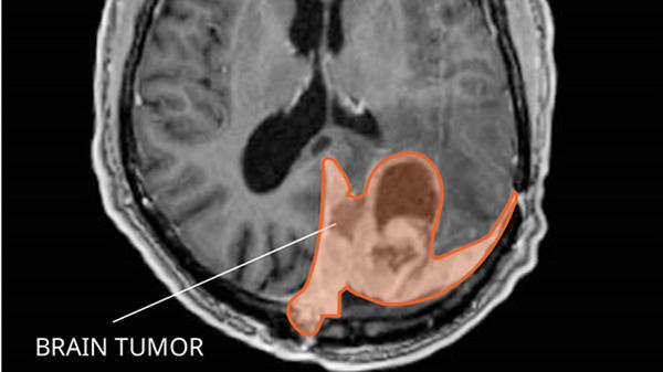
and the infratentorium (the lower back part of the brain). The supratentorium includes the cerebrum, ventricles (fluid-filled spaces), choroid plexus, hypothalamus, pineal gland, pituitary gland, and optic nerve. The infratentorium includes the cerebellum and brain stem (pons and medulla). The spinal cord is also shown.
Anatomy of the inside of the brain. The supratentorium contains the cerebrum, ventricles (with cerebrospinal fluid shown in blue), choroid plexus, hypothalamus, pineal gland, pituitary gland, and optic nerve. The infratentorium contains the cerebellum and brain stem.

Risk Factors

Few definitive observations have been made about environmental or occupational causes of primary CNS tumors.[1]

The following potential risk factors have been considered:

  • Exposure to vinyl chloride may be a risk factor for glioma.
  • Epstein-Barr virus infection has been implicated in the etiology of primary CNS lymphoma.
  • Transplant recipients and patients with AIDS have a substantially increased risk of primary CNS lymphoma.[1,5] For more information, see Primary CNS Lymphoma Treatment.

The familial tumor syndromes and related chromosomal abnormalities that are associated with CNS neoplasms include the following:[6,7]

  • Neurofibromatosis type 1 (17q11).
  • Neurofibromatosis type 2 (22q12).
  • von Hippel-Lindau disease (3p25-26).
  • Tuberous sclerosis (9q34, 16p13).
  • Li-Fraumeni syndrome (17p13).
  • Turcot syndrome type 1 (3p21, 7p22).
  • Turcot syndrome type 2 (5q21).
  • Nevoid basal cell carcinoma syndrome (9q22.3).

Clinical Features

The clinical presentation of various brain tumors is best appreciated by considering the relationship of signs and symptoms to anatomy.[1]

General signs and symptoms include the following:

  • Headaches.
  • Seizures.
  • Visual changes.
  • Gastrointestinal symptoms such as loss of appetite, nausea, and vomiting.
  • Changes in personality, mood, mental capacity, and concentration.

Seizures are a presenting symptom in approximately 20% of patients with supratentorial brain tumors and may antedate the clinical diagnosis by months to years in patients with slow-growing tumors. Among all patients with brain tumors, 70% with primary parenchymal tumors and 40% with metastatic brain tumors develop seizures at some time during the clinical course.[8]

Diagnostic Evaluation

All brain tumors, whether primary, metastatic, malignant, or benign, must be differentiated from other space-occupying lesions that can have similar clinical presentations, such as abscesses, arteriovenous malformations, and infarctions.[9]

Imaging tests

Contrast-enhanced computed tomography (CT) and magnetic resonance imaging (MRI) have complementary roles in the diagnosis of CNS neoplasms.[1,9,10]

  • The speed of CT is desirable for evaluating clinically unstable patients. CT is superior for detecting calcifications, skull lesions, and hyperacute hemorrhages (bleeding less than 24 hours old) and helps direct differential diagnosis and immediate management.
  • MRI has superior soft-tissue resolution. MRI can better detect isodense lesions, tumor enhancements, and associated findings such as edema, all phases of hemorrhagic states (except hyperacute), and infarctions. High-quality MRI is the diagnostic study of choice in the evaluation of intramedullary and extramedullary spinal cord lesions.[1]

In posttherapy imaging, single-photon emission computed tomography (SPECT) and positron emission tomography (PET) may be useful in differentiating tumor recurrence from radiation necrosis.[9]

Biopsy

Biopsy confirmation to corroborate the suspected diagnosis of a primary brain tumor is critical, whether before surgery by needle biopsy or at the time of surgical resection. The exception is cases in which the clinical and radiological evidence clearly points to a benign tumor, which could potentially be managed with active surveillance without biopsy or treatment. For other cases, radiological patterns may be misleading, and a definitive biopsy is needed to rule out other causes of space-occupying lesions, such as metastatic cancer or infection.

CT- or MRI-guided stereotactic techniques can be used to place a needle safely and accurately into almost all locations in the brain.

Prognostic Factors

Several genetic alterations have emerged as powerful prognostic factors in diffuse glioma (astrocytoma, oligodendroglioma, mixed glioma, and glioblastoma), and these alterations may guide patient management. Specific alterations include the following:

  • DNA methylation of the MGMT gene promoter.
  • IDH1 or IDH2 variants.
  • Codeletion of chromosomes 1p and 19q.

Other prognostic factors that confer poor prognosis include the following:[11,12]

  • Age older than 40 years.
  • Progressive disease.
  • Tumor size larger than 5 cm.
  • Tumor crossing the midline.
  • Contrast enhancement on MRI.
  • World Health Organization performance status (≥1).
  • Neurological symptoms.
  • Less than a gross total resection.

In an exploratory analysis of 318 patients with low-grade glioma treated with either radiation therapy alone or temozolomide chemotherapy alone, a combination of these prognostic factors demonstrated the following:[11]

  1. Longer progression-free survival (PFS) in patients with IDH variants without codeletion of 1p/19q when treated with radiation therapy (hazard ratio, 1.86; 95% confidence interval, 1.21–2.87; log-rank P = .0043).
  2. No significant treatment-dependent differences in PFS for patients with IDH variants with codeletion of 1p/19q and IDH wild-type tumors.
  3. Patients with wild-type IDH tumors had the worst prognosis independent of treatment type.
  4. Patients with IDH variants with codeletion of 1p/19q had the best prognosis.
  5. The O6-methylguanine-DNA methyltransferase (MGMT) promoter status in low-grade tumors was methylated in:
    • All IDH variants with codeletion of 1p/19q (45/45).
    • Most, but not all (86%, 62/72), of the IDH variants without codeletion of 1p/19q.
    • Fifty-six percent (5/9) of the IDH wild-type cases.

For more information, see the Treatment of Primary Central Nervous System Tumors by Tumor Type section.

References
  1. Mehta M, Vogelbaum MA, Chang S, et al.: Neoplasms of the central nervous system. In: DeVita VT Jr, Lawrence TS, Rosenberg SA: Cancer: Principles and Practice of Oncology. 9th ed. Lippincott Williams & Wilkins, 2011, pp 1700-49.
  2. American Cancer Society: Cancer Facts and Figures 2025. American Cancer Society, 2025. Available online. Last accessed January 16, 2025.
  3. National Cancer Institute: SEER Cancer Stat Facts: Brain and Other Nervous System Cancer. Bethesda, Md: National Cancer Institute. Available online. Last accessed January 24, 2025.
  4. Bray F, Laversanne M, Sung H, et al.: Global cancer statistics 2022: GLOBOCAN estimates of incidence and mortality worldwide for 36 cancers in 185 countries. CA Cancer J Clin 74 (3): 229-263, 2024. [PUBMED Abstract]
  5. Schabet M: Epidemiology of primary CNS lymphoma. J Neurooncol 43 (3): 199-201, 1999. [PUBMED Abstract]
  6. Behin A, Hoang-Xuan K, Carpentier AF, et al.: Primary brain tumours in adults. Lancet 361 (9354): 323-31, 2003. [PUBMED Abstract]
  7. Kleihues P, Cavenee WK, eds.: Pathology and Genetics of Tumours of the Nervous System. International Agency for Research on Cancer, 2000.
  8. Cloughesy T, Selch MT, Liau L: Brain. In: Haskell CM: Cancer Treatment. 5th ed. WB Saunders Co, 2001, pp 1106-42.
  9. Hutter A, Schwetye KE, Bierhals AJ, et al.: Brain neoplasms: epidemiology, diagnosis, and prospects for cost-effective imaging. Neuroimaging Clin N Am 13 (2): 237-50, x-xi, 2003. [PUBMED Abstract]
  10. Ricci PE: Imaging of adult brain tumors. Neuroimaging Clin N Am 9 (4): 651-69, 1999. [PUBMED Abstract]
  11. Baumert BG, Hegi ME, van den Bent MJ, et al.: Temozolomide chemotherapy versus radiotherapy in high-risk low-grade glioma (EORTC 22033-26033): a randomised, open-label, phase 3 intergroup study. Lancet Oncol 17 (11): 1521-1532, 2016. [PUBMED Abstract]
  12. Reijneveld JC, Taphoorn MJ, Coens C, et al.: Health-related quality of life in patients with high-risk low-grade glioma (EORTC 22033-26033): a randomised, open-label, phase 3 intergroup study. Lancet Oncol 17 (11): 1533-1542, 2016. [PUBMED Abstract]

World Health Organization (WHO) Classification of Adult Primary CNS Tumors

This classification is based on the World Health Organization (WHO) classification of central nervous system (CNS) tumors.[1] The WHO approach incorporates and interrelates morphology, cytogenetics, molecular genetics, and immunological markers in an attempt to construct a cellular classification that is universally applicable and prognostically valid. Earlier attempts to develop a TNM (tumor, node, metastasis)-based classification were dropped for the following reasons:[2]

  • Tumor size (T) is less relevant than are tumor histology and location.
  • Nodal status (N) does not apply because the brain and spinal cord have no lymphatics.
  • Metastatic spread (M) rarely applies because most patients with CNS neoplasms do not live long enough to develop metastatic disease.

The WHO grading of CNS tumors establishes a malignancy scale based on histological features of the tumor.[3] The histological grades are as follows:

  • WHO grade I includes lesions with low proliferative potential, a frequently discrete nature, and the possibility of cure following surgical resection alone.
  • WHO grade II includes lesions that are generally infiltrating and low in mitotic activity but recur more frequently than do grade I malignant tumors after local therapy. Some tumor types tend to progress to higher grades of malignancy.
  • WHO grade III includes lesions with histological evidence of malignancy, including nuclear atypia and increased mitotic activity. These lesions have anaplastic histology and infiltrative capacity. They are usually treated with aggressive adjuvant therapy.
  • WHO grade IV includes lesions that are mitotically active, necrosis prone, and generally associated with a rapid preoperative and postoperative progression and fatal outcomes. The lesions are usually treated with aggressive adjuvant therapy.

Table 1 lists the tumor types and grades.[4] Tumors limited to the peripheral nervous system are not included. Histopathology, grading methods, incidence, and what is known about etiology specific to each tumor type have been described in detail elsewhere.[4,5]

Table 1. WHO Grades of CNS Tumorsa
  I II III IV
aReprinted with permission from Louis, DN, Ohgaki H, Wiestler, OD, Cavenee, WK. World Health Organization Classification of Tumours of the Central Nervous System. IARC, Lyon, 2007.
Astrocytic tumors
Subependymal giant cell astrocytoma X      
Pilocytic astrocytoma X      
Pilomyxoid astrocytoma   X    
Diffuse astrocytoma   X    
Pleomorphic xanthoastrocytoma   X    
Anaplastic astrocytoma     X  
Glioblastoma       X
Giant cell glioblastoma       X
Gliosarcoma       X
Oligodendroglial tumors
Oligodendroglioma   X    
Anaplastic oligodendroglioma     X  
Oligoastrocytic tumors
Oligoastrocytoma   X    
Anaplastic oligoastrocytoma     X  
Ependymal tumors
Subependymoma X      
Myxopapillary ependymoma X      
Ependymoma   X    
Anaplastic ependymoma     X  
Choroid plexus tumors
Choroid plexus papilloma X      
Atypical choroid plexus papilloma   X    
Choroid plexus carcinoma     X  
Other neuroepithelial tumors
Angiocentric glioma X      
Chordoid glioma of the third ventricle   X    
Neuronal and mixed neuronal-glial tumors
Gangliocytoma X      
Ganglioglioma X      
Anaplastic ganglioma     X  
Desmoplastic infantile astrocytoma and ganglioglioma X      
Dysembryoplastic neuroepithelial tumor X      
Central neurocytoma   X    
Extraventricular neurocytoma   X    
Cerebellar liponeurocytoma   X    
Paraganglioma of the spinal cord X      
Papillary glioneuronal tumor X      
Rosette-forming glioneural tumor of the fourth ventricle X      
Pineal tumors
Pineocytoma X      
Pineal parenchymal tumor of intermediate differentiation   X X  
Pineoblastoma       X
Papillary tumor of the pineal region   X X  
Embryonal tumors
Medulloblastoma       X
CNS primitive neuroectodermal tumor       X
Atypical teratoid/rhabdoid tumor       X
Tumors of the cranial and paraspinal nerves
Schwannoma X      
Neurofibroma X      
Perineurioma X X X  
Malignant peripheral nerve sheath tumor   X X X
Meningeal tumors
Meningioma X      
Atypical meningioma   X    
Anaplastic/malignant meningioma     X  
Hemangiopericytoma   X    
Anaplastic hemangiopericytoma     X  
Hemangioblastoma X      
Tumors of the sellar region
Craniopharyngioma X      
Granular cell tumor of the neurohypophysis X      
Pituicytoma X      
Spindle cell oncocytoma of the adenohypophysis X      

Genomic Alterations

Alterations in the BRAF, IDH1, and IDH2 genes, and genomic 1p/19q codeletion, appear to be hallmark aberrations in particular glioma subtypes. Assessment for the presence of these variants aids diagnosis and prognosis and, with regard to 1p/19q codeletion, predicts for response to chemotherapy.

In pilocytic astrocytomas (WHO grade I), tandem duplication at 7q34 leading to a KIAA1549::BRAF gene fusion is found in approximately 70% of pilocytic astrocytomas.[68] Activating single nucleotide variants in BRAF (V600E) are found in an additional 5% to 9% of these tumors. Overall, RAF alterations occur in approximately 80% of pilocytic astrocytomas.

BRAF V600E variants are observed (in about 60%) of other benign gliomas, including pleomorphic xanthoastrocytoma and ganglioglioma, while BRAF tandem duplications are not found in these variant glioma tumors.[911]

Most WHO grade II and III diffuse gliomas (astrocytomas, oligodendrogliomas, and oligoastrocytomas) and 5% to 10% of glioblastomas (WHO grade IV) harbor single nucleotide variants in the R132 position of IDH1 or, rarely, the analogous codon in IDH2 (R172).[1216] The presence of an IDH1 or IDH2 variant is a strong prognostic factor. Patients with these tumor variants have significantly longer survival independent of WHO grade or histological subtype.

Deletion of chromosomes 1p and 19q occurs through a translocation event [17] and is common in oligodendrogliomas. 1p/19q codeletion is a powerful prognostic factor and may predict for response to chemotherapy. For more information, see the Anaplastic oligodendrogliomas treatment section.

These genetic alterations have potential diagnostic utility. Presence of the IDH1 and IDH2 variants may distinguish diffuse gliomas from other gliomas, which often have BRAF genetic alterations, and nonneoplastic reactive astrocytosis.[18] Most (90%) IDH variants in gliomas result in an R132H substitution, which can be detected with a highly sensitive and specific monoclonal antibody. A rapid immunohistochemical analysis using the variant-specific IDH1 antibody can aid diagnostic analysis.[19]

Other CNS tumors are associated with characteristic patterns of altered oncogenes, altered tumor suppressor genes, and chromosomal abnormalities. Familial tumor syndromes with defined chromosomal abnormalities are associated with gliomas.

References
  1. Kleihues P, Cavenee WK, eds.: Pathology and Genetics of Tumours of the Nervous System. International Agency for Research on Cancer, 2000.
  2. Brain and Spinal Cord. In: Amin MB, Edge SB, Greene FL, et al., eds.: AJCC Cancer Staging Manual. 8th ed. Springer; 2017, pp. 857–69.
  3. Kleihues P, Burger PC, Scheithauer BW: The new WHO classification of brain tumours. Brain Pathol 3 (3): 255-68, 1993. [PUBMED Abstract]
  4. Louis DN, Ohgaki H, Wiestler OD, et al.: The 2007 WHO classification of tumours of the central nervous system. Acta Neuropathol 114 (2): 97-109, 2007. [PUBMED Abstract]
  5. Louis DN, Ohgaki H, Wiestler OD, et al., eds.: WHO Classification of Tumours of the Central Nervous System. 4th ed. IARC Press, 2007.
  6. Sievert AJ, Jackson EM, Gai X, et al.: Duplication of 7q34 in pediatric low-grade astrocytomas detected by high-density single-nucleotide polymorphism-based genotype arrays results in a novel BRAF fusion gene. Brain Pathol 19 (3): 449-58, 2009. [PUBMED Abstract]
  7. Pfister S, Janzarik WG, Remke M, et al.: BRAF gene duplication constitutes a mechanism of MAPK pathway activation in low-grade astrocytomas. J Clin Invest 118 (5): 1739-49, 2008. [PUBMED Abstract]
  8. Jones DT, Kocialkowski S, Liu L, et al.: Tandem duplication producing a novel oncogenic BRAF fusion gene defines the majority of pilocytic astrocytomas. Cancer Res 68 (21): 8673-7, 2008. [PUBMED Abstract]
  9. Dias-Santagata D, Lam Q, Vernovsky K, et al.: BRAF V600E mutations are common in pleomorphic xanthoastrocytoma: diagnostic and therapeutic implications. PLoS One 6 (3): e17948, 2011. [PUBMED Abstract]
  10. MacConaill LE, Campbell CD, Kehoe SM, et al.: Profiling critical cancer gene mutations in clinical tumor samples. PLoS One 4 (11): e7887, 2009. [PUBMED Abstract]
  11. Parsons DW, Jones S, Zhang X, et al.: An integrated genomic analysis of human glioblastoma multiforme. Science 321 (5897): 1807-12, 2008. [PUBMED Abstract]
  12. Yan H, Parsons DW, Jin G, et al.: IDH1 and IDH2 mutations in gliomas. N Engl J Med 360 (8): 765-73, 2009. [PUBMED Abstract]
  13. Dubbink HJ, Taal W, van Marion R, et al.: IDH1 mutations in low-grade astrocytomas predict survival but not response to temozolomide. Neurology 73 (21): 1792-5, 2009. [PUBMED Abstract]
  14. Sanson M, Marie Y, Paris S, et al.: Isocitrate dehydrogenase 1 codon 132 mutation is an important prognostic biomarker in gliomas. J Clin Oncol 27 (25): 4150-4, 2009. [PUBMED Abstract]
  15. Hartmann C, Hentschel B, Wick W, et al.: Patients with IDH1 wild type anaplastic astrocytomas exhibit worse prognosis than IDH1-mutated glioblastomas, and IDH1 mutation status accounts for the unfavorable prognostic effect of higher age: implications for classification of gliomas. Acta Neuropathol 120 (6): 707-18, 2010. [PUBMED Abstract]
  16. Hartmann C, Meyer J, Balss J, et al.: Type and frequency of IDH1 and IDH2 mutations are related to astrocytic and oligodendroglial differentiation and age: a study of 1,010 diffuse gliomas. Acta Neuropathol 118 (4): 469-74, 2009. [PUBMED Abstract]
  17. Jenkins RB, Blair H, Ballman KV, et al.: A t(1;19)(q10;p10) mediates the combined deletions of 1p and 19q and predicts a better prognosis of patients with oligodendroglioma. Cancer Res 66 (20): 9852-61, 2006. [PUBMED Abstract]
  18. Camelo-Piragua S, Jansen M, Ganguly A, et al.: A sensitive and specific diagnostic panel to distinguish diffuse astrocytoma from astrocytosis: chromosome 7 gain with mutant isocitrate dehydrogenase 1 and p53. J Neuropathol Exp Neurol 70 (2): 110-5, 2011. [PUBMED Abstract]
  19. Capper D, Weissert S, Balss J, et al.: Characterization of R132H mutation-specific IDH1 antibody binding in brain tumors. Brain Pathol 20 (1): 245-54, 2010. [PUBMED Abstract]

Treatment Option Overview for Adult Primary CNS Tumors

Primary CNS Tumors

This section discusses general treatment modalities for primary central nervous system (CNS) tumors. For a description of specific treatment options for each tumor type, see the Treatment of Primary Central Nervous System Tumors by Tumor Type section.

Radiation therapy and chemotherapy options vary according to histology and anatomical site of the CNS tumor. For glioblastoma, combined modality therapy with resection, radiation, and chemotherapy is standard. Anaplastic astrocytomas, anaplastic oligodendrogliomas, and anaplastic oligoastrocytomas represent only a small proportion of CNS gliomas; therefore, phase III randomized trials restricted to these tumor types are not generally practical. The natural histories of these tumors are variable, depending on histological and molecular factors; therefore, treatment guidelines are evolving. Therapy involving surgically implanted carmustine-impregnated polymer wafers combined with postoperative external-beam radiation therapy (EBRT) may play a role in the treatment of high-grade (grades III and IV) gliomas in some patients.[1]

Treatment options for primary CNS tumors include the following:

Surgery

For most types of CNS tumors in most locations, complete or near-complete surgical removal is generally attempted, within the constraints of preserving neurological function and the patient’s underlying health. This practice is based on observational evidence that survival is better in patients who undergo tumor resection than in those who have closed biopsy alone.[2,3] The benefit of resection has not been tested in randomized trials. Selection bias can enter into observational studies despite attempts to adjust for patient differences that guide the decision to resect the tumor; therefore, the actual difference in outcome between radical surgery and biopsy alone may not be as large as noted in the retrospective studies.[3]

An exception to the use of resection is the case of deep-seated tumors such as pontine gliomas, which are diagnosed on clinical evidence and treated without initial surgery approximately 50% of the time. In most cases, however, diagnosis by biopsy is preferred. Stereotactic biopsy can be used for lesions that are difficult to reach and resect.

The primary goals of surgical resection include the following:[4]

  • To establish a histological diagnosis.
  • To reduce intracranial pressure by removing as much tumor as is safely possible to preserve neurological function.

Total elimination of primary malignant intraparenchymal tumors by surgery alone is rarely achievable. Therefore, intraoperative techniques have been developed to reach a balance between removing as much tumor as is practical and preserving functional status. For example, craniotomies with stereotactic resections of primary gliomas can be performed in cooperative patients while they are awake, with real-time assessment of neurological function.[5] Examples of intraoperative neurological assessment include the following:

  • Resection proceeds until either the magnetic resonance imaging (MRI) signal abnormality being used to monitor the extent of surgery is completely removed or subtle neurological dysfunction appears (e.g., a slight decrease in rapid alternating motor movement or anomia).
  • When the tumor is located in or near language centers in the cortex, intraoperative language mapping can be performed by electrode discharge-induced speech arrest while the patient is asked to count or read.[6]

As is the case with several other specialized operations [7,8] in which postoperative mortality has been associated with the number of procedures performed, postoperative mortality after surgery for primary brain tumors may be associated with hospital and/or surgeon volume.[9] Using the Nationwide Inpatient Sample hospital discharge database for the years 1988 to 2000, which represented 20% of inpatient admissions to nonfederal U.S. hospitals, investigators observed the following:[9]

  • Large-volume hospitals had lower in-hospital mortality rates after craniotomies for primary brain tumors (odds ratio [OR], 0.75 for a tenfold higher caseload; 95% confidence interval [CI], 0.62–0.90) and after needle biopsies (OR, 0.54; 95% CI, 0.35–0.83).
  • Although there was no specific sharp threshold in all-cause mortality outcomes between low-volume hospitals and high-volume hospitals, craniotomy-associated in-hospital mortality was 4.5% for hospitals with 5 or fewer procedures per year and 1.5% for hospitals with at least 42 procedures per year.
  • In-hospital mortality rates decreased over the study years (perhaps because the proportion of elective nonemergent operations increased from 45% to 57%), but the decrease was more rapid in high-volume hospitals than in low-volume hospitals.
  • High-volume surgeons had lower in-hospital patient mortality rates after craniotomy (OR, 0.60; 95% CI, 0.45–0.79).

As with any study of volume-outcome associations, these results may not be causal because of residual confounding factors such as referral patterns, private insurance, and patient selection, despite multivariable adjustment.

Radiation therapy

High-grade tumors

Radiation therapy has a major role in the treatment of patients with high-grade gliomas.

Evidence (postoperative radiation therapy [PORT]):

  1. A systematic review and meta-analysis of five randomized trials (plus one trial with allocation by birth date) comparing PORT with no radiation therapy showed a statistically significant survival advantage with radiation (risk ratio, 0.81; 95% CI, 0.74–0.88).[10][Level of evidence A1]
  2. A randomized trial comparing 60 Gy (in 30 fractions over 6 weeks) with 45 Gy (in 25 fractions over 4 weeks) showed superior survival in the first group (12 months vs. 9 months median survival; hazard ratio [HR], 0.81; 95% CI, 0.66–0.99). The accepted standard dose of EBRT for malignant gliomas is 60 Gy.[11][Level of evidence A1]

EBRT using either 3-dimensional conformal radiation therapy (3D-CRT) or intensity-modulated radiation therapy (IMRT) is considered an acceptable technique in radiation therapy delivery. Typically used are 2- to 3-cm margins on the MRI-based volumes (T1-weighted and fluid-attenuated inversion recovery [FLAIR]) to create the planning target volume.

Dose escalation using radiosurgery has not improved outcomes. A randomized trial tested radiosurgery as a boost added to standard EBRT, but the trial found no improvement in survival, quality of life, or patterns of relapse compared with EBRT without the boost.[12,13]

Brachytherapy has been used to deliver high doses of radiation locally to the tumor while sparing normal brain tissue. However, this approach is technically demanding and is less common since the advent of 3D-CRT and IMRT.

Low-grade tumors

Treatment options for patients with low-grade gliomas (i.e., low-grade astrocytomas, oligodendrogliomas, and mixed oligoastrocytomas) are not as clear as in the case of high-grade tumors and include observation, PORT, and chemotherapy with temozolomide.

Evidence (PORT vs. observation):

  1. The European Organisation for Research and Treatment of Cancer (EORTC) randomly assigned 311 patients with low-grade gliomas to undergo either radiation or observation in the EORTC-22845 trial.[14,15] On review of central pathology, about 25% of patients in the trial were reported to have high-grade tumors. Most of the control patients received radiation therapy at the time of progression.
    • After a median follow-up of 93 months, the median progression-free survival (PFS) was 5.3 years in the radiation arm versus 3.4 years in the control arm (HR, 0.59; 95% CI, 0.45–0.77).[14,15][Level of evidence B1]
    • There was no difference in the overall survival (OS). The median survival was 7.4 years in the radiation arm and 7.2 years in the control arm (HR, 0.97; 95% CI, 0.71–1.34; P = .87).[14,15][Level of evidence A1] This was caused by a longer survival after progression in the control arm (3.4 years) than in the radiation arm (1.0 year) (P < .0001).
    • The investigators did not collect reliable quality-of-life measurements, so it is not clear whether the delay in initial relapse in the radiation therapy arm translated into improved function or quality of life.

Evidence (PORT versus temozolomide for patients with low-grade World Health Organization [WHO] grade II tumors with at least one high-risk feature):

  1. The EORTC 22033-26033 trial (NCT00182819) included 707 patients with low-grade glioma (WHO grade II astrocytoma, oligoastrocytoma, or oligodendroglioma) and at least one high-risk feature (age >40 years, progressive disease, tumor size >5 cm, tumor crossing the midline, or neurological symptoms). Patients were randomly assigned to receive either radiation therapy (n = 240) or temozolomide chemotherapy (n = 237). Radiation therapy consisted of conformal treatment (up to 50.4 Gy; 28 doses of 1.8 Gy daily, 5 days a week, for up to 6.5 weeks). Chemotherapy was dose-dense oral temozolomide (75 mg/m2 daily for 21 days, repeated every 28 days [one cycle], for a maximum of 12 cycles).[16,17]
    1. There was no significant difference in PFS (primary end point) or health-related quality of life (secondary end point).
    2. At a median follow-up of 48 months (interquartile range, 31–56), median PFS was 39 months (95% CI, 35–44) in the temozolomide group and 46 months (95% CI, 40–56) in the radiation therapy group (unadjusted HR, 1.16; 95% CI, 0.9–1.5; P = .22).[16][Level of evidence B1]
    3. An exploratory analysis of 318 molecularly defined patients found that patients with IDH gene variants without codeletion of 1p/19q displayed a significantly longer PFS when treated with radiation therapy (HR, 1.86; 95% CI, 1.21–2.87; log-rank P = .0043).
    4. There were no significant treatment-dependent differences in PFS for patients with IDH variants with codeletion of 1p/19q and IDH wild-type tumors.
    5. Patients with wild-type IDH tumors had the worst prognosis independent of treatment type.
    6. Patients with IDH variants with codeletion of 1p/19q had the best prognosis.
    7. The O6-methylguanine-DNA methyltransferase (MGMT) promoter status was methylated in the following:
      • All IDH variants with codeletion of 1p/19q (45/45).
      • Sixty-two of 72 (86%) of the IDH variants without codeletion of 1p/19q.
      • Five of nine (56%) of the IDH wild-type cases.
Disease progression, subsequent neoplasms, or recurrences

There are no randomized trials to delineate the role of repeat radiation after disease progression or the development of radiation-induced cancers. The literature is limited to small retrospective case series, which makes interpretation difficult.[18] The decision to repeat radiation must be made carefully because of the risk of neurocognitive deficits and radiation-induced necrosis. One advantage of radiosurgery is the ability to deliver therapeutic doses to recurrent tumors that may require the re-irradiation of previously irradiated brain tissue beyond tolerable dose limits.

Chemotherapy

Systemic chemotherapy

For many years, the nitrosourea carmustine ([bis-chloroethylnitrosourea] BCNU) was the standard chemotherapy agent added to surgery and radiation therapy for malignant gliomas, based on the Radiation Therapy Oncology Group’s (RTOG’s) randomized trial (RTOG-8302).[19][Level of evidence A1] A modest impact on survival with the use of nitrosourea-containing chemotherapy regimens for malignant gliomas was confirmed in a patient-level meta-analysis of 12 randomized trials (combined HRdeath, 0.85; 95% CI, 0.78–0.91).[20]

A large multicenter trial (NCT00006353) of patients with glioblastoma, conducted by the EORTC-National Cancer Institute of Canada, reported a survival advantage with the use of temozolomide in addition to radiation therapy.[21,22][Level of evidence A1] On the basis of these results, the oral agent temozolomide has replaced BCNU as the standard systemic chemotherapy for malignant gliomas. For more information, see the Glioblastomas treatment section.

Long-term results of randomized trials in high-risk, low-grade (WHO grade II) gliomas [23][Level of evidence A1] and anaplastic (WHO grade III) oligodendroglial tumors [24,25][Level of evidence A1] have demonstrated that the addition of procarbazine, lomustine, and vincristine (PCV) chemotherapy to radiation therapy after surgery extends survival. Radiation and PCV chemotherapy should be considered for patients deemed appropriate for therapy. For more information, see the Treatment of Primary Central Nervous System Tumors by Tumor Type section.

Localized chemotherapy (carmustine wafer)

The ability to give high doses of chemotherapy while avoiding systemic toxicity is desirable because malignant glioma–related deaths are usually due to uncontrolled intracranial disease rather than distant metastases. A biodegradable carmustine wafer has been developed for that purpose. The wafers contain 3.85% carmustine, and up to eight wafers are implanted into the tumor bed lining at the time of open resection, with an intended total dose of about 7.7 mg per wafer (61.6 mg maximum per patient) over a period of 2 to 3 weeks.

Two randomized placebo-controlled trials of this focal drug-delivery method have shown an OS advantage associated with the carmustine wafers versus radiation therapy alone. In both trials, the upper age limit for patients was 65 years.

Evidence (carmustine wafer):

  1. A small trial was closed because of a lack of continued availability of the carmustine wafers after 32 patients with high-grade gliomas had been entered.[26]
    • Although OS was better in the carmustine-wafer group (median 58.1 vs. 39.9 weeks; P = .012), there was an imbalance in the study arms (only 11 of 16 patients in the carmustine-wafer group vs. 16 of the 16 patients in the placebo-wafer group had grade IV glioblastoma tumors).
  2. A multicenter study of 240 patients with primary malignant gliomas, 207 of whom had glioblastoma, was more informative.[27,28] At initial surgery, patients received either carmustine wafers or placebo wafers, followed by radiation therapy (55–60 Gy). Systemic therapy was not allowed until recurrence, except in the case of anaplastic oligodendrogliomas (n = 9). Unlike the initial trial, patient characteristics were well balanced between the study arms.
    • Median survival in the two groups was 13.8 months in patients treated with carmustine wafers versus 11.6 months in placebo-treated patients (HR, 0.73; 95% CI, 0.56–0.96; P = .017).
  3. A systematic review combining both studies [2628] estimated an HR for overall mortality of 0.65; 95% CI, 0.48–0.86; P = .003.[29][Level of evidence A1]

Active surveillance

Active surveillance is appropriate in some circumstances. With the increasing use of sensitive neuroimaging tools, detection of asymptomatic low-grade meningiomas has increased; most appear to show minimal growth and can often be safely observed, with therapy deferred until the detection of tumor growth or the development of symptoms.[30,31]

Supportive therapy

Dexamethasone, mannitol, and furosemide are used to treat the peritumoral edema associated with brain tumors. The use of anticonvulsants is mandatory for patients with seizures.[4]

References
  1. Lallana EC, Abrey LE: Update on the therapeutic approaches to brain tumors. Expert Rev Anticancer Ther 3 (5): 655-70, 2003. [PUBMED Abstract]
  2. Laws ER, Parney IF, Huang W, et al.: Survival following surgery and prognostic factors for recently diagnosed malignant glioma: data from the Glioma Outcomes Project. J Neurosurg 99 (3): 467-73, 2003. [PUBMED Abstract]
  3. Chang SM, Parney IF, Huang W, et al.: Patterns of care for adults with newly diagnosed malignant glioma. JAMA 293 (5): 557-64, 2005. [PUBMED Abstract]
  4. Cloughesy T, Selch MT, Liau L: Brain. In: Haskell CM: Cancer Treatment. 5th ed. WB Saunders Co, 2001, pp 1106-42.
  5. Meyer FB, Bates LM, Goerss SJ, et al.: Awake craniotomy for aggressive resection of primary gliomas located in eloquent brain. Mayo Clin Proc 76 (7): 677-87, 2001. [PUBMED Abstract]
  6. Sanai N, Mirzadeh Z, Berger MS: Functional outcome after language mapping for glioma resection. N Engl J Med 358 (1): 18-27, 2008. [PUBMED Abstract]
  7. Begg CB, Cramer LD, Hoskins WJ, et al.: Impact of hospital volume on operative mortality for major cancer surgery. JAMA 280 (20): 1747-51, 1998. [PUBMED Abstract]
  8. Birkmeyer JD, Finlayson EV, Birkmeyer CM: Volume standards for high-risk surgical procedures: potential benefits of the Leapfrog initiative. Surgery 130 (3): 415-22, 2001. [PUBMED Abstract]
  9. Barker FG, Curry WT, Carter BS: Surgery for primary supratentorial brain tumors in the United States, 1988 to 2000: the effect of provider caseload and centralization of care. Neuro Oncol 7 (1): 49-63, 2005. [PUBMED Abstract]
  10. Laperriere N, Zuraw L, Cairncross G, et al.: Radiotherapy for newly diagnosed malignant glioma in adults: a systematic review. Radiother Oncol 64 (3): 259-73, 2002. [PUBMED Abstract]
  11. Bleehen NM, Stenning SP: A Medical Research Council trial of two radiotherapy doses in the treatment of grades 3 and 4 astrocytoma. The Medical Research Council Brain Tumour Working Party. Br J Cancer 64 (4): 769-74, 1991. [PUBMED Abstract]
  12. Tsao MN, Mehta MP, Whelan TJ, et al.: The American Society for Therapeutic Radiology and Oncology (ASTRO) evidence-based review of the role of radiosurgery for malignant glioma. Int J Radiat Oncol Biol Phys 63 (1): 47-55, 2005. [PUBMED Abstract]
  13. Souhami L, Seiferheld W, Brachman D, et al.: Randomized comparison of stereotactic radiosurgery followed by conventional radiotherapy with carmustine to conventional radiotherapy with carmustine for patients with glioblastoma multiforme: report of Radiation Therapy Oncology Group 93-05 protocol. Int J Radiat Oncol Biol Phys 60 (3): 853-60, 2004. [PUBMED Abstract]
  14. Karim AB, Afra D, Cornu P, et al.: Randomized trial on the efficacy of radiotherapy for cerebral low-grade glioma in the adult: European Organization for Research and Treatment of Cancer Study 22845 with the Medical Research Council study BRO4: an interim analysis. Int J Radiat Oncol Biol Phys 52 (2): 316-24, 2002. [PUBMED Abstract]
  15. van den Bent MJ, Afra D, de Witte O, et al.: Long-term efficacy of early versus delayed radiotherapy for low-grade astrocytoma and oligodendroglioma in adults: the EORTC 22845 randomised trial. Lancet 366 (9490): 985-90, 2005. [PUBMED Abstract]
  16. Baumert BG, Hegi ME, van den Bent MJ, et al.: Temozolomide chemotherapy versus radiotherapy in high-risk low-grade glioma (EORTC 22033-26033): a randomised, open-label, phase 3 intergroup study. Lancet Oncol 17 (11): 1521-1532, 2016. [PUBMED Abstract]
  17. Reijneveld JC, Taphoorn MJ, Coens C, et al.: Health-related quality of life in patients with high-risk low-grade glioma (EORTC 22033-26033): a randomised, open-label, phase 3 intergroup study. Lancet Oncol 17 (11): 1533-1542, 2016. [PUBMED Abstract]
  18. Paulino AC, Mai WY, Chintagumpala M, et al.: Radiation-induced malignant gliomas: is there a role for reirradiation? Int J Radiat Oncol Biol Phys 71 (5): 1381-7, 2008. [PUBMED Abstract]
  19. Walker MD, Green SB, Byar DP, et al.: Randomized comparisons of radiotherapy and nitrosoureas for the treatment of malignant glioma after surgery. N Engl J Med 303 (23): 1323-9, 1980. [PUBMED Abstract]
  20. Stewart LA: Chemotherapy in adult high-grade glioma: a systematic review and meta-analysis of individual patient data from 12 randomised trials. Lancet 359 (9311): 1011-8, 2002. [PUBMED Abstract]
  21. Stupp R, Mason WP, van den Bent MJ, et al.: Radiotherapy plus concomitant and adjuvant temozolomide for glioblastoma. N Engl J Med 352 (10): 987-96, 2005. [PUBMED Abstract]
  22. Stupp R, Hegi ME, Mason WP, et al.: Effects of radiotherapy with concomitant and adjuvant temozolomide versus radiotherapy alone on survival in glioblastoma in a randomised phase III study: 5-year analysis of the EORTC-NCIC trial. Lancet Oncol 10 (5): 459-66, 2009. [PUBMED Abstract]
  23. Buckner JC, Pugh SL, Shaw EG, et al.: Phase III study of radiation therapy with or without procarbazine, CCNU, and vincristine (PCV) in low-grade glioma: RTOG 9802 with Alliance, ECOG, and SWOG. [Abstract] J Clin Oncol 32 (Suppl 5): A-2000, 2014.
  24. van den Bent MJ, Brandes AA, Taphoorn MJ, et al.: Adjuvant procarbazine, lomustine, and vincristine chemotherapy in newly diagnosed anaplastic oligodendroglioma: long-term follow-up of EORTC brain tumor group study 26951. J Clin Oncol 31 (3): 344-50, 2013. [PUBMED Abstract]
  25. Cairncross G, Wang M, Shaw E, et al.: Phase III trial of chemoradiotherapy for anaplastic oligodendroglioma: long-term results of RTOG 9402. J Clin Oncol 31 (3): 337-43, 2013. [PUBMED Abstract]
  26. Valtonen S, Timonen U, Toivanen P, et al.: Interstitial chemotherapy with carmustine-loaded polymers for high-grade gliomas: a randomized double-blind study. Neurosurgery 41 (1): 44-8; discussion 48-9, 1997. [PUBMED Abstract]
  27. Westphal M, Hilt DC, Bortey E, et al.: A phase 3 trial of local chemotherapy with biodegradable carmustine (BCNU) wafers (Gliadel wafers) in patients with primary malignant glioma. Neuro-oncol 5 (2): 79-88, 2003. [PUBMED Abstract]
  28. Westphal M, Ram Z, Riddle V, et al.: Gliadel wafer in initial surgery for malignant glioma: long-term follow-up of a multicenter controlled trial. Acta Neurochir (Wien) 148 (3): 269-75; discussion 275, 2006. [PUBMED Abstract]
  29. Hart MG, Grant R, Garside R, et al.: Chemotherapeutic wafers for high grade glioma. Cochrane Database Syst Rev (3): CD007294, 2008. [PUBMED Abstract]
  30. Nakamura M, Roser F, Michel J, et al.: The natural history of incidental meningiomas. Neurosurgery 53 (1): 62-70; discussion 70-1, 2003. [PUBMED Abstract]
  31. Yano S, Kuratsu J; Kumamoto Brain Tumor Research Group: Indications for surgery in patients with asymptomatic meningiomas based on an extensive experience. J Neurosurg 105 (4): 538-43, 2006. [PUBMED Abstract]

Treatment of Primary CNS Tumors by Tumor Type

Table 2. Treatment of Primary Central Nervous System Tumors by Tumor Type
Tumor Type Treatment Options
Astrocytic tumors
—Brain stem gliomas Radiation therapy
—Pineal astrocytic tumors Surgery plus radiation therapy
Surgery plus radiation therapy and chemotherapy for higher-grade tumors
—Pilocytic astrocytomas Surgery alone
Surgery followed by radiation therapy
—Diffuse astrocytomas (WHO grade II) Surgery with or without radiation therapy
Surgery followed by radiation therapy and chemotherapy
—Anaplastic astrocytomas (WHO grade III) Surgery plus radiation therapy with or without chemotherapy
Surgery plus chemotherapy
—Glioblastomas Surgery plus radiation therapy and chemotherapy
Surgery plus radiation therapy
Carmustine-impregnated polymer implant
Radiation therapy and concurrent chemotherapy
Oligodendroglial tumors
—Oligodendrogliomas Surgery with or without radiation therapy
Surgery with radiation therapy and chemotherapy
—Anaplastic oligodendrogliomas Surgery plus radiation therapy with or without chemotherapy
Mixed gliomas Surgery plus radiation therapy with or without chemotherapy
Ependymal tumors
—Grades I and II ependymal tumors Surgery alone
Surgery followed by radiation therapy
—Anaplastic ependymoma Surgery plus radiation therapy
Embryonal cell tumors
—Medulloblastomas Surgery plus craniospinal radiation therapy
Pineal parenchymal tumors Surgery plus radiation therapy (for pineocytoma)
Surgery plus radiation therapy and chemotherapy (for pineoblastoma)
Meningeal tumors
—Grade I meningiomas Active surveillance with deferred treatment
Surgery
Stereotactic radiosurgery
Surgery plus radiation therapy
Fractionated radiation therapy
—Grades II and III meningiomas and hemangiopericytomas Surgery plus radiation therapy
Germ cell tumors Depends on multiple factors
Tumors of the sellar region
—Craniopharyngiomas Surgery alone
Debulking surgery plus radiation therapy

Astrocytic Tumors Treatment

Brain stem gliomas treatment

Patients with brain stem gliomas have relatively poor prognoses that correlate with histology (when biopsies are performed), location, and extent of tumor. The overall median survival time of patients in studies has been 44 to 74 weeks.

Treatment options for brain stem gliomas include the following:

  1. Radiation therapy.

Pineal astrocytic tumors treatment

Depending on the degree of anaplasia, patients with pineal astrocytomas have variable prognoses. Patients with higher-grade tumors have worse prognoses.

Treatment options for pineal astrocytic tumors include the following:

  1. Surgery plus radiation therapy for pineal astrocytoma.
  2. Surgery plus radiation therapy and chemotherapy for higher-grade tumors.

Pilocytic astrocytomas treatment

This astrocytic tumor is classified as a World Health Organization (WHO) grade I tumor and is often curable.

Treatment options for pilocytic astrocytomas include the following:

  1. Surgery alone if the tumor is totally resectable.
  2. Surgery followed by radiation therapy to known or suspected residual tumor.

Diffuse astrocytomas treatment

This WHO grade II astrocytic tumor is less often curable than is a pilocytic astrocytoma.

Treatment options for diffuse astrocytomas (WHO grade II) include the following:

  1. Surgery with or without radiation therapy.
  2. Surgery followed by radiation therapy and chemotherapy.

Controversy exists about the timing of radiation therapy after surgery. For more information, see the Low-grade tumors section.

  • Radiation therapy improved progression-free survival (PFS) in patients who received early radiation therapy in the European Organisation for Research and Treatment of Cancer (EORTC) EORTC-22845 trial. For more information, see the Oligodendrogliomas treatment section.[1][Level of evidence A1]
  • In the same trial, there was no difference in overall survival (OS) between patients who had radiation therapy after surgery and those who were treated with radiation therapy at the time of progression.[1][Level of evidence A1]

Some physicians use surgery alone if a patient has clinical factors that are considered low risk, such as age younger than 40 years and the lack of contrast enhancement on a computed tomography scan.[2]

Evidence (surgery followed by radiation therapy and chemotherapy):

  1. For patients with low-grade (WHO grade II) tumors, which are considered high risk, radiation therapy followed by six cycles of vincristine (PCV) chemotherapy is a recommended option. This recommendation is based on the long-term follow-up results of the Radiation Therapy Oncology Group’s (RTOG’s) 1986-initiated randomized trial (RTOG 9802 [NCT00003375]).[3][Level of evidence A1] In this trial, patients with high-risk, low-grade glioma, defined as patients aged 18 to 39 years with biopsy or subtotal resection, or patients aged 40 years or older, were randomly assigned to either 54 Gy of radiation therapy or radiation therapy followed by six cycles of PCV chemotherapy.
    1. The addition of PCV to radiation therapy increased median PFS from 4.0 years to 10.4 years (hazard ratio [HR], 0.50; P = .002) and median OS from 7.8 years to 13.3 years (HR, 0.59; P = .03).
    2. Notably, the RTOG 9802 study enrolled patients with a variety of tumors, including astrocytomas, oligodendrogliomas, and mixed oligoastrocytomas.
      • In a risk-adjusted multivariate analysis, patients treated with PCV and patients with an oligodendroglial histology had better survival outcomes. A subset analysis of histological type suggested that the addition of PCV mainly benefited patients with oligodendroglial tumors, although this data is yet to be validated.[4]
      • Median OS for PCV versus the control arm was not reached versus 10.8 years for oligodendrogliomas (P = .008), 11.4 years versus 5.9 years for oligoastrocytomas (P = .05), and 7.7 years versus 4.4 years for astrocytomas (P = .31).

The discovery of the IDH1 and IDH2 variants in diffuse gliomas has greatly helped to identify patients with high-risk disease. Large retrospective studies have demonstrated that IDH1 and IDH2 variants are powerful independent prognostic factors for improved survival.[59] Most WHO grade II and III gliomas harbor IDH1 and IDH2 variants,[6,10,11] and, therefore, those variants should be included in the assessment of high risk. Molecular correlative data from the RTOG 98-02 trial, which would be informative about which patients benefited the most from the addition of PCV, have not been reported.

Anaplastic astrocytomas treatment

Patients with anaplastic astrocytomas (WHO grade III) have a low cure rate with standard local treatment.

Treatment options for anaplastic astrocytomas include the following:

  1. Surgery plus radiation therapy with or without chemotherapy.
  2. Surgery plus chemotherapy.

A subset of anaplastic astrocytomas is aggressive; these tumors are frequently managed in the same way as glioblastomas, with surgery and radiation, and often with chemotherapy. However, the optimal treatment for these tumors is not established. Two phase III randomized trials restricted to patients with anaplastic gliomas (NCT00626990 and NCT00887146) are active, but efficacy data are not available. It is not known whether the improved survival of patients with chemotherapy-treated glioblastoma can be extrapolated to patients with anaplastic astrocytomas.

IDH1 and IDH2 variants are present in 50% to 70% of anaplastic astrocytomas and are independently associated with significantly improved survival.[6,9] Assessment of IDH1 and IDH2 variant status may guide decisions about treatment options.

Evidence (surgery plus radiation therapy or chemotherapy):

  1. Postoperative radiation alone has been compared with postoperative chemotherapy alone in patients with anaplastic gliomas (i.e., 144 astrocytomas, 91 oligoastrocytomas, and 39 oligodendrogliomas), with crossover to the other modality at the time of tumor progression. Of the 139 patients randomly assigned to undergo radiation therapy, 135 were randomly assigned to receive chemotherapy, with a 32-week course of either PCV or single-agent temozolomide (2:1:1 randomization).[12][Levels of evidence A1 and B1]
    • The order of the modalities did not affect time-to-treatment failure (TTF) or OS.
    • Neither TTF nor OS differed across the treatment arms.

Patients with anaplastic astrocytomas are appropriate candidates for clinical trials designed to improve local control by adding newer forms of treatment to standard treatment. Information about ongoing clinical trials is available from the NCI website.

Glioblastomas treatment

For patients with glioblastoma (WHO grade IV), the cure rate is very low with standard local treatment.

Methylation of the promoter of the MGMT DNA repair enzyme gene is an independent prognostic factor for improved survival in newly diagnosed glioblastoma.[13,14] MGMT promoter methylation and concomitant inactivation of the DNA repair enzyme activities may also predict for response to temozolomide chemotherapy.[13] However, the clinical data that MGMT promoter methylation is a predictive marker is less certain.

Treatment options for patients with newly diagnosed glioblastoma include the following:

  1. Surgery plus radiation therapy and chemotherapy.
  2. Surgery plus radiation therapy.
  3. Carmustine-impregnated polymer implanted during initial surgery.
  4. Radiation therapy and concurrent chemotherapy.

The standard treatment for patients with newly diagnosed glioblastoma is surgery followed by concurrent radiation therapy and daily temozolomide, and then followed by six cycles of temozolomide. The addition of bevacizumab to radiation therapy and temozolomide did not improve OS.

Evidence (surgery plus radiation therapy and chemotherapy):

  1. Standard therapy is based on a large, multicenter, randomized trial (NCT00006353) conducted by the EORTC and National Cancer Institute of Canada (NCIC). This trial reported a survival benefit with concurrent radiation therapy and temozolomide, compared with radiation therapy alone.[15,16][Level of evidence A1] In this study, 573 patients with glioblastoma were randomly assigned to receive standard radiation to the tumor volume with a 2- to 3-cm margin (60 Gy, 2 Gy per fraction, over 6 weeks) alone or with temozolomide (75 mg/m2 orally per day during radiation therapy for up to 49 days, followed by a 4-week break and then up to six cycles of five daily doses every 28 days at a dose of 150 mg/m2, increasing to 200 mg/m2 after the first cycle).
    1. OS was statistically significantly better in the combined radiation therapy–temozolomide group (HRdeath, 0.6; 95% confidence interval [CI], 0.5–0.7; OS rate at 3 years was 16.0% for the radiation therapy–temozolomide group vs. 4.4% in the radiation therapy–alone group).
    2. A companion molecular correlation subset study to the EORTC-NCIC trial provided strong evidence that epigenetic silencing of the MGMT DNA-repair gene by promoter DNA methylation was associated with increased OS in patients with newly diagnosed glioblastoma.[13]
      • MGMT promoter methylation was an independent favorable prognostic factor (HR, 0.45; 95% CI, 0.32–0.61; log-rank P < .001).
      • The median OS for patients with MGMT methylation was 18.2 months (95% CI, 15.5–22.0), compared with 12.2 months (95% CI, 11.4–13.5) for patients without MGMT methylation.
  2. To test whether protracted (dose-dense) temozolomide enhances treatment response in patients with newly diagnosed glioblastoma, a multicenter, randomized, phase III trial conducted by the RTOG, EORTC, and the North Central Cancer Therapy Group, RTOG 0525 (NCT00304031), compared standard adjuvant temozolomide treatment (days 1–5 of a 28-day cycle) with a dose-dense schedule (days 1–21 of a 28-day cycle). All patients were treated with surgery followed by radiation therapy and concurrent daily temozolomide. Patients were then randomly assigned to receive either standard adjuvant temozolomide or dose-dense temozolomide.[14][Level of evidence A1]
    • Among 833 randomly assigned patients, no statistically significant difference between standard and dose-dense temozolomide was observed for median OS (16.6 months for standard temozolomide vs. 14.9 months for dose-dense temozolomide; HR, 1.03; P = .63) or for median PFS (5.5 vs. 6.7 months; HR, 0.87; P = .06).
    • Protracted temozolomide, which depletes intracellular MGMT, was predicted to have greater efficacy in tumors with MGMT-promoter methylation. To test this retrospectively, MGMT status was determined in 86% of randomly assigned patients. No difference in efficacy was observed in either the MGMT-methylated or MGMT-unmethylated subsets. There was no survival advantage for the use of dose-dense temozolomide versus standard-dose temozolomide in newly diagnosed glioblastoma patients, regardless of MGMT status. However, this study confirmed the strong prognostic effect of MGMT methylation because the median OS was 21.2 months (95% CI, 17.9–24.8) for patients with methylation versus 14 months (HR, 1.74; 95% CI, 12.9–14.7; P < .001) for patients without methylation.
    • The efficacy of dose-dense temozolomide for patients who have recurrent glioblastoma, however, is yet to be determined.

Evidence (surgery and chemoradiation therapy with or without bevacizumab):

In 2013, final data from two multicenter, phase III, randomized, double-blind, placebo-controlled trials of bevacizumab in patients who had newly diagnosed glioblastoma were reported: RTOG 0825 (NCT00884741) and the Roche-sponsored AVAglio (NCT00943826).[17,18][Level of evidence A1] Bevacizumab did not improve OS in either trial.

There was significant crossover in both trials. Approximately 40% of RTOG 0825 patients and approximately 30% of AVAglio patients received bevacizumab at the first sign of disease progression.

  1. RTOG 0825 (NCT00884741): Patients were randomly assigned to receive standard therapy (chemoradiation therapy with temozolomide) or standard therapy plus bevacizumab. OS and PFS were coprimary end points.[17][Level of evidence A1]
    • Bevacizumab did not improve OS (median OS was 16–17 months for each arm). However, it increased median PFS (10.7 months in the bevacizumab arm vs. 7.3 months in the placebo arm; HR, 0.79; P = .007).
    • The PFS result in the RTOG 0825 trial did not meet the prespecified significance level (P = .004).
  2. AVAglio (NCT00943826): Patients were randomly assigned to receive standard therapy (chemoradiation therapy with temozolomide) or standard therapy plus bevacizumab. OS and PFS were coprimary end points.[18][Level of evidence A1]
    • Bevacizumab did not improve OS (median OS was 16–17 months for each arm). However, it increased median PFS (10.6 months in the bevacizumab arm vs. 6.2 months in the placebo arm; HR, 0.64; P < .0001).
    • The PFS result was statistically significant and associated with clinical benefit because patients who received bevacizumab remained functionally independent longer (9.0 months in the bevacizumab arm vs. 6.0 months in the standard therapy arm) and had a longer time until their Karnofsky Performance status deteriorated (HR, 0.65; P < .0001).
    • Patients who received bevacizumab also had delayed initiation of corticosteroids (12.3 months vs. 3.7 months; HR, 0.71; P = .002), and more patients were able to discontinue corticosteroids if they were already taking them (66% in the bevacizumab arm vs. 47% in the standard therapy arm).

The two trials had contradictory results in health-related quality of life (HRQOL) and neurocognitive outcomes studies. In the mandatory HRQOL studies in the AVAglio trial, bevacizumab-treated patients experienced improved HRQOL, but bevacizumab-treated patients in the elective RTOG 0825 studies showed more decline in patient-reported HRQOL and neurocognitive function. The reasons for these discrepancies are unclear.

Based on these results, there is no definite evidence that the addition of bevacizumab to standard therapy is beneficial for all newly diagnosed glioblastoma patients. Certain subgroups may benefit from the addition of bevacizumab, but this is not yet known.

Patients with glioblastoma are appropriate candidates for clinical trials designed to improve local control by adding newer forms of treatment to standard treatment. Information about ongoing clinical trials is available from the NCI website.

Oligodendroglial Tumors Treatment

Oligodendrogliomas treatment

Patients who have oligodendrogliomas (WHO grade II) generally have better prognoses than do patients who have diffuse astrocytomas. In particular, patients who have oligodendrogliomas with 1p/19q codeletion have a much longer survival.[3] Most of the oligodendrogliomas eventually progress.

Treatment options for oligodendrogliomas include the following:

  1. Surgery with or without radiation therapy.
  2. Surgery with radiation therapy and chemotherapy.

Controversy exists concerning the timing of radiation therapy after surgery. A study (EORTC-22845) of 300 patients with low-grade gliomas who had surgery and were randomly assigned to either radiation therapy or watchful waiting, did not show a difference in OS between the two groups.[1][Level of evidence A1] For more information, see the Low-grade tumors section.

For low-grade (WHO grade II) tumors that are considered high risk, radiation therapy followed by six cycles of PCV chemotherapy is a recommended option based on the long-term follow-up results of RTOG-9802, a randomized trial for high-risk, low-grade gliomas.[3][Level of evidence A1] Notably, RTOG-9802 enrolled patients with a variety of tumors, including astrocytomas, oligodendrogliomas, and mixed oligoastrocytomas. In a retrospective subset analysis, only the oligodendroglial tumors appeared to benefit from the addition of PCV.[4]. For more information, see the Diffuse astrocytomas treatment section.

The discovery of the IDH1 and IDH2 variants, which are independent prognostic factors for significantly improved survival in diffuse gliomas, has greatly helped to identify patients with high-risk disease. For more information, see the Diffuse astrocytomas treatment section. In addition, a high proportion of WHO grade II oligodendrogliomas have 1p/19q codeletion, which is a powerful prognostic factor for improved survival.[1921] Therefore, the presence of IDH1 and IDH2 variants and 1p/19q codeletion should be included in the assessment of high risk. Molecular correlative data from the RTOG-9802 trial, which would be informative about which patients benefited most from the addition of PCV, have not been reported.

Anaplastic oligodendrogliomas treatment

Patients with anaplastic oligodendrogliomas (WHO grade III) have a low cure rate with standard local treatment, but their prognoses are generally better than are the prognoses of patients with anaplastic astrocytomas. Prognoses are particularly better for patients with 1p/19q codeletion, which occurs in most of these tumors. Two phase III randomized trials restricted to patients with anaplastic gliomas (NCT00626990 and NCT00887146) are active; however, efficacy data are not yet available. For more information, see the Anaplastic astrocytomas treatment section. These patients are appropriate candidates for clinical trials designed to improve local control by adding newer forms of treatment.

Information about ongoing clinical trials is available from the NCI website.

Treatment options for anaplastic oligodendrogliomas include the following:

  1. Surgery plus radiation therapy with or without chemotherapy.[22]

Evidence (surgery followed by radiation therapy with or without chemotherapy):

  1. Mature results from the EORTC Brain Tumor Group Study 26951 (NCT00002840), a phase III randomized study with 11.7 years of follow-up, demonstrated increased OS and PFS in patients with anaplastic oligodendroglial tumors with six cycles of adjuvant PCV chemotherapy after radiation therapy, compared with radiation therapy alone.[23][Level of evidence A1]
    • OS was significantly longer in the radiation therapy and PCV arm (42.3 months vs. 30.6 months; HR, 0.75; 95% CI, 0.60–0.95).
    • Patients with 1p/19q-codeleted tumors derived more benefit from adjuvant PCV chemotherapy than did those with non–1p/19q-deleted tumors.[23]
  2. In contrast, the RTOG trial (RTOG-9402 [NCT00002569]) demonstrated no differences in median survival by treatment arm between an 8-week, intensive PCV chemotherapy regimen followed by immediate involved-field-plus-radiation therapy and radiation therapy alone.[24]
    • In an unplanned subgroup analysis, patients with 1p/19q-codeleted anaplastic oligodendrogliomas and mixed anaplastic astrocytomas demonstrated a median survival of 14.7 years versus 7.3 years (HR, 0.59; 95% CI, 0.37–0.95; P = .03).
    • For patients with non-codeleted tumors, there was no difference in median survival by treatment arm (2.6 vs. 2.7 years; HR, 0.85; 95% CI, 0.58–1.23; P = .39).[24][Level of evidence A1]
  3. Postoperative radiation therapy alone has been compared with postoperative chemotherapy alone in patients with anaplastic gliomas (including 144 astrocytomas, 91 oligoastrocytomas, and 39 oligodendrogliomas) with crossover to the other modality at the time of tumor progression. Of the 139 patients randomly assigned to undergo radiation therapy, 135 were randomly assigned to receive chemotherapy, with a 32-week course of either PCV or single-agent temozolomide (2:1:1 randomization).[12][Levels of evidence A1 and B1]
    • TTF or OS did not differ across the treatment arms and were not affected by the order of the modalities.

On the basis of these data, CODEL (NCT00887146), a study that randomly assigned patients to receive radiation therapy alone (control arm), radiation therapy with temozolomide, and temozolomide alone (exploratory arm), was halted because radiation therapy alone was no longer considered adequate treatment in patients with anaplastic oligodendroglioma with 1p/19q-codeletions.[25] Temozolomide and PCV chemotherapy in anaplastic oligodendroglioma have not been compared, although in the setting of grade III anaplastic gliomas, no survival difference was seen between PCV chemotherapy and temozolomide.[12,26]

The combination of radiation and chemotherapy is not known to be superior in outcome to sequential modality therapy.

A high proportion of anaplastic oligodendrogliomas have IDH1 andIDH2 variants and 1p/19q codeletion, both powerful prognostic factors for improved survival. For more information, see the Diffuse astrocytomas treatment section.[23,24] In addition, PCV chemotherapy has been shown to be predictive in a retrospective analysis of the phase III trials described earlier. Therefore, assessment of these molecular markers may aid management decisions for anaplastic oligodendrogliomas.

Mixed Gliomas Treatment

Patients with mixed glial tumors, which include oligoastrocytoma (WHO grade II) and anaplastic oligoastrocytoma (WHO grade III), have highly variable prognoses based on their status of the IDH1 and IDH2 genes and 1p/19q chromosomes.[2729] Therefore, the optimal treatment for these tumors as a group is uncertain. Often, they are treated similarly to astrocytic tumors because a subset of tumors may have outcomes similar to WHO grade III astrocytic or WHO grade IV glioblastoma tumors. Testing for these known, strong prognostic molecular markers should be performed, which may help to guide the assessment of risk and subsequent management.

Treatment options for mixed gliomas include the following:

  1. Surgery plus radiation therapy with or without chemotherapy.

For more information, see the Astrocytic Tumors Treatment section.

Ependymal Tumors Treatment

Ependymal tumors (WHO grade I) and ependymomas (WHO grade II)—i.e., subependymomas and myxopapillary ependymomas—are often curable.

Treatment options for grades I and II ependymal tumors include the following:

  1. Surgery alone if the tumor is totally resectable.
  2. Surgery followed by radiation therapy to known or suspected residual tumor.

Patients with anaplastic ependymomas (WHO grade III) have variable prognoses that depend on the location and extent of disease. Frequently, but not invariably, patients with anaplastic ependymomas have worse prognoses than do those patients with lower-grade ependymal tumors.

Treatment options for anaplastic ependymomas include the following:

  1. Surgery plus radiation therapy.[30]

Embryonal Cell Tumors (Medulloblastomas) Treatment

Medulloblastoma occurs primarily in children but may also occur in adults.[31] For more information, see Childhood Medulloblastoma and Other Central Nervous System Embryonal Tumors Treatment.

Treatment options for medulloblastomas include the following:

  1. Surgery plus craniospinal radiation therapy for patients with good-risk disease.[32]
  2. Surgery plus craniospinal radiation therapy and various chemotherapy regimens for patients with poor-risk disease (under clinical evaluation).[32]

Pineal Parenchymal Tumors Treatment

Pineocytomas (WHO grade II), pineoblastomas (WHO grade IV), and pineal parenchymal tumors of intermediate differentiation are diverse tumors that require special consideration. Pineocytomas are slow-growing tumors and prognosis varies.

Pineoblastomas grow more rapidly and patients with these tumors have worse prognoses. Pineal parenchymal tumors of intermediate differentiation have unpredictable growth and clinical behavior.

Treatment options for pineal parenchymal tumors include the following:

  1. Surgery plus radiation therapy for pineocytoma.
  2. Surgery plus radiation therapy and chemotherapy for pineoblastoma.

Meningeal Tumors Treatment

WHO grade I meningiomas are usually curable when they are resectable. With the increasing use of sensitive neuroimaging tools, there has been more detection of asymptomatic low-grade meningiomas. Most appear to show minimal growth and can often be safely observed while therapy is deferred until growth or the development of symptoms.[33,34]

Treatment options for meningeal tumors include the following:

  1. Active surveillance with deferred treatment, especially for incidentally discovered asymptomatic tumors.[33,34]
  2. Surgery.
  3. Stereotactic radiosurgery for tumors smaller than 3 cm.
  4. Surgery plus radiation therapy in selected cases, such as for patients with known or suspected residual disease or with recurrence after previous surgery.
  5. Fractionated radiation therapy for patients with unresectable tumors.[35]

The prognoses for patients with WHO grade II meningiomas (atypical, clear cell, and chordoid), WHO grade III meningiomas (anaplastic/malignant, rhabdoid, and papillary), and hemangiopericytomas are worse than the prognoses for patients with low-grade meningiomas because complete resections are less commonly feasible, and the proliferative capacity is greater.

Treatment options for grades II and III meningiomas and hemangiopericytomas include the following:

  1. Surgery plus radiation therapy.

Germ Cell Tumors Treatment

The prognoses and treatment of patients with germ cell tumors—which include germinomas, embryonal carcinomas, choriocarcinomas, and teratomas—depend on tumor histology, tumor location, presence and levels of biological markers, and surgical resectability.

Treatment of Tumors of the Sellar Region

Craniopharyngiomas (WHO grade I) are often curable.

Treatment options for craniopharyngiomas include the following:

  1. Surgery alone if the tumor is totally resectable.
  2. Debulking surgery plus radiation therapy if the tumor is unresectable.

Treatment Options Under Clinical Evaluation for Primary CNS Tumors

Patients who have central nervous system (CNS) tumors that are either infrequently curable or unresectable should consider enrollment in clinical trials. Information about ongoing clinical trials is available from the NCI website.

Heavy-particle radiation, such as proton-beam therapy, carries the theoretical advantage of delivering high doses of ionizing radiation to the tumor bed while sparing surrounding brain tissue. The data are preliminary for this investigational technique and are not widely available.

Novel biological therapies under clinical evaluation for patients with CNS tumors include the following:[36]

  • Dendritic cell vaccination.[37]
  • Tyrosine kinase receptor inhibitors.[38]
  • Farnesyl transferase inhibitors.
  • Viral-based gene therapy.[39,40]
  • Oncolytic viruses.
  • Epidermal growth factor-receptor inhibitors.
  • Vascular endothelial growth factor inhibitors.[36]
  • Other antiangiogenesis agents.

Current Clinical Trials

Use our advanced clinical trial search to find NCI-supported cancer clinical trials that are now enrolling patients. The search can be narrowed by location of the trial, type of treatment, name of the drug, and other criteria. General information about clinical trials is also available.

References
  1. van den Bent MJ, Afra D, de Witte O, et al.: Long-term efficacy of early versus delayed radiotherapy for low-grade astrocytoma and oligodendroglioma in adults: the EORTC 22845 randomised trial. Lancet 366 (9490): 985-90, 2005. [PUBMED Abstract]
  2. Kaye AH, Walker DG: Low grade astrocytomas: controversies in management. J Clin Neurosci 7 (6): 475-83, 2000. [PUBMED Abstract]
  3. Buckner JC, Pugh SL, Shaw EG, et al.: Phase III study of radiation therapy with or without procarbazine, CCNU, and vincristine (PCV) in low-grade glioma: RTOG 9802 with Alliance, ECOG, and SWOG. [Abstract] J Clin Oncol 32 (Suppl 5): A-2000, 2014.
  4. Buckner JC, Shaw E, Pugh S, et al.: R9802: Phase III study of radiation therapy with or without procarbazine, CCNU, and vincristine (PCV) in low-grade glioma: Results by histologic type. [Abstract] Neuro Oncol 16 (Suppl 5): A-AT-13, v11, 2014.
  5. Parsons DW, Jones S, Zhang X, et al.: An integrated genomic analysis of human glioblastoma multiforme. Science 321 (5897): 1807-12, 2008. [PUBMED Abstract]
  6. Yan H, Parsons DW, Jin G, et al.: IDH1 and IDH2 mutations in gliomas. N Engl J Med 360 (8): 765-73, 2009. [PUBMED Abstract]
  7. Dubbink HJ, Taal W, van Marion R, et al.: IDH1 mutations in low-grade astrocytomas predict survival but not response to temozolomide. Neurology 73 (21): 1792-5, 2009. [PUBMED Abstract]
  8. Sanson M, Marie Y, Paris S, et al.: Isocitrate dehydrogenase 1 codon 132 mutation is an important prognostic biomarker in gliomas. J Clin Oncol 27 (25): 4150-4, 2009. [PUBMED Abstract]
  9. Hartmann C, Hentschel B, Wick W, et al.: Patients with IDH1 wild type anaplastic astrocytomas exhibit worse prognosis than IDH1-mutated glioblastomas, and IDH1 mutation status accounts for the unfavorable prognostic effect of higher age: implications for classification of gliomas. Acta Neuropathol 120 (6): 707-18, 2010. [PUBMED Abstract]
  10. Hartmann C, Meyer J, Balss J, et al.: Type and frequency of IDH1 and IDH2 mutations are related to astrocytic and oligodendroglial differentiation and age: a study of 1,010 diffuse gliomas. Acta Neuropathol 118 (4): 469-74, 2009. [PUBMED Abstract]
  11. Watanabe T, Nobusawa S, Kleihues P, et al.: IDH1 mutations are early events in the development of astrocytomas and oligodendrogliomas. Am J Pathol 174 (4): 1149-53, 2009. [PUBMED Abstract]
  12. Wick W, Hartmann C, Engel C, et al.: NOA-04 randomized phase III trial of sequential radiochemotherapy of anaplastic glioma with procarbazine, lomustine, and vincristine or temozolomide. J Clin Oncol 27 (35): 5874-80, 2009. [PUBMED Abstract]
  13. Hegi ME, Diserens AC, Gorlia T, et al.: MGMT gene silencing and benefit from temozolomide in glioblastoma. N Engl J Med 352 (10): 997-1003, 2005. [PUBMED Abstract]
  14. Gilbert MR, Wang M, Aldape KD, et al.: Dose-dense temozolomide for newly diagnosed glioblastoma: a randomized phase III clinical trial. J Clin Oncol 31 (32): 4085-91, 2013. [PUBMED Abstract]
  15. Stupp R, Mason WP, van den Bent MJ, et al.: Radiotherapy plus concomitant and adjuvant temozolomide for glioblastoma. N Engl J Med 352 (10): 987-96, 2005. [PUBMED Abstract]
  16. Stupp R, Hegi ME, Mason WP, et al.: Effects of radiotherapy with concomitant and adjuvant temozolomide versus radiotherapy alone on survival in glioblastoma in a randomised phase III study: 5-year analysis of the EORTC-NCIC trial. Lancet Oncol 10 (5): 459-66, 2009. [PUBMED Abstract]
  17. Gilbert MR, Dignam JJ, Armstrong TS, et al.: A randomized trial of bevacizumab for newly diagnosed glioblastoma. N Engl J Med 370 (8): 699-708, 2014. [PUBMED Abstract]
  18. Chinot OL, Wick W, Mason W, et al.: Bevacizumab plus radiotherapy-temozolomide for newly diagnosed glioblastoma. N Engl J Med 370 (8): 709-22, 2014. [PUBMED Abstract]
  19. Fallon KB, Palmer CA, Roth KA, et al.: Prognostic value of 1p, 19q, 9p, 10q, and EGFR-FISH analyses in recurrent oligodendrogliomas. J Neuropathol Exp Neurol 63 (4): 314-22, 2004. [PUBMED Abstract]
  20. Smith JS, Perry A, Borell TJ, et al.: Alterations of chromosome arms 1p and 19q as predictors of survival in oligodendrogliomas, astrocytomas, and mixed oligoastrocytomas. J Clin Oncol 18 (3): 636-45, 2000. [PUBMED Abstract]
  21. Okamoto Y, Di Patre PL, Burkhard C, et al.: Population-based study on incidence, survival rates, and genetic alterations of low-grade diffuse astrocytomas and oligodendrogliomas. Acta Neuropathol 108 (1): 49-56, 2004. [PUBMED Abstract]
  22. van den Bent MJ, Chinot O, Boogerd W, et al.: Second-line chemotherapy with temozolomide in recurrent oligodendroglioma after PCV (procarbazine, lomustine and vincristine) chemotherapy: EORTC Brain Tumor Group phase II study 26972. Ann Oncol 14 (4): 599-602, 2003. [PUBMED Abstract]
  23. van den Bent MJ, Brandes AA, Taphoorn MJ, et al.: Adjuvant procarbazine, lomustine, and vincristine chemotherapy in newly diagnosed anaplastic oligodendroglioma: long-term follow-up of EORTC brain tumor group study 26951. J Clin Oncol 31 (3): 344-50, 2013. [PUBMED Abstract]
  24. Cairncross G, Wang M, Shaw E, et al.: Phase III trial of chemoradiotherapy for anaplastic oligodendroglioma: long-term results of RTOG 9402. J Clin Oncol 31 (3): 337-43, 2013. [PUBMED Abstract]
  25. Gilbert MR: Minding the Ps and Qs: perseverance and quality studies lead to major advances in patients with anaplastic oligodendroglioma. J Clin Oncol 31 (3): 299-300, 2013. [PUBMED Abstract]
  26. Brada M, Stenning S, Gabe R, et al.: Temozolomide versus procarbazine, lomustine, and vincristine in recurrent high-grade glioma. J Clin Oncol 28 (30): 4601-8, 2010. [PUBMED Abstract]
  27. Jiao Y, Killela PJ, Reitman ZJ, et al.: Frequent ATRX, CIC, FUBP1 and IDH1 mutations refine the classification of malignant gliomas. Oncotarget 3 (7): 709-22, 2012. [PUBMED Abstract]
  28. Killela PJ, Reitman ZJ, Jiao Y, et al.: TERT promoter mutations occur frequently in gliomas and a subset of tumors derived from cells with low rates of self-renewal. Proc Natl Acad Sci U S A 110 (15): 6021-6, 2013. [PUBMED Abstract]
  29. Killela PJ, Pirozzi CJ, Healy P, et al.: Mutations in IDH1, IDH2, and in the TERT promoter define clinically distinct subgroups of adult malignant gliomas. Oncotarget 5 (6): 1515-25, 2014. [PUBMED Abstract]
  30. Oya N, Shibamoto Y, Nagata Y, et al.: Postoperative radiotherapy for intracranial ependymoma: analysis of prognostic factors and patterns of failure. J Neurooncol 56 (1): 87-94, 2002. [PUBMED Abstract]
  31. Brandes AA, Ermani M, Amista P, et al.: The treatment of adults with medulloblastoma: a prospective study. Int J Radiat Oncol Biol Phys 57 (3): 755-61, 2003. [PUBMED Abstract]
  32. Brandes AA, Franceschi E, Tosoni A, et al.: Long-term results of a prospective study on the treatment of medulloblastoma in adults. Cancer 110 (9): 2035-41, 2007. [PUBMED Abstract]
  33. Nakamura M, Roser F, Michel J, et al.: The natural history of incidental meningiomas. Neurosurgery 53 (1): 62-70; discussion 70-1, 2003. [PUBMED Abstract]
  34. Yano S, Kuratsu J; Kumamoto Brain Tumor Research Group: Indications for surgery in patients with asymptomatic meningiomas based on an extensive experience. J Neurosurg 105 (4): 538-43, 2006. [PUBMED Abstract]
  35. Debus J, Wuendrich M, Pirzkall A, et al.: High efficacy of fractionated stereotactic radiotherapy of large base-of-skull meningiomas: long-term results. J Clin Oncol 19 (15): 3547-53, 2001. [PUBMED Abstract]
  36. Fine HA: Promising new therapies for malignant gliomas. Cancer J 13 (6): 349-54, 2007 Nov-Dec. [PUBMED Abstract]
  37. Fecci PE, Mitchell DA, Archer GE, et al.: The history, evolution, and clinical use of dendritic cell-based immunization strategies in the therapy of brain tumors. J Neurooncol 64 (1-2): 161-76, 2003 Aug-Sep. [PUBMED Abstract]
  38. Newton HB: Molecular neuro-oncology and development of targeted therapeutic strategies for brain tumors. Part 1: Growth factor and Ras signaling pathways. Expert Rev Anticancer Ther 3 (5): 595-614, 2003. [PUBMED Abstract]
  39. Kew Y, Levin VA: Advances in gene therapy and immunotherapy for brain tumors. Curr Opin Neurol 16 (6): 665-70, 2003. [PUBMED Abstract]
  40. Chiocca EA, Aghi M, Fulci G: Viral therapy for glioblastoma. Cancer J 9 (3): 167-79, 2003 May-Jun. [PUBMED Abstract]

Treatment of Primary Tumors of the Spinal Axis

Surgery and radiation therapy are the primary modalities used to treat tumors of the spinal axis. Therapeutic options vary according to the histology of the tumor.[1] The experience with chemotherapy for primary spinal cord tumors is limited; no reports of controlled clinical trials are available for these types of tumors.[1,2] Chemotherapy is indicated for most patients with leptomeningeal involvement from a primary or metastatic tumor and positive cerebrospinal fluid cytology.[1] Most patients require treatment with corticosteroids, particularly if they are receiving radiation therapy.

Patients who have spinal axis tumors that are either infrequently curable or unresectable should consider enrollment in clinical trials. Information about ongoing clinical trials is available from the NCI website.

References
  1. Cloughesy T, Selch MT, Liau L: Brain. In: Haskell CM: Cancer Treatment. 5th ed. WB Saunders Co, 2001, pp 1106-42.
  2. Mehta M, Vogelbaum MA, Chang S, et al.: Neoplasms of the central nervous system. In: DeVita VT Jr, Lawrence TS, Rosenberg SA: Cancer: Principles and Practice of Oncology. 9th ed. Lippincott Williams & Wilkins, 2011, pp 1700-49.

Metastatic Brain Tumors

General Information About Metastatic Brain Tumors

Brain metastases outnumber primary neoplasms by at least 10 to 1, and they occur in 20% to 40% of cancer patients, with subsequent median survival generally less than 6 months.[1] The exact incidence is unknown because no national cancer registry documents brain metastases, but it has been estimated that 98,000 to 170,000 new cases are diagnosed in the United States each year.[2,3] This number may be increasing because of the capacity of magnetic resonance imaging (MRI) to detect small metastases and because of prolonged survival resulting from improved systemic therapy.[1,2]

The most common primary tumors with brain metastases and the percentage of patients affected are as follows:[1,2]

  • Lung (18%–64%).
  • Breast (2%–21%).
  • Cancer of unknown primary (1%–18%).
  • Melanoma (4%–16%).
  • Colorectal (2%–12%).
  • Kidney (1%–8%).

Eighty percent of brain metastases occur in the cerebral hemispheres, 15% occur in the cerebellum, and 5% occur in the brain stem.[2] Metastases to the brain are multiple in more than 70% of cases, but solitary metastases also occur.[1]

Brain involvement can occur with cancers of the nasopharyngeal region by direct extension along the cranial nerves or through the foramina at the base of the skull. Dural metastases may constitute as much as 9% of total brain metastases.

Clinical Features

The diagnosis of brain metastases in cancer patients is based on the following:

  • Patient history.
  • Neurological examination.
  • Diagnostic procedures, including a contrast MRI of the brain.

Patients may describe any of the following:

  • Headaches.
  • Weakness.
  • Seizures.
  • Sensory defects.
  • Gait problems.

Often, family members or friends may notice the following:

  • Lethargy.
  • Emotional lability.
  • Personality change.

Diagnostic Evaluation

A physical examination may show objective neurological findings or only minor cognitive changes. The presence of multiple lesions and a high predilection of primary tumor metastasis may be sufficient to make the diagnosis of brain metastasis.

A lesion in the brain should not be assumed to be a metastasis just because a patient has had a previous cancer; such an assumption could result in overlooking appropriate treatment of a curable tumor.

Imaging tests

Computed tomography scans with contrast or MRIs with gadolinium are quite sensitive in diagnosing the presence of metastases. Positron emission tomography scanning and spectroscopic evaluation are new strategies to diagnose cerebral metastases and to differentiate the metastases from other intracranial lesions.[4]

Biopsy

In the case of a solitary lesion or a questionable relationship to the primary tumor, a brain biopsy (via resection or stereotactic biopsy) may be necessary.

Treatment of Metastatic Brain Tumors

The optimal therapy for patients with brain metastases continues to evolve.[1,2,5] The following treatments have been used in the management of metastatic brain tumors:

  • Radiation therapy.
  • Radiosurgery.
  • Surgical resection.
  • Corticosteroids.
  • Anticonvulsants.

Because most cases of brain metastases involve multiple metastases, a mainstay of therapy has historically been whole-brain radiation therapy (WBRT). However, stereotactic radiosurgery has become increasingly common. The role of radiosurgery continues to be defined. Stereotactic radiosurgery in combination with WBRT has been assessed.

Surgery is indicated to obtain tissue from a metastasis with an unknown primary tumor or to decompress a symptomatic dominant lesion that is causing significant mass effect.

Chemotherapy is usually not the primary therapy for most patients; however, it may have a role in the treatment of patients with brain metastases from chemosensitive tumors and can even be curative when combined with radiation for metastatic testicular germ cell tumors.[1,6] Intrathecal chemotherapy is also used for meningeal spread of metastatic tumors.

Treatment for patients with one to four metastases

Treatment options for patients with one to four metastases

About 10% to 15% of patients with cancer will have a single brain metastasis. Radiation therapy is the mainstay of palliation for these patients. The extent of extracranial disease can influence treatment of the brain lesions. In the presence of extensive active systemic disease, surgery provides little benefit for overall survival (OS). In patients with stable minimal extracranial disease, combined modality treatment may be considered, using surgical resection followed by radiation therapy. However, the published literature does not provide clear guidance.

Treatment options for patients with one to four metastases include the following:

  1. WBRT with or without surgical resection.
  2. WBRT with or without stereotactic radiosurgery.
  3. Focal therapy alone (surgical resection or stereotactic radiosurgery).

Evidence (treatment for one to four metastases):

  1. Three randomized trials examined resection of solitary brain metastases followed by WBRT versus WBRT alone, totaling 195 randomly assigned patients.[79] The process that necessarily goes into selecting appropriate patients for surgical resection may account for the small numbers in each trial. In the first trial,[7][Level of evidence B1] performed at a single center, all patients were selected and operated upon by one surgeon.
    1. The first two trials showed an improvement in survival in the surgery group,[7,8] but the third trial showed a trend in favor of the WBRT-only group.[9]
    2. The three trials were combined in a trial-level meta-analysis.[10] The combined analysis showed the following:
      • The combined analysis did not show a statistically significant difference in OS (hazard ratio [HR], 0.72; 95% confidence interval [CI], 0.34–1.53; P = .4); or in death from neurological causes (relative riskdeath, 0.68; 95% CI, 0.43–1.09; P = .11).[10]
      • One of the trials reported that combined therapy increased the duration of functionally independent survival.[7][Level of evidence B1]
      • None of the trials assessed or reported quality of life.
  2. The need for WBRT after resection of solitary brain metastases has been studied.[11] Patients were randomly assigned to either undergo postoperative WBRT or receive no further treatment after resection.
    • Patients in the WBRT group were less likely to have tumor progression in the brain and were significantly less likely to die of neurological causes.
    • OS was the same in each group, and there was no difference in duration of functional independence.
  3. One additional randomized study of observation versus WBRT after either surgery or stereotactic radiosurgery for solitary brain metastases was closed after 19 patients had been entered because of slow accrual; therefore, little can be deduced from the trial.[12]
  4. A Radiation Therapy Oncology Group (RTOG) study (RTOG-9508) randomly assigned 333 patients with one to three metastases with a maximum diameter of 4 cm to WBRT (37.5 Gy over 3 weeks) with or without a stereotactic boost.[13] Patients with active systemic disease requiring therapy were excluded. The primary end point was OS with predefined hypotheses in both the full study population and the 186 patients with a solitary metastasis (and no statistical adjustment of P values for the two separate hypotheses).[13][Levels of evidence B1 for the full study population and A1 for patients with solitary metastases]
    1. Mean OS in the combined-therapy group was 5.7 months, and mean OS in the WBRT–alone group was 6.5 months (P = .14).
      • In the subgroup with solitary metastases, OS was better in the combined-therapy group (6.5 months vs. 4.9 months; P = .039 in univariate analysis; P = .053 in a multivariable analysis adjusting for baseline prognostic factors).
      • In patients with multiple metastases, survival was 5.8 months in the combined-therapy group versus 6.7 months in the WBRT–only group (P = .98).
      • The combined-treatment group had a survival advantage of 2.5 months in patients with a single metastasis but not in patients with multiple lesions.
    2. Local control was better in the full population with combined therapy.
    3. At the 6-month follow-up, Karnofsky Performance status (considered a soft end point because of its imprecision and subjectivity) was better in the combined-therapy group, but there was no difference in mental status between the treatment groups. Acute and late toxicities were similar in both treatment arms. Quality of life was not assessed.
  5. A phase III randomized trial compared adjuvant WBRT with observation after surgery or radiosurgery for a limited number of brain metastases in patients with stable solid tumors.[14][Level of evidence A3]
    • Health-related quality of life was improved in the observation-only arm, compared with WBRT.
    • Patients in the observation arm had better mean scores in physical, role, and cognitive functioning at 9 months.
    • In an exploratory analysis, statistically significant worse scores for bladder control, communication deficit, drowsiness, hair loss, motor dysfunction, leg weakness, appetite loss, constipation, nausea/vomiting, pain, and social functioning were observed in patients who underwent WBRT, compared with those who underwent observation only.
  6. A meta-analysis of two trials with a total of 358 participants found no statistically significant difference in OS between the WBRT plus stereotactic radiosurgery group and the WBRT-alone group (HR, 0.82; 95% CI, 0.65–1.02).[15][Level of evidence B1]
    • Patients in the WBRT plus stereotactic radiosurgery group had decreased local failure, compared with patients who received WBRT alone (HR, 0.27; 95% CI, 0.14–0.52).
    • Unchanged or improved Karnofsky Performance status at 6 months was seen in 43% of patients in the combined-therapy group versus 28% in the WBRT-alone group (P = .03).

A study that had a primary end point of learning and neurocognition, using a standardized test for total recall, was stopped by the Data and Safety Monitoring Board because of worse outcomes in the WBRT group.[16][Level of evidence B1]

Given this body of information, focal therapy plus WBRT or focal therapy alone, with close follow-up with serial MRIs and initiation of salvage therapy when clinically indicated, appear to be reasonable treatment options. The pros and cons of each approach should be discussed with the patient.

Several randomized trials have been performed that were designed with varying primary end points to address whether WBRT is necessary after focal treatment. The results can be summarized as follows:[1618]

  1. Studies consistently show that the addition of WBRT to focal therapy decreases the risk of progression and new metastases in the brain.
  2. The addition of WBRT does not improve OS.
  3. The decrease in risk of intracranial disease progression does not translate into improved functional or neurological status, nor does it appear to decrease the risk of death from neurological deterioration.
  4. About one-half or more of the patients who receive focal therapy alone ultimately require salvage therapy, such as WBRT or radiosurgery, compared with about one-quarter of the patients who are given up-front WBRT.
  5. The impact of better local control associated with WBRT on quality of life has not been reported and remains an open question.

Leptomeningeal Carcinomatosis (LC)

LC occurs in about 5% of all cancer patients. The most common types of cancer to spread to the leptomeninges are:

  • Breast tumors (35%).
  • Lung tumors (24%).
  • Hematologic malignancies (16%).

Diagnosis includes a combination of neurospinal axis imaging and cerebrospinal fluid (CSF) cytology. Median OS is in the range of 10 to 12 weeks.

The management of LC includes the following:

  • Intrathecal chemotherapy.
  • Intrathecal chemotherapy and systemic chemotherapy.
  • Intrathecal chemotherapy and radiation therapy.
  • Supportive care.

In a series of 149 patients with metastatic non-small cell lung carcinoma, cytologically proven LC, poor performance status, high protein level in the CSF, and a high initial CSF white blood cell count were significant poor prognostic factors for survival.[19] Patients received active treatment, including intrathecal chemotherapy, WBRT, or epidermal growth factor receptor-tyrosine kinase inhibitors, or underwent a ventriculoperitoneal shunt procedure.

In a retrospective series of 38 patients with metastatic breast cancer and LC, the proportion of LC cases varied by breast cancer subtype:[20]

  • Luminal A (18.4%).
  • Luminal B (31.6%).
  • Human epidermal growth factor receptor 2 (HER2) positive (26.3%).
  • Triple-negative breast cancer subtype (23.7%).

Patients with triple-negative breast cancer had a shorter interval between metastatic breast cancer diagnosis and the development of LC. Median survival did not differ across breast cancer subtypes. Consideration of intrathecal administration of trastuzumab in patients with HER2-positive LC has also been described in case reports.[21]

References
  1. Patchell RA: The management of brain metastases. Cancer Treat Rev 29 (6): 533-40, 2003. [PUBMED Abstract]
  2. Mehta M, Vogelbaum MA, Chang S, et al.: Neoplasms of the central nervous system. In: DeVita VT Jr, Lawrence TS, Rosenberg SA: Cancer: Principles and Practice of Oncology. 9th ed. Lippincott Williams & Wilkins, 2011, pp 1700-49.
  3. Hutter A, Schwetye KE, Bierhals AJ, et al.: Brain neoplasms: epidemiology, diagnosis, and prospects for cost-effective imaging. Neuroimaging Clin N Am 13 (2): 237-50, x-xi, 2003. [PUBMED Abstract]
  4. Schaefer PW, Budzik RF, Gonzalez RG: Imaging of cerebral metastases. Neurosurg Clin N Am 7 (3): 393-423, 1996. [PUBMED Abstract]
  5. Soffietti R, Cornu P, Delattre JY, et al.: EFNS Guidelines on diagnosis and treatment of brain metastases: report of an EFNS Task Force. Eur J Neurol 13 (7): 674-81, 2006. [PUBMED Abstract]
  6. Ogawa K, Yoshii Y, Nishimaki T, et al.: Treatment and prognosis of brain metastases from breast cancer. J Neurooncol 86 (2): 231-8, 2008. [PUBMED Abstract]
  7. Patchell RA, Tibbs PA, Walsh JW, et al.: A randomized trial of surgery in the treatment of single metastases to the brain. N Engl J Med 322 (8): 494-500, 1990. [PUBMED Abstract]
  8. Vecht CJ, Haaxma-Reiche H, Noordijk EM, et al.: Treatment of single brain metastasis: radiotherapy alone or combined with neurosurgery? Ann Neurol 33 (6): 583-90, 1993. [PUBMED Abstract]
  9. Mintz AH, Kestle J, Rathbone MP, et al.: A randomized trial to assess the efficacy of surgery in addition to radiotherapy in patients with a single cerebral metastasis. Cancer 78 (7): 1470-6, 1996. [PUBMED Abstract]
  10. Hart MG, Grant R, Garside R, et al.: Chemotherapeutic wafers for high grade glioma. Cochrane Database Syst Rev (3): CD007294, 2008. [PUBMED Abstract]
  11. Patchell RA, Tibbs PA, Regine WF, et al.: Postoperative radiotherapy in the treatment of single metastases to the brain: a randomized trial. JAMA 280 (17): 1485-9, 1998. [PUBMED Abstract]
  12. Roos DE, Wirth A, Burmeister BH, et al.: Whole brain irradiation following surgery or radiosurgery for solitary brain metastases: mature results of a prematurely closed randomized Trans-Tasman Radiation Oncology Group trial (TROG 98.05). Radiother Oncol 80 (3): 318-22, 2006. [PUBMED Abstract]
  13. Andrews DW, Scott CB, Sperduto PW, et al.: Whole brain radiation therapy with or without stereotactic radiosurgery boost for patients with one to three brain metastases: phase III results of the RTOG 9508 randomised trial. Lancet 363 (9422): 1665-72, 2004. [PUBMED Abstract]
  14. Soffietti R, Kocher M, Abacioglu UM, et al.: A European Organisation for Research and Treatment of Cancer phase III trial of adjuvant whole-brain radiotherapy versus observation in patients with one to three brain metastases from solid tumors after surgical resection or radiosurgery: quality-of-life results. J Clin Oncol 31 (1): 65-72, 2013. [PUBMED Abstract]
  15. Patil CG, Pricola K, Sarmiento JM, et al.: Whole brain radiation therapy (WBRT) alone versus WBRT and radiosurgery for the treatment of brain metastases. Cochrane Database Syst Rev 9: CD006121, 2012. [PUBMED Abstract]
  16. Chang EL, Wefel JS, Hess KR, et al.: Neurocognition in patients with brain metastases treated with radiosurgery or radiosurgery plus whole-brain irradiation: a randomised controlled trial. Lancet Oncol 10 (11): 1037-44, 2009. [PUBMED Abstract]
  17. Aoyama H, Shirato H, Tago M, et al.: Stereotactic radiosurgery plus whole-brain radiation therapy vs stereotactic radiosurgery alone for treatment of brain metastases: a randomized controlled trial. JAMA 295 (21): 2483-91, 2006. [PUBMED Abstract]
  18. Kocher M, Soffietti R, Abacioglu U, et al.: Adjuvant whole-brain radiotherapy versus observation after radiosurgery or surgical resection of one to three cerebral metastases: results of the EORTC 22952-26001 study. J Clin Oncol 29 (2): 134-41, 2011. [PUBMED Abstract]
  19. Lee SJ, Lee JI, Nam DH, et al.: Leptomeningeal carcinomatosis in non-small-cell lung cancer patients: impact on survival and correlated prognostic factors. J Thorac Oncol 8 (2): 185-91, 2013. [PUBMED Abstract]
  20. Torrejón D, Oliveira M, Cortes J, et al.: Implication of breast cancer phenotype for patients with leptomeningeal carcinomatosis. Breast 22 (1): 19-23, 2013. [PUBMED Abstract]
  21. Bartsch R, Berghoff AS, Preusser M: Optimal management of brain metastases from breast cancer. Issues and considerations. CNS Drugs 27 (2): 121-34, 2013. [PUBMED Abstract]

Treatment of Recurrent Adult CNS Tumors

Patients who have recurrent CNS tumors are rarely curable and should consider enrollment in clinical trials. Information about ongoing clinical trials is available from the NCI website.

Treatment options for recurrent CNS tumors include the following:

Chemotherapy

Localized chemotherapy (carmustine wafer)

Carmustine wafers have been investigated for the treatment of recurrent malignant gliomas, but the impact on survival is less clear than at the time of initial diagnosis and resection.

Evidence (localized chemotherapy):

  1. In a multicenter, randomized, placebo-controlled trial, 222 patients with recurrent malignant primary brain tumors requiring reoperation were randomly assigned to receive implanted carmustine wafers or placebo biodegradable wafers.[1][Level of evidence A1] Approximately one-half of the patients had received previous systemic chemotherapy. The two treatment groups were well balanced at baseline.
    • Median survival was 31 weeks in the group receiving carmustine wafers versus 23 weeks in the group receiving placebo wafers. The statistical significance between the two overall survival curves depended on the method of analysis.
    • The hazard ratio (HR) for risk of dying in the direct intention-to-treat comparison between the two groups was 0.83 (95% confidence interval [CI], 0.63–1.10; P = .19). The baseline characteristics were similar in the two groups, but the investigators performed an additional analysis, adjusting for prognostic factors, because they felt that even small differences in baseline characteristics could have a powerful influence on outcomes. In the adjusted proportional hazards model, the HR for risk of death was 0.67 (95% CI, 0.51–0.90; P = .006). The investigators emphasized this latter analysis and reported this as a positive trial.[1][Level of evidence A1]
  2. A Cochrane Collaboration systematic review of chemotherapeutic wafers for high-grade glioma focused on the unadjusted analysis and reported the same trial as negative.[2]

Systemic chemotherapy

Systemic therapy (e.g., temozolomide, lomustine, or the combination of procarbazine, a nitrosourea, and vincristine [PCV] in patients who have not previously received the drugs) has been used at the time of recurrence of primary malignant brain tumors. However, this approach has not been tested in controlled studies. Patient-selection factors likely play a strong role in determining outcomes, so the impact of therapy on survival is not clear.

Antiangiogenesis Therapy

In 2009, the U.S. Food and Drug Administration (FDA) granted accelerated approval of bevacizumab monotherapy for patients with progressive glioblastoma. The indication was granted under the FDA’s accelerated approval program that permits the use of certain surrogate end points or an effect on a clinical end point other than survival or irreversible morbidity as bases for approvals of products intended for serious or life-threatening illnesses or conditions.

The approval was based on the demonstration of improved objective response rates observed in two historically controlled, single-arm, or noncomparative phase II trials.[3,4][Level of evidence C3] Based on these data and the FDA approval, bevacizumab monotherapy has become standard therapy for recurrent glioblastoma.

Evidence (antiangiogenesis therapy):

  1. The FDA independently reviewed an open-label, multicenter, noncomparative phase II study that randomly assigned 167 patients with recurrent glioblastoma multiforme (GBM) to receive bevacizumab alone or bevacizumab in combination with irinotecan.[3] However, only efficacy data from the bevacizumab monotherapy arm (n = 85) were used to support drug approval.
    • Tumor responses were observed in 26% of patients treated with bevacizumab alone, and the median duration of response in these patients was 4.2 months.
    • Based on this externally controlled trial, the incidence of adverse events associated with bevacizumab did not appear to be significantly increased in GBM patients.
  2. The FDA independently assessed another single-arm, single-institution trial in which 56 patients with recurrent glioblastoma were treated with bevacizumab alone.[4]
    • Responses were observed in 20% of patients, and the median duration of response was 3.9 months.

No data are available from prospective randomized controlled trials demonstrating improvement in health outcomes, such as disease-related symptoms or increased survival with the use of bevacizumab to treat glioblastoma.

Radiation Therapy

Because there are no randomized trials, the role of repeat radiation after disease progression or the development of radiation-induced cancers is also ill defined. Interpretation is difficult because the literature is limited to small retrospective case series.[5] The decision must be made carefully because of the risk of neurocognitive deficits and radiation necrosis.

Surgery

Re-resection of recurrent CNS tumors is an option for some patients. However, most patients do not qualify because of a deteriorating condition or technically inoperable tumors. The evidence is limited to noncontrolled studies and case series of patients who are healthy enough and have tumors that are small enough to technically debulk. The impact on survival of reoperation versus patient selection is not known.

Current Clinical Trials

Use our advanced clinical trial search to find NCI-supported cancer clinical trials that are now enrolling patients. The search can be narrowed by location of the trial, type of treatment, name of the drug, and other criteria. General information about clinical trials is also available.

References
  1. Brem H, Piantadosi S, Burger PC, et al.: Placebo-controlled trial of safety and efficacy of intraoperative controlled delivery by biodegradable polymers of chemotherapy for recurrent gliomas. The Polymer-brain Tumor Treatment Group. Lancet 345 (8956): 1008-12, 1995. [PUBMED Abstract]
  2. Hart MG, Grant R, Garside R, et al.: Chemotherapeutic wafers for high grade glioma. Cochrane Database Syst Rev (3): CD007294, 2008. [PUBMED Abstract]
  3. Friedman HS, Prados MD, Wen PY, et al.: Bevacizumab alone and in combination with irinotecan in recurrent glioblastoma. J Clin Oncol 27 (28): 4733-40, 2009. [PUBMED Abstract]
  4. Kreisl TN, Kim L, Moore K, et al.: Phase II trial of single-agent bevacizumab followed by bevacizumab plus irinotecan at tumor progression in recurrent glioblastoma. J Clin Oncol 27 (5): 740-5, 2009. [PUBMED Abstract]
  5. Paulino AC, Mai WY, Chintagumpala M, et al.: Radiation-induced malignant gliomas: is there a role for reirradiation? Int J Radiat Oncol Biol Phys 71 (5): 1381-7, 2008. [PUBMED Abstract]

Latest Updates to This Summary (02/12/2025)

The PDQ cancer information summaries are reviewed regularly and updated as new information becomes available. This section describes the latest changes made to this summary as of the date above.

General Information About Adult Central Nervous System Tumors

Updated statistics with estimated new cases and deaths for 2025 (cited American Cancer Society as reference 2).

Updated statistics about incidence and mortality rates for the United States. Also updated statistics with worldwide cases and deaths (cited Bray et al. as reference 4).

This summary is written and maintained by the PDQ Adult Treatment Editorial Board, which is editorially independent of NCI. The summary reflects an independent review of the literature and does not represent a policy statement of NCI or NIH. More information about summary policies and the role of the PDQ Editorial Boards in maintaining the PDQ summaries can be found on the About This PDQ Summary and PDQ® Cancer Information for Health Professionals pages.

About This PDQ Summary

Purpose of This Summary

This PDQ cancer information summary for health professionals provides comprehensive, peer-reviewed, evidence-based information about the treatment of adult central nervous system tumors. It is intended as a resource to inform and assist clinicians in the care of their patients. It does not provide formal guidelines or recommendations for making health care decisions.

Reviewers and Updates

This summary is reviewed regularly and updated as necessary by the PDQ Adult Treatment Editorial Board, which is editorially independent of the National Cancer Institute (NCI). The summary reflects an independent review of the literature and does not represent a policy statement of NCI or the National Institutes of Health (NIH).

Board members review recently published articles each month to determine whether an article should:

  • be discussed at a meeting,
  • be cited with text, or
  • replace or update an existing article that is already cited.

Changes to the summaries are made through a consensus process in which Board members evaluate the strength of the evidence in the published articles and determine how the article should be included in the summary.

The lead reviewers for Adult Central Nervous System Tumors Treatment are:

  • Solmaz Sahebjam, MD ()
  • Minh Tam Truong, MD (Boston University Medical Center)

Any comments or questions about the summary content should be submitted to Cancer.gov through the NCI website’s Email Us. Do not contact the individual Board Members with questions or comments about the summaries. Board members will not respond to individual inquiries.

Levels of Evidence

Some of the reference citations in this summary are accompanied by a level-of-evidence designation. These designations are intended to help readers assess the strength of the evidence supporting the use of specific interventions or approaches. The PDQ Adult Treatment Editorial Board uses a formal evidence ranking system in developing its level-of-evidence designations.

Permission to Use This Summary

PDQ is a registered trademark. Although the content of PDQ documents can be used freely as text, it cannot be identified as an NCI PDQ cancer information summary unless it is presented in its entirety and is regularly updated. However, an author would be permitted to write a sentence such as “NCI’s PDQ cancer information summary about breast cancer prevention states the risks succinctly: [include excerpt from the summary].”

The preferred citation for this PDQ summary is:

PDQ® Adult Treatment Editorial Board. PDQ Adult Central Nervous System Tumors Treatment. Bethesda, MD: National Cancer Institute. Updated <MM/DD/YYYY>. Available at: /types/brain/hp/adult-brain-treatment-pdq. Accessed <MM/DD/YYYY>. [PMID: 26389419]

Images in this summary are used with permission of the author(s), artist, and/or publisher for use within the PDQ summaries only. Permission to use images outside the context of PDQ information must be obtained from the owner(s) and cannot be granted by the National Cancer Institute. Information about using the illustrations in this summary, along with many other cancer-related images, is available in Visuals Online, a collection of over 2,000 scientific images.

Disclaimer

Based on the strength of the available evidence, treatment options may be described as either “standard” or “under clinical evaluation.” These classifications should not be used as a basis for insurance reimbursement determinations. More information on insurance coverage is available on Cancer.gov on the Managing Cancer Care page.

Contact Us

More information about contacting us or receiving help with the Cancer.gov website can be found on our Contact Us for Help page. Questions can also be submitted to Cancer.gov through the website’s Email Us.

Gastrointestinal Neuroendocrine Tumors—Health Professional Version

Gastrointestinal Neuroendocrine Tumors—Health Professional Version

Causes & Prevention

NCI does not have PDQ evidence-based information about prevention of gastrointestinal neuroendocrine tumors.

Screening

NCI does not have PDQ evidence-based information about screening for gastrointestinal neuroendocrine tumors.

Supportive & Palliative Care

We offer evidence-based supportive and palliative care information for health professionals on the assessment and management of cancer-related symptoms and conditions.

Cancer Pain Nausea and Vomiting Nutrition in Cancer Care Transition to End-of-Life Care Last Days of Life View all Supportive and Palliative Care Summaries