Vulvar Cancer Treatment (PDQ®)–Health Professional Version

Vulvar Cancer Treatment (PDQ®)–Health Professional Version

General Information About Vulvar Cancer

This summary addresses squamous cell cancer of the vulva and vulvar intraepithelial neoplasia (VIN). VIN may be a precursor to invasive squamous cell cancer.

About 50% of vulvar carcinomas arise in the labia majora, the most common site. The labia minora are the site of 15% to 20% of vulvar carcinoma cases. The clitoris and Bartholin glands are less frequently involved.[1] Lesions are multifocal in about 5% of cases. More than 90% of invasive vulvar cancers are squamous cell carcinomas.[2]

Incidence and Mortality

Vulvar cancer accounts for about 6% of cancers of the female genital system in the United States.[3]

Estimated new cases and deaths from vulvar cancer in the United States in 2025:[3]

  • New cases: 7,480.
  • Deaths: 1,770.

Anatomy

The vulva is the area immediately external to the vagina, including the mons pubis, labia, clitoris, and Bartholin glands.

EnlargeAnatomy of the vulva; drawing shows the mons pubis, clitoris, urethral opening, inner and outer lips of the vagina, and the vaginal opening. Also shown are the perineum and anus.
Anatomy of the vulva. The vulva includes the mons pubis, clitoris, inner and outer lips of the vagina, and the openings of the urethra and vagina.

Risk Factors

Increasing age is the most important risk factor for most cancers. Other risk factors associated with vulvar cancer include:

  • Human papillomavirus (HPV) infection: In many cases, the development of vulvar cancer is preceded by condyloma or squamous dysplasia. The prevailing evidence favors HPV infection as a causative factor in many genital tract carcinomas.[4]

    HPV-associated VIN, termed usual-type VIN when high grades 2 and 3, is most common in women younger than 50 years, whereas non–HPV-associated VIN, termed differentiated-type VIN when high grade 3, is most common in older women.[5,6]

    The former lesion-type VIN grade 1 is no longer classified as a true VIN.[5,6] The HPV-related basaloid and warty types are associated with VIN. About 75% to 100% of basaloid and warty carcinomas harbor HPV infection. In addition to the much higher prevalence of HPV in these subtypes than in the keratinizing subtypes, the basaloid and warty subtypes also share many common risk factors with cervical cancers, including:

    • High number of sexual partners.[7]
    • Initiation of sexual intercourse at an early age.[7]
    • History of abnormal Pap smears.[7]

For more information, see Cervical Cancer Treatment.

Clinical Features

Women with VIN may not present with symptoms at diagnosis.

Possible signs and symptoms of invasive squamous cell cancers of the vulva include:

  • Vulvar lesion.
  • Vulvar pruritus.
  • Bleeding.
  • Pain.

Diagnostic and Staging Evaluation

The following procedures may be used to diagnose and stage vulvar cancer:

  • Physical examination and history.
  • Pelvic examination.
  • Pap smear.
  • HPV testing.
  • Biopsy. The patient may be examined under anesthesia.
  • Colposcopy.
  • Imaging studies (magnetic resonance imaging, computed tomography [CT], and positron emission tomography-CT).

Prognosis

Prognosis depends on the pathological status of the inguinal lymph nodes and whether spread to adjacent structures has occurred.[8] In patients with operable disease without lymph node involvement, the overall survival (OS) rate is 90%. However, in patients with nodal involvement, the 5-year OS rate is approximately 50% to 60%.[9]

The size of the primary tumor is less important in defining prognosis.[8]

Follow-Up After Treatment

Invasive and preinvasive neoplasms of the vulva may be HPV-induced, and the carcinogenic effect may be widespread in the vulvar epithelium. As a result, patients are monitored regularly for signs or symptoms of recurrence.

References
  1. Macnab JC, Walkinshaw SA, Cordiner JW, et al.: Human papillomavirus in clinically and histologically normal tissue of patients with genital cancer. N Engl J Med 315 (17): 1052-8, 1986. [PUBMED Abstract]
  2. Eifel PJ, Klopp AH, Berek JS, et al.: Cancer of the cervix, vagina, and vulva. In: DeVita VT Jr, Lawrence TS, Rosenberg SA, et al., eds.: DeVita, Hellman, and Rosenberg’s Cancer: Principles & Practice of Oncology. 11th ed. Wolters Kluwer, 2019, pp 1171-1210.
  3. American Cancer Society: Cancer Facts and Figures 2025. American Cancer Society, 2025. Available online. Last accessed January 16, 2025.
  4. Hampl M, Sarajuuri H, Wentzensen N, et al.: Effect of human papillomavirus vaccines on vulvar, vaginal, and anal intraepithelial lesions and vulvar cancer. Obstet Gynecol 108 (6): 1361-8, 2006. [PUBMED Abstract]
  5. Pepas L, Kaushik S, Bryant A, et al.: Medical interventions for high grade vulval intraepithelial neoplasia. Cochrane Database Syst Rev (4): CD007924, 2011. [PUBMED Abstract]
  6. Sideri M, Jones RW, Wilkinson EJ, et al.: Squamous vulvar intraepithelial neoplasia: 2004 modified terminology, ISSVD Vulvar Oncology Subcommittee. J Reprod Med 50 (11): 807-10, 2005. [PUBMED Abstract]
  7. Schiffman M, Kjaer SK: Chapter 2: Natural history of anogenital human papillomavirus infection and neoplasia. J Natl Cancer Inst Monogr (31): 14-9, 2003. [PUBMED Abstract]
  8. Olawaiye AB, Hagemann I, Bhoshale P, et al.: Vulva. In: Goodman KA, Gollub M, Eng C, et al.: AJCC Cancer Staging System. Version 9. American Joint Committee on Cancer; American College of Surgeons, 2023.
  9. Homesley HD, Bundy BN, Sedlis A, et al.: Assessment of current International Federation of Gynecology and Obstetrics staging of vulvar carcinoma relative to prognostic factors for survival (a Gynecologic Oncology Group study). Am J Obstet Gynecol 164 (4): 997-1003; discussion 1003-4, 1991. [PUBMED Abstract]

Cellular Classification of Vulvar Cancer

The histological classification of vulvar disease and precursor lesions of cancer of the vulva was developed by the International Society for the Study of Vulvovaginal Disease (ISSVD).[1]

Nonneoplastic epithelial disorders of vulvar skin and mucosa

  • Lichen sclerosus (lichen sclerosus et atrophicus).
  • Squamous cell hyperplasia (formerly hyperplastic dystrophy).
  • Other dermatoses.

Vulvar intraepithelial neoplasia (VIN)

  • Low-grade squamous intraepithelial lesion (SIL) of the vulva (vulvar LSIL) encompasses flat condyloma or human papillomavirus effect.
  • High-grade SIL (vulvar HSIL) was termed VIN, usual type in the 2004 ISSVD terminology.
  • VIN, differentiated type.

Paget disease of the vulva

  • Characteristic large pale cells in the epithelium and skin adnexa.

Other histologies

  • Basal cell carcinoma.
  • Langerhans cell histiocytosis.
  • Malignant melanoma.
  • Sarcoma.
  • Verrucous carcinoma.
References
  1. Bornstein J, Bogliatto F, Haefner HK, et al.: The 2015 International Society for the Study of Vulvovaginal Disease (ISSVD) Terminology of Vulvar Squamous Intraepithelial Lesions. J Low Genit Tract Dis 20 (1): 11-4, 2016. [PUBMED Abstract]

Stage Information for Vulvar Cancer

The staging evaluation for vulvar cancer may include the following procedures:

  • Cystoscopy.
  • Proctoscopy.
  • X-ray examination of the lungs.
  • Intravenous (IV) urography (also known as IV pyelography).

Suspected bladder or rectal involvement must be confirmed by biopsy.[1]

The Fédération Internationale de Gynécologie et d’Obstétrique (FIGO) Staging

FIGO and the American Joint Committee on Cancer have designated staging to define vulvar cancer; the FIGO system is most commonly used.[1,2] Stage is based on pathological staging at the time of surgery or before any radiation or chemotherapy, if they are the initial treatment modalities.[3]

The staging system does not apply to malignant melanoma of the vulva, which is staged like melanoma of the skin.[1] For more information, see the Stage Information for Melanoma section in Melanoma Treatment.

Table 1. Definitions of FIGO Stage I, IA, and IBa
Stage Definition
FIGO = Fédération Internationale de Gynécologie et d’Obstétrique.
aAdapted from FIGO Committee on Gynecologic Oncology.[2]
bDepth of invasion is measured from the basement membrane of the deepest, adjacent, dysplastic, tumor-free rete ridge (or nearest dysplastic rete peg) to the deepest point of invasion.
I Tumor confined to the vulva.
IA Tumor size ≤2 cm and stromal invasion ≤1 mmb.
IB Tumor size >2 cm or stromal invasion >1 mmb.
Table 2. Definition of FIGO Stage IIa
Stage Definition
FIGO = Fédération Internationale de Gynécologie et d’Obstétrique.
aAdapted from FIGO Committee on Gynecologic Oncology.[2]
II Tumor of any size with extension to lower one-third of the urethra, lower one-third of the vagina, lower one-third of the anus with negative nodes.
Table 3. Definitions of FIGO Stage III, IIIA, IIIB, and IIICa
Stage Definition
FIGO = Fédération Internationale de Gynécologie et d’Obstétrique.
aAdapted from FIGO Committee on Gynecologic Oncology.[2]
bRegional refers to inguinal and femoral lymph nodes.
III Tumor of any size with extension to upper part of adjacent perineal structures, or with any number of nonfixed, nonulcerated lymph nodes.
IIIA Tumor of any size with disease extension to upper two-thirds of the urethra, upper two-thirds of the vagina, bladder mucosa, rectal mucosa, or regional lymph node metastases ≤5 mm.
IIIB Regionalb lymph node metastases >5 mm.
IIIC Regionalb lymph node metastases with extracapsular spread.
Table 4. Definitions of FIGO Stage IV, IVA, and IVBa
Stage Definition
FIGO = Fédération Internationale de Gynécologie et d’Obstétrique.
aAdapted from FIGO Committee on Gynecologic Oncology.[2]
bRegional refers to inguinal and femoral lymph nodes.
IV Tumor of any size fixed to bone, or fixed, ulcerated lymph node metastases, or distant metastases.
IVA Disease fixed to pelvic bone, or fixed or ulcerated regionalb lymph node metastases.
IVB Distant metastases.

Grade is reported in registry systems and may differ between systems; a two-, three-, or four-grade system may be applied. If not specified, the following system is generally used:[1]

  • GX: Grade cannot be assessed.
  • G1: Well differentiated.
  • G2: Moderately differentiated.
  • G3: Poorly differentiated.

Overall, about 30% of patients with operable disease have lymph node spread. The pattern of spread is influenced by the histology. Risk factors for lymph node metastasis include:[48]

  • Clinical node status.
  • Age.
  • Degree of tumor differentiation.
  • Tumor stage.
  • Tumor thickness.
  • Depth of stromal invasion.
  • Presence of capillary-lymphatic space invasion.

Well-differentiated lesions tend to spread along the surface with minimal invasion, whereas anaplastic lesions are more likely to be deeply invasive. Spread beyond the vulva is either to adjacent organs such as the vagina, urethra, and anus, or via the lymphatics to the inguinal and femoral lymph nodes, followed by the deep pelvic nodes. Hematogenous spread appears to be uncommon.

References
  1. Olawaiye AB, Hagemann I, Bhoshale P, et al.: Vulva. In: Goodman KA, Gollub M, Eng C, et al.: AJCC Cancer Staging System. Version 9. American Joint Committee on Cancer; American College of Surgeons, 2023.
  2. Olawaiye AB, Cuello MA, Rogers LJ: Cancer of the vulva: 2021 update. Int J Gynaecol Obstet 155 (Suppl 1): 7-18, 2021. [PUBMED Abstract]
  3. Hopkins MP, Reid GC, Johnston CM, et al.: A comparison of staging systems for squamous cell carcinoma of the vulva. Gynecol Oncol 47 (1): 34-7, 1992. [PUBMED Abstract]
  4. Homesley HD, Bundy BN, Sedlis A, et al.: Assessment of current International Federation of Gynecology and Obstetrics staging of vulvar carcinoma relative to prognostic factors for survival (a Gynecologic Oncology Group study). Am J Obstet Gynecol 164 (4): 997-1003; discussion 1003-4, 1991. [PUBMED Abstract]
  5. Boyce J, Fruchter RG, Kasambilides E, et al.: Prognostic factors in carcinoma of the vulva. Gynecol Oncol 20 (3): 364-77, 1985. [PUBMED Abstract]
  6. Sedlis A, Homesley H, Bundy BN, et al.: Positive groin lymph nodes in superficial squamous cell vulvar cancer. A Gynecologic Oncology Group Study. Am J Obstet Gynecol 156 (5): 1159-64, 1987. [PUBMED Abstract]
  7. Binder SW, Huang I, Fu YS, et al.: Risk factors for the development of lymph node metastasis in vulvar squamous cell carcinoma. Gynecol Oncol 37 (1): 9-16, 1990. [PUBMED Abstract]
  8. Homesley HD, Bundy BN, Sedlis A, et al.: Prognostic factors for groin node metastasis in squamous cell carcinoma of the vulva (a Gynecologic Oncology Group study) Gynecol Oncol 49 (3): 279-83, 1993. [PUBMED Abstract]

Treatment Option Overview for Vulvar Cancer

The primary treatment for vulvar cancer is surgery. Radiation therapy is also given to patients with stage III or IV disease.[13] Newer strategies have integrated surgery, radiation therapy, and chemotherapy and tailor the treatment to the extent of clinical and pathological disease. Patterns of practice in combining these treatments vary.[4]

Because there are few patients with advanced disease (stages III and IV), only limited data are available on treatment efficacy in this setting, and there is no standard chemotherapy regimen for these patients. Physicians may offer eligible patients with stage III or IV disease participation in clinical trials.

Information about ongoing clinical trials is available from the NCI website.

Table 5. Treatment Options for Vulvar Cancer
Stage (FIGO Staging Criteria) Treatment Options
FIGO = Fédération Internationale de Gynécologie et d’Obstétrique; VIN = vulvar intraepithelial neoplasia.
VIN (this stage is not recognized by FIGO) Surgery
Topical imiquimod
Stages I and II vulvar cancer Surgery
Surgery and radiation therapy
Radiation therapy alone
Stage III vulvar cancer Surgery with or without radiation therapy
Radiation therapy or chemoradiation therapy followed by surgery
Radiation therapy with or without chemotherapy
Stage IVA vulvar cancer Surgery
Surgery and radiation therapy
Radiation therapy or chemoradiation therapy followed by surgery
Radiation therapy with or without chemotherapy
Stage IVB vulvar cancer Chemotherapy
Recurrent vulvar cancer Wide local excision with or without radiation therapy
Radical vulvectomy and pelvic exenteration
Synchronous radiation therapy and cytotoxic chemotherapy with or without surgery

Surgery

Surgical resection

Since the 1980s, the trend of surgical resection in patients with vulvar cancer has been toward more limited surgery, often combined with radiation therapy to minimize morbidity.[5] In tumors clinically confined to the vulva or perineum, radical local excision with a margin of at least 1 cm has generally replaced radical vulvectomy; separate incision has replaced en bloc inguinal lymph node dissection; ipsilateral inguinal node dissection has replaced bilateral dissection for laterally localized tumors; and femoral lymph node dissection has been omitted in many cases.[2,57] However, the different surgical techniques have not been directly compared in randomized controlled trials. In addition, nonrandomized studies lack uniform staging definitions and clear descriptions of lymph node dissection or ancillary radiation.[8][Levels of evidence C2 and C3] The evidence base is, therefore, limited.

Sentinel lymph node dissection (SLND)

Another strategy to minimize the morbidity incurred by groin lymph node dissection in patients with early clinical-stage disease is SLND, reserving groin dissection for those with metastases to the sentinel node(s).

Evidence (SLND):

  1. In a multicenter case series, 403 patients with primary vulvar squamous cell cancers smaller than 4 cm and clinically negative groin lymph nodes underwent 623 SLNDs (using radioactive tracer and blue dye for sentinel node identification).[9] All patients had radical resection of the primary tumor. Node metastases were identified in 26% of SLND procedures, and these patients went on to full inguinofemoral lymphadenectomy. The patients with negative sentinel nodes received no further therapy. After two local recurrences in 17 patients with multifocal primary tumors, the protocol was amended to allow only patients with unifocal tumors into the study.[9][Level of evidence C3]
    • Local morbidity was much lower in patients who underwent SLND than in patients with positive sentinel nodes who also underwent inguinofemoral lymphadenectomy (see Table 6).
      Table 6. Comparisona of Local Morbidity in Patients Treated With SLND Versus SLND and Inguinofemoral Lymphadenectomy
      Complications Local Morbidity From SLND (%) Local Morbidity From SLND and Inguinofemoral Lymphadenectomy (%)
      SLND = Sentinel lymph node dissection.
      aP < .0001 for all comparisons.
      Wound breakdown 11.7 34
      Cellulitis 4.5 21.3
      Chronic lymphedema 1.9 25.2
    • The mean hospital stay was 8.4 days for patients who underwent SLND and 13.7 days for patients who underwent SLND and inguinofemoral lymphadenectomy (P < .0001).
    • The actuarial groin recurrence rate for all patients with negative SLND results at 2 years was 3% (95% confidence interval [CI], 1%–6%) and 2% (95% CI, 1%–5%) for those with unifocal primary tumors.

SLND may be useful when performed by a surgeon experienced in the procedure, and it may avoid the need for full groin lymph node dissection or radiation therapy in patients with clinically nonsuspicious lymph nodes.

Radiation Therapy

Radical radiation therapy can be used for patients unable to tolerate surgery or when surgery is not an option because of the site or extent of disease.[1013]

Groin lymph node metastases are present in approximately 20% to 35% of patients with tumors clinically confined to the vulva and with clinically negative nodes.[9,14] Lymph node dissection is traditionally part of the primary surgical therapy in all but the smallest tumors. Some investigators recommend radiation therapy as a means to avoid the morbidity of lymph node dissection, but it is not clear whether radiation therapy can achieve the same local control rates or survival rates as lymph node dissection in early-stage disease.

Localized node-negative disease

A randomized trial to address the efficacy of radiation therapy in patients with clinically localized vulvar cancer has been reported.[14,15] In that study, women with disease clinically confined to the vulva, who did not have clinically suspicious groin lymph node metastases, underwent radical vulvectomy followed by either groin radiation (50 Gy at 2 Gy per fraction) or groin dissection (plus groin radiation if nodes were pathologically involved). Although the planned accrual was 300 patients, the study was stopped after 58 women were randomly assigned because of worse outcomes in the radiation therapy arm.

  • Five of 27 (18.5%) women in the radiation therapy arm and 0 of 25 women in the groin dissection arm had a groin recurrence, but this difference was not statistically significant (relative risk [RR], 10.21; 95% CI, 0.59–175.78).
  • There were ten deaths in the radiation therapy arm and three deaths in the groin dissection arm (RR, 4.31; 95% CI, 1.03–18.15).
  • Disease-specific mortality was not statistically significantly different between the two arms. However, there were eight vulvar cancer–related deaths in the radiation therapy arm versus two vulvar cancer–related deaths in the groin dissection arm (including one related to the groin dissection surgery) (RR, 3.70; 95% CI, 0.87–15.80).[14,15][Level of evidence A1]
  • There were fewer cases of lymphedema (none in the radiation therapy arm vs. seven in the groin dissection arm) and shorter hospital stays in the radiation therapy arm. The dose penetration of the radiation (3 cm for full dose) has been criticized as inadequate.[14]

In summary, the trial was stopped prematurely, and little can be said about the relative efficacy of the two treatment approaches.[14]

Pelvic node–positive disease

Pelvic radiation therapy has been compared with pelvic node resection in patients with documented groin node–positive disease.

Evidence (pelvic node resection vs. pelvic radiation therapy):

  1. Patients with clinical stage I to stage IV primary squamous cell carcinoma of the vulva in whom groin node metastases were found at radical vulvectomy and bilateral groin lymph node dissection were randomly assigned (during the surgical procedure) to receive either ipsilateral pelvic node resection or pelvic radiation therapy (45 Gy–50 Gy at 1.8 Gy–2.0 Gy per fraction).[16] Because of a perceived emerging benefit of radiation therapy, the planned accrual of 152 patients was stopped after 114 patients were randomly assigned. However, the apparent benefit of radiation was subsequently attenuated with further follow-up.[16][Level of evidence A1]
    • After a median follow-up of 74 months, the 6-year overall survival (OS) rate was 51% in the pelvic radiation therapy arm versus 41% in the pelvic node resection arm (hazard ratio [HR], 0.61; 95% CI, 0.3–1.3; P = .18).
    • Vulvar cancer–specific mortality was statistically significantly lower in the pelvic radiation therapy arm (29% in the pelvic radiation therapy arm vs. 51% in the pelvic node resection arm) (HR, 0.49; 95% CI, 0.28–0.87; P = .015). However, there were 14 intercurrent deaths in the pelvic radiation therapy arm versus two deaths in the pelvic node resection arm.
    • Late chronic lymphedema was similar in both trial arms with 16% in the pelvic radiation therapy arm and 22% in the pelvic node resection arm.

Chemotherapy

There is no standard chemotherapy for vulvar cancer, and reports describing the use of this modality in the setting of metastatic or recurrent disease are anecdotal.[5]

Extrapolating from regimens used for anal or cervical squamous cell cancers, chemotherapy has been studied in combination with radiation in the neoadjuvant setting or as primary therapy in advanced disease. Chemotherapy regimens have included various combinations of fluorouracil (5-FU), cisplatin, mitomycin, or bleomycin.[5]

There is no clear evidence of improvement in survival or palliation. Given the advanced age and comorbidities of many patients with advanced or recurrent vulvar cancer, patient tolerance is a major consideration in the use of these agents.

Systemic treatment for inoperable patients

A systematic review of the use of neoadjuvant chemoradiation therapy in patients who were considered inoperable or who would have required extensive surgery, such as pelvic exenteration, colostomy, or urinary diversion, revealed no randomized trials.[17] Five nonrandomized studies that met the inclusion criteria of neoadjuvant chemoradiation therapy administered in this population with an intent to permit curative surgery were reviewed.[1822] The five studies used four different chemoradiation therapy schedules and different radiation therapy dose-fractionation techniques. In the four studies using 5-FU with either cisplatin or mitomycin, the operability rate after chemoradiation therapy ranged from 63% to 92%.[1821] In the one study using bleomycin, the operability rate was only 20%.[22]

In summary, there is evidence that neoadjuvant chemoradiation therapy with 5-FU plus either cisplatin or mitomycin may convert patients to a more operable status, but the evidence base is limited by study design. In addition to a paucity of randomized trials, interpretation of these studies is complicated by the lack of a standard definition of operability.[4][Level of evidence C3] Treatment-related toxicity is substantial.

Systemic treatment for operable patients

There is limited evidence about the use of neoadjuvant chemoradiation therapy in advanced operable vulvar cancer, and the available data do not suggest an advantage to this approach. A systematic review found only one randomized trial that addressed this issue, and it was published only in abstract form.[4,23] In that trial, 68 patients with advanced vulvar cancer (T2* >4 cm, T3*, any case with positive lymph nodes) were randomly assigned to either receive preoperative neoadjuvant radiation therapy (50 Gy) concomitantly with 5-FU and mitomycin or primary surgery. Neoadjuvant therapy–related serious toxicity was high (13 of 24 patients; 10 patients had wound diastasis). After a mean follow-up of 42 months, the 5-year OS rate was 30% in the neoadjuvant chemoradiation therapy arm and 49% in the primary surgery arm (RRdeath, 1.39; 95% CI, 0.94–2.06; P = .19).[4,23][Level of evidence A1] [Note: *T2 is defined as tumor confined to the vulva and/or perineum, more than 2 cm in greatest dimension, and T3 is defined as tumor that invades any of the following: the lower urethra, vagina, or anus.]

Fluorouracil dosing

The DPYD gene encodes an enzyme that catabolizes pyrimidines and fluoropyrimidines, like capecitabine and fluorouracil. An estimated 1% to 2% of the population has germline pathogenic variants in DPYD, which lead to reduced DPD protein function and an accumulation of pyrimidines and fluoropyrimidines in the body.[24,25] Patients with the DPYD*2A variant who receive fluoropyrimidines may experience severe, life-threatening toxicities that are sometimes fatal. Many other DPYD variants have been identified, with a range of clinical effects.[2426] Fluoropyrimidine avoidance or a dose reduction of 50% may be recommended based on the patient’s DPYD genotype and number of functioning DPYD alleles.[2729] DPYD genetic testing costs less than $200, but insurance coverage varies due to a lack of national guidelines.[30] In addition, testing may delay therapy by 2 weeks, which would not be advisable in urgent situations. This controversial issue requires further evaluation.[31]

References
  1. Hacker NF, Van der Velden J: Conservative management of early vulvar cancer. Cancer 71 (4 Suppl): 1673-7, 1993. [PUBMED Abstract]
  2. Thomas GM, Dembo AJ, Bryson SC, et al.: Changing concepts in the management of vulvar cancer. Gynecol Oncol 42 (1): 9-21, 1991. [PUBMED Abstract]
  3. Homesley HD, Bundy BN, Sedlis A, et al.: Radiation therapy versus pelvic node resection for carcinoma of the vulva with positive groin nodes. Obstet Gynecol 68 (6): 733-40, 1986. [PUBMED Abstract]
  4. Shylasree TS, Bryant A, Howells RE: Chemoradiation for advanced primary vulval cancer. Cochrane Database Syst Rev (4): CD003752, 2011. [PUBMED Abstract]
  5. Eifel PJ, Klopp AH, Berek JS, et al.: Cancer of the cervix, vagina, and vulva. In: DeVita VT Jr, Lawrence TS, Rosenberg SA, et al., eds.: DeVita, Hellman, and Rosenberg’s Cancer: Principles & Practice of Oncology. 11th ed. Wolters Kluwer, 2019, pp 1171-1210.
  6. Hoffman MS, Roberts WS, Lapolla JP, et al.: Recent modifications in the treatment of invasive squamous cell carcinoma of the vulva. Obstet Gynecol Surv 44 (4): 227-33, 1989. [PUBMED Abstract]
  7. Heaps JM, Fu YS, Montz FJ, et al.: Surgical-pathologic variables predictive of local recurrence in squamous cell carcinoma of the vulva. Gynecol Oncol 38 (3): 309-14, 1990. [PUBMED Abstract]
  8. Ansink A, van der Velden J: Surgical interventions for early squamous cell carcinoma of the vulva. Cochrane Database Syst Rev (2): CD002036, 2000. [PUBMED Abstract]
  9. Van der Zee AG, Oonk MH, De Hullu JA, et al.: Sentinel node dissection is safe in the treatment of early-stage vulvar cancer. J Clin Oncol 26 (6): 884-9, 2008. [PUBMED Abstract]
  10. Petereit DG, Mehta MP, Buchler DA, et al.: Inguinofemoral radiation of N0,N1 vulvar cancer may be equivalent to lymphadenectomy if proper radiation technique is used. Int J Radiat Oncol Biol Phys 27 (4): 963-7, 1993. [PUBMED Abstract]
  11. Slevin NJ, Pointon RC: Radical radiotherapy for carcinoma of the vulva. Br J Radiol 62 (734): 145-7, 1989. [PUBMED Abstract]
  12. Perez CA, Grigsby PW, Galakatos A, et al.: Radiation therapy in management of carcinoma of the vulva with emphasis on conservation therapy. Cancer 71 (11): 3707-16, 1993. [PUBMED Abstract]
  13. Kumar PP, Good RR, Scott JC: Techniques for management of vulvar cancer by irradiation alone. Radiat Med 6 (4): 185-91, 1988 Jul-Aug. [PUBMED Abstract]
  14. Stehman FB, Bundy BN, Thomas G, et al.: Groin dissection versus groin radiation in carcinoma of the vulva: a Gynecologic Oncology Group study. Int J Radiat Oncol Biol Phys 24 (2): 389-96, 1992. [PUBMED Abstract]
  15. van der Velden J, Fons G, Lawrie TA: Primary groin irradiation versus primary groin surgery for early vulvar cancer. Cochrane Database Syst Rev (5): CD002224, 2011. [PUBMED Abstract]
  16. Kunos C, Simpkins F, Gibbons H, et al.: Radiation therapy compared with pelvic node resection for node-positive vulvar cancer: a randomized controlled trial. Obstet Gynecol 114 (3): 537-46, 2009. [PUBMED Abstract]
  17. van Doorn HC, Ansink A, Verhaar-Langereis M, et al.: Neoadjuvant chemoradiation for advanced primary vulvar cancer. Cochrane Database Syst Rev 3: CD003752, 2006. [PUBMED Abstract]
  18. Eifel PJ, Morris M, Burke TW, et al.: Prolonged continuous infusion cisplatin and 5-fluorouracil with radiation for locally advanced carcinoma of the vulva. Gynecol Oncol 59 (1): 51-6, 1995. [PUBMED Abstract]
  19. Landoni F, Maneo A, Zanetta G, et al.: Concurrent preoperative chemotherapy with 5-fluorouracil and mitomycin C and radiotherapy (FUMIR) followed by limited surgery in locally advanced and recurrent vulvar carcinoma. Gynecol Oncol 61 (3): 321-7, 1996. [PUBMED Abstract]
  20. Montana GS, Thomas GM, Moore DH, et al.: Preoperative chemo-radiation for carcinoma of the vulva with N2/N3 nodes: a gynecologic oncology group study. Int J Radiat Oncol Biol Phys 48 (4): 1007-13, 2000. [PUBMED Abstract]
  21. Moore DH, Thomas GM, Montana GS, et al.: Preoperative chemoradiation for advanced vulvar cancer: a phase II study of the Gynecologic Oncology Group. Int J Radiat Oncol Biol Phys 42 (1): 79-85, 1998. [PUBMED Abstract]
  22. Scheiströen M, Tropé C: Combined bleomycin and irradiation in preoperative treatment of advanced squamous cell carcinoma of the vulva. Acta Oncol 32 (6): 657-61, 1993. [PUBMED Abstract]
  23. Maneo A, Landoni F, Colombo A, et al.: Randomised study between neoadjuvant chemoradiotherapy and primary surgery for the treatment of advanced vulvar cancer. [Abstract] Int J Gynecol Cancer 13 (Suppl 1): A-PL19, 6, 2003.
  24. Sharma BB, Rai K, Blunt H, et al.: Pathogenic DPYD Variants and Treatment-Related Mortality in Patients Receiving Fluoropyrimidine Chemotherapy: A Systematic Review and Meta-Analysis. Oncologist 26 (12): 1008-1016, 2021. [PUBMED Abstract]
  25. Lam SW, Guchelaar HJ, Boven E: The role of pharmacogenetics in capecitabine efficacy and toxicity. Cancer Treat Rev 50: 9-22, 2016. [PUBMED Abstract]
  26. Shakeel F, Fang F, Kwon JW, et al.: Patients carrying DPYD variant alleles have increased risk of severe toxicity and related treatment modifications during fluoropyrimidine chemotherapy. Pharmacogenomics 22 (3): 145-155, 2021. [PUBMED Abstract]
  27. Amstutz U, Henricks LM, Offer SM, et al.: Clinical Pharmacogenetics Implementation Consortium (CPIC) Guideline for Dihydropyrimidine Dehydrogenase Genotype and Fluoropyrimidine Dosing: 2017 Update. Clin Pharmacol Ther 103 (2): 210-216, 2018. [PUBMED Abstract]
  28. Henricks LM, Lunenburg CATC, de Man FM, et al.: DPYD genotype-guided dose individualisation of fluoropyrimidine therapy in patients with cancer: a prospective safety analysis. Lancet Oncol 19 (11): 1459-1467, 2018. [PUBMED Abstract]
  29. Lau-Min KS, Varughese LA, Nelson MN, et al.: Preemptive pharmacogenetic testing to guide chemotherapy dosing in patients with gastrointestinal malignancies: a qualitative study of barriers to implementation. BMC Cancer 22 (1): 47, 2022. [PUBMED Abstract]
  30. Brooks GA, Tapp S, Daly AT, et al.: Cost-effectiveness of DPYD Genotyping Prior to Fluoropyrimidine-based Adjuvant Chemotherapy for Colon Cancer. Clin Colorectal Cancer 21 (3): e189-e195, 2022. [PUBMED Abstract]
  31. Baker SD, Bates SE, Brooks GA, et al.: DPYD Testing: Time to Put Patient Safety First. J Clin Oncol 41 (15): 2701-2705, 2023. [PUBMED Abstract]

Treatment of Vulvar Intraepithelial Neoplasia

Treatment Options for Vulvar Intraepithelial Neoplasia (VIN)

Treatment options for VIN include:

  1. Surgery.
    • Separate excision of focal lesions.[1]
    • Wide local excision.[1]
    • Carbon dioxide (CO2) laser surgery and vaporization.[2,3] A disadvantage of vaporization is that it does not provide tissue for histological examination to confirm complete removal of the lesion and the absence of invasive disease.
    • Ultrasonic surgical aspiration.[2,3]
    • Superficial skinning vulvectomy with or without grafting.[1]
  2. Topical imiquimod for patients who want to avoid surgery.[48]

Traditionally, there were three grades of VIN, however, there is little evidence that all three grades are part of the same biological continuum or that grade 1 is even a cancer precursor. In 2004, the International Society for the Study of Vulvovaginal Disease (ISSVD) changed its terminology, reserving the designation VIN for two categories of lesions based on morphological appearance.[9] In 2015, the ISSVD developed terminology for vulvar squamous intraepithelial lesions (SIL), which includes:[10]

  • Low-grade SIL of the vulva (vulvar LSIL) encompasses flat condyloma or human papillomavirus effect.
  • High-grade SIL (vulvar HSIL) was termed VIN, usual type in the 2004 ISSVD terminology.
  • VIN, differentiated type.

High-grade VIN is usually managed with active therapy because of a higher risk for progression to invasive disease.[2] Estimates of progression rates are imprecise. A systematic literature review that included 88 untreated patients with VIN 3 reported a 9% progression rate (8 of 88 patients) to invasive vulvar cancer during 12 to 96 months of observation. In the same review, the spontaneous regression rate was 1.2%, all of which occurred in women younger than 35 years.[1] However, in a single-center study, 10 of 63 (16%) untreated women with VIN 2 or VIN 3 progressed to invasive cancer after a mean of 3.9 years.[11]

VIN lesions may be multifocal or confluent and extensive. It is important to perform multiple biopsies in treatment planning to exclude occult invasive disease. VIN located in nonhairy areas can be considered an epithelial disease; however, VIN found in hairy sites usually involves the pilosebaceous apparatus and requires a greater depth of excision because it can track along hair roots.

Surgery

The principal treatment approach is surgery, but there is no consensus on the optimal surgical procedure. The goal is to remove or destroy the entire VIN lesion while preserving vulvar anatomy and function. Simple vulvectomy yields a 5-year survival rate of nearly 100% but is seldom indicated. Other more limited surgical procedures, including separate excision of multiple lesions, are less deforming.[12] The choice of treatment depends on the extent of the disease and the experience of the treating physician. There are no reliable data comparing the efficacy and safety of the various surgical approaches.

A systematic literature review identified only a single randomized trial comparing any of the surgical approaches.[2] In that trial, 30 women with high-grade VIN were randomly assigned to either receive CO2 laser ablation or ultrasound surgical aspiration.[3] There were no statistically significant differences in disease recurrence, painful dysuria or burning, adhesions, or eschar formation between the two treatments after 1 year of follow-up. Scarring was observed in 5 of 16 women treated with laser ablation and 0 of 14 women treated with ultrasound surgical aspiration (P < .01), but consequences of the scarring on sexual function or quality of life were not reported.[3][Level of evidence B1] The trial was too small to draw reliable conclusions about the relative efficacy of these surgical techniques. The remainder of the surgical literature is derived from case series and is prone to important study biases.[Level of evidence C2]

Whatever procedure is used, patients are at substantial risk of recurrence, particularly when the lesions are high grade or multifocal.[13] The most common sites of recurrence are the perianal skin, presacral area, and clitoral hood. About 4% of patients treated for VIN subsequently develop invasive cancer.[14,15]

Nonsurgical interventions

Topical imiquimod

Among women with high-grade VIN, substantial response rates and acceptable tolerability were reported for topical imiquimod 5%, an immune-response modifier with activity in human papillomavirus types 6- and 11-associated vulvar condylomata.

Evidence (imiquimod):

  1. Three randomized placebo-controlled trials (including a total of 104 patients) with clinical response as the primary end points have been reported in either peer-reviewed-journals or in abstract format.[7];[46][Level of evidence B3] The results of these trials were summarized in a systematic review.[8]
    • At 5 to 6 months, the complete response rates in patients were 36 of 62 in the combined imiquimod arm versus 0 of 42 in the combined placebo arm, and the partial response rates were 18 of 62 in the combined imiquimod arm versus 1 of 42 in the combined placebo arm (relative risk, 11.95; 95% confidence interval, 3.21–44.51).
    • In the only trial reporting progression to cancer at 12 months, there was no difference in progression rate, but the trial was severely underpowered because only 3 of the total 52 women developed invasive disease by 12 months.[6]
    • The only trial reporting quality of life [6] showed no difference between imiquimod and placebo.
    • Local side effects of imiquimod included pain, edema, erythema, and a single case of erosion. However, no patients had to discontinue treatment as a result of toxicity.
Other nonsurgical interventions

Nonsurgical approaches have been studied because of the physical and psychosexual morbidity associated with many vulvar surgical procedures. Some of these approaches, including topical fluorouracil, recombinant interferon gamma, bleomycin, and trinitrochlorobenzene, have been largely abandoned because of high recurrence rates or intolerable local side effects, such as pain, irritation, and ulceration.[8,16]

Photodynamic therapy, using topically applied 5-aminolevulinic acid as the sensitizing agent for 635 nm laser light, has also been studied. However, data are limited to small case series with variable response rates.[17,18][Level of evidence C3]

Current Clinical Trials

Use our advanced clinical trial search to find NCI-supported cancer clinical trials that are now enrolling patients. The search can be narrowed by location of the trial, type of treatment, name of the drug, and other criteria. General information about clinical trials is also available.

References
  1. van Seters M, van Beurden M, de Craen AJ: Is the assumed natural history of vulvar intraepithelial neoplasia III based on enough evidence? A systematic review of 3322 published patients. Gynecol Oncol 97 (2): 645-51, 2005. [PUBMED Abstract]
  2. Kaushik S, Pepas L, Nordin A, et al.: Surgical interventions for high grade vulval intraepithelial neoplasia. Cochrane Database Syst Rev (1): CD007928, 2011. [PUBMED Abstract]
  3. von Gruenigen VE, Gibbons HE, Gibbins K, et al.: Surgical treatments for vulvar and vaginal dysplasia: a randomized controlled trial. Obstet Gynecol 109 (4): 942-7, 2007. [PUBMED Abstract]
  4. Sterling JC, Smith NA, Loo WJ, et al.: Randomized, doubleblind, placebo-controlled trial for treatment of high grade vulval intraepithelial neoplasia with imiquimod. [Abstract] J Eur Acad Derm Venereol 19 (Suppl 2): A-FC06.1, 22, 2005.
  5. Mathiesen O, Buus SK, Cramers M: Topical imiquimod can reverse vulvar intraepithelial neoplasia: a randomised, double-blinded study. Gynecol Oncol 107 (2): 219-22, 2007. [PUBMED Abstract]
  6. van Seters M, van Beurden M, ten Kate FJ, et al.: Treatment of vulvar intraepithelial neoplasia with topical imiquimod. N Engl J Med 358 (14): 1465-73, 2008. [PUBMED Abstract]
  7. Terlou A, van Seters M, Ewing PC, et al.: Treatment of vulvar intraepithelial neoplasia with topical imiquimod: seven years median follow-up of a randomized clinical trial. Gynecol Oncol 121 (1): 157-62, 2011. [PUBMED Abstract]
  8. Pepas L, Kaushik S, Bryant A, et al.: Medical interventions for high grade vulval intraepithelial neoplasia. Cochrane Database Syst Rev (4): CD007924, 2011. [PUBMED Abstract]
  9. Sideri M, Jones RW, Wilkinson EJ, et al.: Squamous vulvar intraepithelial neoplasia: 2004 modified terminology, ISSVD Vulvar Oncology Subcommittee. J Reprod Med 50 (11): 807-10, 2005. [PUBMED Abstract]
  10. Bornstein J, Bogliatto F, Haefner HK, et al.: The 2015 International Society for the Study of Vulvovaginal Disease (ISSVD) Terminology of Vulvar Squamous Intraepithelial Lesions. J Low Genit Tract Dis 20 (1): 11-4, 2016. [PUBMED Abstract]
  11. Jones RW, Rowan DM, Stewart AW: Vulvar intraepithelial neoplasia: aspects of the natural history and outcome in 405 women. Obstet Gynecol 106 (6): 1319-26, 2005. [PUBMED Abstract]
  12. Eifel PJ, Klopp AH, Berek JS, et al.: Cancer of the cervix, vagina, and vulva. In: DeVita VT Jr, Lawrence TS, Rosenberg SA, et al., eds.: DeVita, Hellman, and Rosenberg’s Cancer: Principles & Practice of Oncology. 11th ed. Wolters Kluwer, 2019, pp 1171-1210.
  13. Küppers V, Stiller M, Somville T, et al.: Risk factors for recurrent VIN. Role of multifocality and grade of disease. J Reprod Med 42 (3): 140-4, 1997. [PUBMED Abstract]
  14. Buscema J, Woodruff JD, Parmley TH, et al.: Carcinoma in situ of the vulva. Obstet Gynecol 55 (2): 225-30, 1980. [PUBMED Abstract]
  15. Jones RW, Rowan DM: Vulvar intraepithelial neoplasia III: a clinical study of the outcome in 113 cases with relation to the later development of invasive vulvar carcinoma. Obstet Gynecol 84 (5): 741-5, 1994. [PUBMED Abstract]
  16. Sillman FH, Sedlis A, Boyce JG: A review of lower genital intraepithelial neoplasia and the use of topical 5-fluorouracil. Obstet Gynecol Surv 40 (4): 190-220, 1985. [PUBMED Abstract]
  17. Hillemanns P, Untch M, Dannecker C, et al.: Photodynamic therapy of vulvar intraepithelial neoplasia using 5-aminolevulinic acid. Int J Cancer 85 (5): 649-53, 2000. [PUBMED Abstract]
  18. Fehr MK, Hornung R, Schwarz VA, et al.: Photodynamic therapy of vulvar intraepithelial neoplasia III using topically applied 5-aminolevulinic acid. Gynecol Oncol 80 (1): 62-6, 2001. [PUBMED Abstract]

Treatment of Stages I and II Vulvar Cancer

Treatment Options for Stages I and II Vulvar Cancer

Treatment options for stage I and stage II vulvar cancer include:

Surgery

Radical local excision with ipsilateral or bilateral inguinal and femoral lymph node dissection may be indicated. For stage I microinvasive lesions (<1 mm invasion) with no associated severe vulvar dystrophy, a wide (1 cm margin) excision (without lymph node dissection) may be done. For all other stage I lesions, if well lateralized, without diffuse severe dystrophy, and with clinically negative nodes, a radical local excision with complete unilateral lymphadenectomy may be done.[1] Candidates for this procedure should have lesions 2 cm or smaller in diameter with 5 mm or less invasion, no capillary-lymphatic space invasion, and clinically uninvolved nodes.[2,3]

For stage II disease, large T2* tumors may require modified radical vulvectomy or radical vulvectomy.[4] [Note: *T2 is defined as tumor confined to the vulva and/or perineum, more than 2 cm in greatest dimension.]

For both stage I and stage II disease, radical local excision and sentinel node dissection is indicated and groin dissection is reserved for those with metastasis to the sentinel node(s).[5]

Surgery and radiation therapy

Some investigators recommend radical excision and groin nodal radiation therapy as a means to avoid the morbidity of lymph node dissection. However, it is not clear whether radiation therapy can achieve the same local control rates or survival rates as lymph node dissection in early-stage disease. A randomized trial to address this issue in patients with clinically localized vulvar disease was stopped early as a result of early emergence of worse outcomes in the radiation therapy arm.[6,7] For stage II disease, adjuvant local radiation therapy may be indicated for surgical margins smaller than 8 mm, capillary-lymphatic space invasion, and thickness greater than 5 mm.[8,9]

Radiation therapy alone

For patients unable to tolerate radical surgery or deemed ineligible for surgery because of the site or extent of disease, radical radiation therapy may be associated with favorable survival.[1013]

Current Clinical Trials

Use our advanced clinical trial search to find NCI-supported cancer clinical trials that are now enrolling patients. The search can be narrowed by location of the trial, type of treatment, name of the drug, and other criteria. General information about clinical trials is also available.

References
  1. Malfetano JH, Piver MS, Tsukada Y, et al.: Univariate and multivariate analyses of 5-year survival, recurrence, and inguinal node metastases in stage I and II vulvar carcinoma. J Surg Oncol 30 (2): 124-31, 1985. [PUBMED Abstract]
  2. Stehman FB, Bundy BN, Dvoretsky PM, et al.: Early stage I carcinoma of the vulva treated with ipsilateral superficial inguinal lymphadenectomy and modified radical hemivulvectomy: a prospective study of the Gynecologic Oncology Group. Obstet Gynecol 79 (4): 490-7, 1992. [PUBMED Abstract]
  3. Hacker NF, Van der Velden J: Conservative management of early vulvar cancer. Cancer 71 (4 Suppl): 1673-7, 1993. [PUBMED Abstract]
  4. Eifel PJ, Klopp AH, Berek JS, et al.: Cancer of the cervix, vagina, and vulva. In: DeVita VT Jr, Lawrence TS, Rosenberg SA, et al., eds.: DeVita, Hellman, and Rosenberg’s Cancer: Principles & Practice of Oncology. 11th ed. Wolters Kluwer, 2019, pp 1171-1210.
  5. Van der Zee AG, Oonk MH, De Hullu JA, et al.: Sentinel node dissection is safe in the treatment of early-stage vulvar cancer. J Clin Oncol 26 (6): 884-9, 2008. [PUBMED Abstract]
  6. Stehman FB, Bundy BN, Thomas G, et al.: Groin dissection versus groin radiation in carcinoma of the vulva: a Gynecologic Oncology Group study. Int J Radiat Oncol Biol Phys 24 (2): 389-96, 1992. [PUBMED Abstract]
  7. van der Velden J, Fons G, Lawrie TA: Primary groin irradiation versus primary groin surgery for early vulvar cancer. Cochrane Database Syst Rev (5): CD002224, 2011. [PUBMED Abstract]
  8. Thomas GM, Dembo AJ, Bryson SC, et al.: Changing concepts in the management of vulvar cancer. Gynecol Oncol 42 (1): 9-21, 1991. [PUBMED Abstract]
  9. Faul CM, Mirmow D, Huang Q, et al.: Adjuvant radiation for vulvar carcinoma: improved local control. Int J Radiat Oncol Biol Phys 38 (2): 381-9, 1997. [PUBMED Abstract]
  10. Petereit DG, Mehta MP, Buchler DA, et al.: Inguinofemoral radiation of N0,N1 vulvar cancer may be equivalent to lymphadenectomy if proper radiation technique is used. Int J Radiat Oncol Biol Phys 27 (4): 963-7, 1993. [PUBMED Abstract]
  11. Slevin NJ, Pointon RC: Radical radiotherapy for carcinoma of the vulva. Br J Radiol 62 (734): 145-7, 1989. [PUBMED Abstract]
  12. Perez CA, Grigsby PW, Galakatos A, et al.: Radiation therapy in management of carcinoma of the vulva with emphasis on conservation therapy. Cancer 71 (11): 3707-16, 1993. [PUBMED Abstract]
  13. Kumar PP, Good RR, Scott JC: Techniques for management of vulvar cancer by irradiation alone. Radiat Med 6 (4): 185-91, 1988 Jul-Aug. [PUBMED Abstract]

Treatment of Stage III Vulvar Cancer

Treatment Options for Stage III Vulvar Cancer

Treatment options for stage III vulvar cancer include:

Surgery with or without radiation therapy

Modified radical or radical vulvectomy with inguinal and femoral lymphadenectomy is the standard therapy.[1] Nodal involvement is a key determinant of survival. Radiation therapy is given to patients with large primary lesions and narrow margins. Radiation therapy to the pelvis and groin is given if inguinal lymph nodes are positive.[2] Radiation therapy to the pelvis and groin is usually given if two or more groin nodes are involved.[2,3]

Localized adjuvant radiation therapy consisting of 45 Gy to 50 Gy may also be indicated when there is capillary-lymphatic space invasion and a thickness of greater than 5 mm, particularly if the nodes are involved.[1]

Radiation therapy or chemoradiation therapy followed by surgery

Preoperative neoadjuvant radiation therapy or chemoradiation therapy may be used to improve operability and even decrease the extent of surgery required.[410]

Radiation therapy with or without chemotherapy

For patients unable to tolerate radical surgery or deemed ineligible for surgery because of the site or extent of disease, radical radiation therapy may be associated with long-term survival.[11,12] Some physicians prefer to add concurrent fluorouracil (5-FU) or 5-FU and cisplatin.[1,13]

Current Clinical Trials

Use our advanced clinical trial search to find NCI-supported cancer clinical trials that are now enrolling patients. The search can be narrowed by location of the trial, type of treatment, name of the drug, and other criteria. General information about clinical trials is also available.

References
  1. Thomas GM, Dembo AJ, Bryson SC, et al.: Changing concepts in the management of vulvar cancer. Gynecol Oncol 42 (1): 9-21, 1991. [PUBMED Abstract]
  2. Kunos C, Simpkins F, Gibbons H, et al.: Radiation therapy compared with pelvic node resection for node-positive vulvar cancer: a randomized controlled trial. Obstet Gynecol 114 (3): 537-46, 2009. [PUBMED Abstract]
  3. Homesley HD, Bundy BN, Sedlis A, et al.: Prognostic factors for groin node metastasis in squamous cell carcinoma of the vulva (a Gynecologic Oncology Group study) Gynecol Oncol 49 (3): 279-83, 1993. [PUBMED Abstract]
  4. Boronow RC, Hickman BT, Reagan MT, et al.: Combined therapy as an alternative to exenteration for locally advanced vulvovaginal cancer. II. Results, complications, and dosimetric and surgical considerations. Am J Clin Oncol 10 (2): 171-81, 1987. [PUBMED Abstract]
  5. Anderson JM, Cassady JR, Shimm DS, et al.: Vulvar carcinoma. Int J Radiat Oncol Biol Phys 32 (5): 1351-7, 1995. [PUBMED Abstract]
  6. van Doorn HC, Ansink A, Verhaar-Langereis M, et al.: Neoadjuvant chemoradiation for advanced primary vulvar cancer. Cochrane Database Syst Rev 3: CD003752, 2006. [PUBMED Abstract]
  7. Eifel PJ, Morris M, Burke TW, et al.: Prolonged continuous infusion cisplatin and 5-fluorouracil with radiation for locally advanced carcinoma of the vulva. Gynecol Oncol 59 (1): 51-6, 1995. [PUBMED Abstract]
  8. Landoni F, Maneo A, Zanetta G, et al.: Concurrent preoperative chemotherapy with 5-fluorouracil and mitomycin C and radiotherapy (FUMIR) followed by limited surgery in locally advanced and recurrent vulvar carcinoma. Gynecol Oncol 61 (3): 321-7, 1996. [PUBMED Abstract]
  9. Montana GS, Thomas GM, Moore DH, et al.: Preoperative chemo-radiation for carcinoma of the vulva with N2/N3 nodes: a gynecologic oncology group study. Int J Radiat Oncol Biol Phys 48 (4): 1007-13, 2000. [PUBMED Abstract]
  10. Moore DH, Thomas GM, Montana GS, et al.: Preoperative chemoradiation for advanced vulvar cancer: a phase II study of the Gynecologic Oncology Group. Int J Radiat Oncol Biol Phys 42 (1): 79-85, 1998. [PUBMED Abstract]
  11. Perez CA, Grigsby PW, Galakatos A, et al.: Radiation therapy in management of carcinoma of the vulva with emphasis on conservation therapy. Cancer 71 (11): 3707-16, 1993. [PUBMED Abstract]
  12. Slevin NJ, Pointon RC: Radical radiotherapy for carcinoma of the vulva. Br J Radiol 62 (734): 145-7, 1989. [PUBMED Abstract]
  13. Eifel PJ, Klopp AH, Berek JS, et al.: Cancer of the cervix, vagina, and vulva. In: DeVita VT Jr, Lawrence TS, Rosenberg SA, et al., eds.: DeVita, Hellman, and Rosenberg’s Cancer: Principles & Practice of Oncology. 11th ed. Wolters Kluwer, 2019, pp 1171-1210.

Treatment of Stage IVA Vulvar Cancer

Treatment Options for Stage IVA Vulvar Cancer

Treatment options for stage IVA vulvar cancer include:

Surgery

Radical vulvectomy and pelvic exenteration may be indicated for patients with stage IVA vulvar cancer.

Surgery and radiation therapy

Surgery followed by radiation therapy may be done for large resected lesions with narrow margins. Localized adjuvant radiation therapy consisting of 45 Gy to 50 Gy may also be indicated when there is capillary-lymphatic space invasion and thickness greater than 5 mm.[1] Radiation therapy to the pelvis and groin is given if two or more groin lymph nodes are involved.[2,3]

Radiation therapy or chemoradiation therapy followed by surgery

Neoadjuvant radiation therapy or chemoradiation therapy of large primary lesions (to improve operability) may be done, followed by radical surgery.[410]

Radiation therapy with or without chemotherapy

For patients unable to tolerate radical vulvectomy or who are deemed ineligible for surgery because of the site or extent of disease, radical radiation therapy may be associated with long-term survival.[11,12] When radiation therapy is used for primary definitive treatment of vulvar cancer, some physicians prefer to add concurrent fluorouracil (5-FU) or 5-FU and cisplatin.[1,1317]

Current Clinical Trials

Use our advanced clinical trial search to find NCI-supported cancer clinical trials that are now enrolling patients. The search can be narrowed by location of the trial, type of treatment, name of the drug, and other criteria. General information about clinical trials is also available.

References
  1. Thomas GM, Dembo AJ, Bryson SC, et al.: Changing concepts in the management of vulvar cancer. Gynecol Oncol 42 (1): 9-21, 1991. [PUBMED Abstract]
  2. Homesley HD, Bundy BN, Sedlis A, et al.: Radiation therapy versus pelvic node resection for carcinoma of the vulva with positive groin nodes. Obstet Gynecol 68 (6): 733-40, 1986. [PUBMED Abstract]
  3. Kunos C, Simpkins F, Gibbons H, et al.: Radiation therapy compared with pelvic node resection for node-positive vulvar cancer: a randomized controlled trial. Obstet Gynecol 114 (3): 537-46, 2009. [PUBMED Abstract]
  4. Boronow RC, Hickman BT, Reagan MT, et al.: Combined therapy as an alternative to exenteration for locally advanced vulvovaginal cancer. II. Results, complications, and dosimetric and surgical considerations. Am J Clin Oncol 10 (2): 171-81, 1987. [PUBMED Abstract]
  5. Anderson JM, Cassady JR, Shimm DS, et al.: Vulvar carcinoma. Int J Radiat Oncol Biol Phys 32 (5): 1351-7, 1995. [PUBMED Abstract]
  6. van Doorn HC, Ansink A, Verhaar-Langereis M, et al.: Neoadjuvant chemoradiation for advanced primary vulvar cancer. Cochrane Database Syst Rev 3: CD003752, 2006. [PUBMED Abstract]
  7. Eifel PJ, Morris M, Burke TW, et al.: Prolonged continuous infusion cisplatin and 5-fluorouracil with radiation for locally advanced carcinoma of the vulva. Gynecol Oncol 59 (1): 51-6, 1995. [PUBMED Abstract]
  8. Landoni F, Maneo A, Zanetta G, et al.: Concurrent preoperative chemotherapy with 5-fluorouracil and mitomycin C and radiotherapy (FUMIR) followed by limited surgery in locally advanced and recurrent vulvar carcinoma. Gynecol Oncol 61 (3): 321-7, 1996. [PUBMED Abstract]
  9. Montana GS, Thomas GM, Moore DH, et al.: Preoperative chemo-radiation for carcinoma of the vulva with N2/N3 nodes: a gynecologic oncology group study. Int J Radiat Oncol Biol Phys 48 (4): 1007-13, 2000. [PUBMED Abstract]
  10. Moore DH, Thomas GM, Montana GS, et al.: Preoperative chemoradiation for advanced vulvar cancer: a phase II study of the Gynecologic Oncology Group. Int J Radiat Oncol Biol Phys 42 (1): 79-85, 1998. [PUBMED Abstract]
  11. Slevin NJ, Pointon RC: Radical radiotherapy for carcinoma of the vulva. Br J Radiol 62 (734): 145-7, 1989. [PUBMED Abstract]
  12. Perez CA, Grigsby PW, Galakatos A, et al.: Radiation therapy in management of carcinoma of the vulva with emphasis on conservation therapy. Cancer 71 (11): 3707-16, 1993. [PUBMED Abstract]
  13. Russell AH, Mesic JB, Scudder SA, et al.: Synchronous radiation and cytotoxic chemotherapy for locally advanced or recurrent squamous cancer of the vulva. Gynecol Oncol 47 (1): 14-20, 1992. [PUBMED Abstract]
  14. Berek JS, Heaps JM, Fu YS, et al.: Concurrent cisplatin and 5-fluorouracil chemotherapy and radiation therapy for advanced-stage squamous carcinoma of the vulva. Gynecol Oncol 42 (3): 197-201, 1991. [PUBMED Abstract]
  15. Koh WJ, Wallace HJ, Greer BE, et al.: Combined radiotherapy and chemotherapy in the management of local-regionally advanced vulvar cancer. Int J Radiat Oncol Biol Phys 26 (5): 809-16, 1993. [PUBMED Abstract]
  16. Thomas G, Dembo A, DePetrillo A, et al.: Concurrent radiation and chemotherapy in vulvar carcinoma. Gynecol Oncol 34 (3): 263-7, 1989. [PUBMED Abstract]
  17. Eifel PJ, Klopp AH, Berek JS, et al.: Cancer of the cervix, vagina, and vulva. In: DeVita VT Jr, Lawrence TS, Rosenberg SA, et al., eds.: DeVita, Hellman, and Rosenberg’s Cancer: Principles & Practice of Oncology. 11th ed. Wolters Kluwer, 2019, pp 1171-1210.

Treatment of Stage IVB Vulvar Cancer

Treatment Options for Stage IVB Vulvar Cancer

There is no standard treatment approach in the management of stage IVB vulvar cancer.

Local therapy must be individualized depending on the extent of local and metastatic disease.

There is no standard chemotherapy for metastatic disease, and reports describing the use of this modality are anecdotal.[1] However, by largely extrapolating from regimens used for anal or cervical cancer, chemotherapy has been studied. Regimens have included various combinations of fluorouracil, cisplatin, mitomycin, or bleomycin.[13] Given the advanced age and comorbidity of many patients with advanced or recurrent vulvar cancer, patient tolerance is a major consideration in the use of these agents.

Current Clinical Trials

Use our advanced clinical trial search to find NCI-supported cancer clinical trials that are now enrolling patients. The search can be narrowed by location of the trial, type of treatment, name of the drug, and other criteria. General information about clinical trials is also available.

References
  1. Eifel PJ, Klopp AH, Berek JS, et al.: Cancer of the cervix, vagina, and vulva. In: DeVita VT Jr, Lawrence TS, Rosenberg SA, et al., eds.: DeVita, Hellman, and Rosenberg’s Cancer: Principles & Practice of Oncology. 11th ed. Wolters Kluwer, 2019, pp 1171-1210.
  2. van Doorn HC, Ansink A, Verhaar-Langereis M, et al.: Neoadjuvant chemoradiation for advanced primary vulvar cancer. Cochrane Database Syst Rev 3: CD003752, 2006. [PUBMED Abstract]
  3. Cormio G, Loizzi V, Gissi F, et al.: Cisplatin and vinorelbine chemotherapy in recurrent vulvar carcinoma. Oncology 77 (5): 281-4, 2009. [PUBMED Abstract]

Treatment of Recurrent Vulvar Cancer

Treatment Options for Recurrent Vulvar Cancer

Treatment options for recurrent vulvar cancer include:

  1. Wide local excision with or without radiation therapy in patients with local recurrence.
  2. Radical vulvectomy and pelvic exenteration in patients with local recurrence.
  3. Synchronous radiation therapy and cytotoxic chemotherapy with or without surgery.[1]

Treatment and outcome depend on the site and extent of recurrence.[2] Radical excision of localized recurrence may be considered if technically feasible.[3] Palliative radiation therapy is used for some patients. Radiation therapy with or without chemotherapy may be associated with substantial disease-free periods in some patients with a small local recurrence.[1,4,5] When local recurrence occurs more than 2 years after primary treatment, a combination of radiation therapy and surgery may result in a 5-year survival rate of greater than 50%.[6,7]

Current Clinical Trials

Use our advanced clinical trial search to find NCI-supported cancer clinical trials that are now enrolling patients. The search can be narrowed by location of the trial, type of treatment, name of the drug, and other criteria. General information about clinical trials is also available.

References
  1. Russell AH, Mesic JB, Scudder SA, et al.: Synchronous radiation and cytotoxic chemotherapy for locally advanced or recurrent squamous cancer of the vulva. Gynecol Oncol 47 (1): 14-20, 1992. [PUBMED Abstract]
  2. Piura B, Masotina A, Murdoch J, et al.: Recurrent squamous cell carcinoma of the vulva: a study of 73 cases. Gynecol Oncol 48 (2): 189-95, 1993. [PUBMED Abstract]
  3. Hopkins MP, Reid GC, Morley GW: The surgical management of recurrent squamous cell carcinoma of the vulva. Obstet Gynecol 75 (6): 1001-5, 1990. [PUBMED Abstract]
  4. Miyazawa K, Nori D, Hilaris BS, et al.: Role of radiation therapy in the treatment of advanced vulvar carcinoma. J Reprod Med 28 (8): 539-41, 1983. [PUBMED Abstract]
  5. Thomas G, Dembo A, DePetrillo A, et al.: Concurrent radiation and chemotherapy in vulvar carcinoma. Gynecol Oncol 34 (3): 263-7, 1989. [PUBMED Abstract]
  6. Podratz KC, Symmonds RE, Taylor WF, et al.: Carcinoma of the vulva: analysis of treatment and survival. Obstet Gynecol 61 (1): 63-74, 1983. [PUBMED Abstract]
  7. Shimm DS, Fuller AF, Orlow EL, et al.: Prognostic variables in the treatment of squamous cell carcinoma of the vulva. Gynecol Oncol 24 (3): 343-58, 1986. [PUBMED Abstract]

Latest Updates to This Summary (05/14/2025)

The PDQ cancer information summaries are reviewed regularly and updated as new information becomes available. This section describes the latest changes made to this summary as of the date above.

Editorial changes were made to this summary.

This summary is written and maintained by the PDQ Adult Treatment Editorial Board, which is editorially independent of NCI. The summary reflects an independent review of the literature and does not represent a policy statement of NCI or NIH. More information about summary policies and the role of the PDQ Editorial Boards in maintaining the PDQ summaries can be found on the About This PDQ Summary and PDQ® Cancer Information for Health Professionals pages.

About This PDQ Summary

Purpose of This Summary

This PDQ cancer information summary for health professionals provides comprehensive, peer-reviewed, evidence-based information about the treatment of vulvar cancer. It is intended as a resource to inform and assist clinicians in the care of their patients. It does not provide formal guidelines or recommendations for making health care decisions.

Reviewers and Updates

This summary is reviewed regularly and updated as necessary by the PDQ Adult Treatment Editorial Board, which is editorially independent of the National Cancer Institute (NCI). The summary reflects an independent review of the literature and does not represent a policy statement of NCI or the National Institutes of Health (NIH).

Board members review recently published articles each month to determine whether an article should:

  • be discussed at a meeting,
  • be cited with text, or
  • replace or update an existing article that is already cited.

Changes to the summaries are made through a consensus process in which Board members evaluate the strength of the evidence in the published articles and determine how the article should be included in the summary.

The lead reviewers for Vulvar Cancer Treatment are:

  • Olga T. Filippova, MD (Lenox Hill Hospital)
  • Marina Stasenko, MD (New York University Medical Center)

Any comments or questions about the summary content should be submitted to Cancer.gov through the NCI website’s Email Us. Do not contact the individual Board Members with questions or comments about the summaries. Board members will not respond to individual inquiries.

Levels of Evidence

Some of the reference citations in this summary are accompanied by a level-of-evidence designation. These designations are intended to help readers assess the strength of the evidence supporting the use of specific interventions or approaches. The PDQ Adult Treatment Editorial Board uses a formal evidence ranking system in developing its level-of-evidence designations.

Permission to Use This Summary

PDQ is a registered trademark. Although the content of PDQ documents can be used freely as text, it cannot be identified as an NCI PDQ cancer information summary unless it is presented in its entirety and is regularly updated. However, an author would be permitted to write a sentence such as “NCI’s PDQ cancer information summary about breast cancer prevention states the risks succinctly: [include excerpt from the summary].”

The preferred citation for this PDQ summary is:

PDQ® Adult Treatment Editorial Board. PDQ Vulvar Cancer Treatment. Bethesda, MD: National Cancer Institute. Updated <MM/DD/YYYY>. Available at: /types/vulvar/hp/vulvar-treatment-pdq. Accessed <MM/DD/YYYY>. [PMID: 26389203]

Images in this summary are used with permission of the author(s), artist, and/or publisher for use within the PDQ summaries only. Permission to use images outside the context of PDQ information must be obtained from the owner(s) and cannot be granted by the National Cancer Institute. Information about using the illustrations in this summary, along with many other cancer-related images, is available in Visuals Online, a collection of over 2,000 scientific images.

Disclaimer

Based on the strength of the available evidence, treatment options may be described as either “standard” or “under clinical evaluation.” These classifications should not be used as a basis for insurance reimbursement determinations. More information on insurance coverage is available on Cancer.gov on the Managing Cancer Care page.

Contact Us

More information about contacting us or receiving help with the Cancer.gov website can be found on our Contact Us for Help page. Questions can also be submitted to Cancer.gov through the website’s Email Us.
Soft Tissue Sarcoma Research

Childhood Vascular Tumors (PDQ®)–Patient Version

Childhood Vascular Tumors (PDQ®)–Patient Version

What are childhood vascular tumors?

Childhood vascular tumors are abnormal growths of blood vessels or lymph vessels that can occur anywhere in the body. These tumors may be benign (which means they are not cancer) or cancer. There are many types of vascular tumors. The most common type is infantile hemangioma, which is a benign tumor that usually goes away on its own.

Tests to diagnose childhood vascular tumors

If your child has symptoms, such as discoloration on or under the skin, that suggest a vascular tumor, the doctor will need to find out if these are due to a vascular tumor or another problem. The doctor will ask when the symptoms started and how often your child has been having them. They will also ask about your child’s personal and family health history and do a physical exam. Depending on these results, they may recommend other tests. If your child is diagnosed with a vascular tumor, the results of these tests will help you and your child’s doctor plan treatment.

The tests used to diagnose a vascular tumor in children may include:

Ultrasound exam

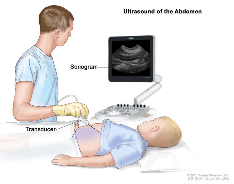
An ultrasound uses high-energy sound waves (ultrasound) that bounce off internal tissues or organs and make echoes. The echoes form a picture of body tissues called a sonogram.

EnlargeAbdominal ultrasound; drawing shows a child lying on an exam table during an abdominal ultrasound procedure. A technician is shown pressing a transducer (a device that makes sound waves that bounce off tissues inside the body) against the skin of the abdomen. A computer screen shows a sonogram (picture).
Abdominal ultrasound. An ultrasound transducer connected to a computer is pressed against the skin of the abdomen. The transducer bounces sound waves off internal organs and tissues to make echoes that form a sonogram (computer picture).

CT scan (CAT scan)

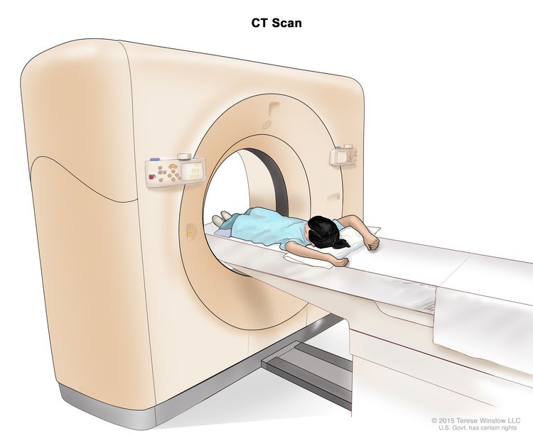
A CT scan uses a computer linked to an x-ray machine to make a series of detailed pictures of areas inside the body. The pictures are taken from different angles and are used to create 3-D views of tissues and organs. A dye may be injected into a vein or swallowed to help the organs or tissues show up more clearly. This procedure is also called computed tomography, computerized tomography, or computerized axial tomography. Learn more about Computed Tomography (CT) Scans and Cancer.

EnlargeComputed tomography (CT) scan; drawing shows a child lying on a table that slides through the CT scanner, which takes a series of detailed x-ray pictures of areas inside the body.
Computed tomography (CT) scan. The child lies on a table that slides through the CT scanner, which takes a series of detailed x-ray pictures of areas inside the body.

Magnetic resonance imaging (MRI)

MRI uses a magnet, radio waves, and a computer to make a series of detailed pictures of areas inside the body. This procedure is also called nuclear magnetic resonance imaging (NMRI).

EnlargeMagnetic resonance imaging (MRI) scan; drawing shows a child lying on a table that slides into the MRI machine, which takes a series of detailed pictures of areas inside the body.
Magnetic resonance imaging (MRI) scan. The child lies on a table that slides into the MRI machine, which takes a series of detailed pictures of areas inside the body. The positioning of the child on the table depends on the part of the body being imaged.

Chest x-ray

An x-ray is a type of radiation that can go through the body and make pictures. A chest x-ray makes pictures of the organs and bones inside the chest.

Biopsy

Biopsy is a procedure in which a sample of tissue is removed from the tumor so that a pathologist can view it under a microscope to check for cancer. While a biopsy is not always needed to diagnose a vascular tumor, it may help find gene mutations that will help with treatment planning.

Getting a second opinion

You may want to get a second opinion to confirm your child’s diagnosis and treatment plan. If you seek a second opinion, you will need to get medical test results and reports from the first doctor to share with the second doctor. The second doctor will review the pathology report, slides, and scans. This doctor may agree with the first doctor, suggest changes to the treatment plan, or provide more information about your child’s tumor.

To learn more about choosing a doctor and getting a second opinion, visit Finding Health Care Services. You can contact NCI’s Cancer Information Service via chat, email, or phone (both in English and Spanish) for help finding a doctor or hospital that can provide a second opinion. For questions you might want to ask at your child’s appointments, visit Questions to Ask Your Doctor.

Types of childhood vascular tumors

Benign tumors

Benign vascular tumors are not cancer.

Infantile hemangioma

Infantile hemangioma (also called a strawberry mark) is the most common type of benign vascular tumor in children. It occurs when immature cells that are meant to form blood vessels form a tumor instead. It tends to appear between the ages of 3 to 6 weeks and is usually not seen at birth. The hemangioma often gets bigger for about 5 months and then stops growing. It slowly fades over the next several years, but a red mark or loose or wrinkled skin may remain. It is rare for an infantile hemangioma to come back after it has faded away.

Infantile hemangioma can develop anywhere on the body, including the skin, the tissue below the skin, or within an organ. It most commonly appears on the skin on the head and neck. Hemangioma may be a single lesion, one or more lesions spread over a larger area, or multiple lesions in different parts of the body. A hemangioma that covers a larger area, involves an organ, or has multiple lesions is more likely to cause problems.

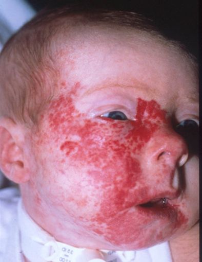
  • Hemangioma in the airway usually occurs along with a large hemangioma on the face that looks like a beard. Airway hemangioma may cause the airway to narrow, leading to trouble breathing.
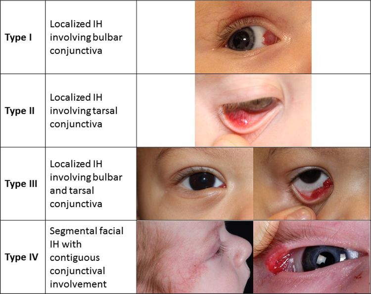
  • Periocular hemangioma involves the eye or tissues around the eye. It may cause vision problems or blindness and is sometimes linked with other eye problems.
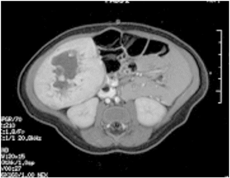
  • Having more than five hemangiomas on the skin is a sign that there may be hemangiomas in an organ, such as the liver, heart, muscle, or thyroid gland. The liver is affected most often.

Some hemangiomas appear between the ages of 3 to 6 weeks but do not grow bigger. This type of hemangioma is called infantile hemangioma with minimal or arrested growth. The lesion appears as light and dark areas of redness on the skin of the lower body or the head and neck. Hemangiomas of this type go away over time without treatment.

Causes and risk factors for infantile hemangioma

Infantile hemangioma is caused by certain changes to how the vascular cells function, especially how they grow and divide into new cells. Often, the exact cause of these changes is unknown.

A risk factor is anything that increases the chance of getting a disease. Not every child with a risk factor will develop an infantile hemangioma. And it can develop in some children who don’t have a known risk factor.

Infantile hemangioma is more common in:

  • girls
  • White people
  • premature babies
  • twins, triplets, or other multiple births
  • babies conceived using in vitro fertilization
  • babies of mothers who are older at the time of the pregnancy, have pre-eclampsia (high blood pressure during pregnancy), or who have problems with the placenta during pregnancy

Other risk factors for infantile hemangioma include:

  • Having a family history of infantile hemangioma, usually in a mother, father, or sibling.
  • Having PHACE syndrome, a rare disorder marked by problems that affect the large blood vessels, heart, eyes, and/or brain. PHACE syndrome increases the risk of a hemangioma that spreads across a large area of the head or face and sometimes the neck, chest, or arm.
  • Having LUMBAR/PELVIS/SACRAL syndrome, a rare disorder marked by problems that affect the urinary system, genitals, rectum, anus, brain, spinal cord, and nerve functions. This syndrome increases the risk of a hemangioma that spreads across a large area of the lower back, arms, chest, or legs.

Talk with your child’s doctor if you think your child may be at risk.

Symptoms of infantile hemangioma

Infantile hemangioma may cause any of the following symptoms. It’s important to check with your child’s doctor if your child has a:

  • Lesion on the skin: An area of spidery veins or lightened or discolored skin may be the first sign of a hemangioma. This may develop into a firm, warm, bright red-to-crimson lesion on the skin that may look like a bruise. A lesion may also form an ulcer that is painful and can lead to bleeding, infection, and scarring. Later, as the hemangioma goes away, it becomes softer and begins fading in the center before flattening and losing color.
  • Lesion below the skin: A lesion that grows under the skin in the fat may appear blue or purple. If the lesion is deep enough under the skin surface, it may not be seen.
  • Lesion in an organ: There may be no symptoms if a hemangioma forms on an organ.

These symptoms may be caused by problems other than a hemangioma. The only way to know is for your child to see a doctor.

Diagnosis of infantile hemangioma

A physical exam and personal and family health history are usually all that are needed to diagnose infantile hemangioma. If the hemangioma looks unusual, a biopsy may be done. An ultrasound may be done if the hemangioma is deeper inside the body with no change to the skin or if the lesion covers a large area of the body. Infants with five or more hemangiomas on the skin should have an ultrasound of the liver to check for a liver hemangioma.

If the hemangioma is part of a syndrome, more tests, such as an echocardiogram, MRI, magnetic resonance angiogram, and eye exam, may be done.

Learn more about these tests in Tests to diagnose childhood vascular tumors.

Treatment of infantile hemangioma

Most hemangiomas fade and shrink without treatment. If a hemangioma is large, causing other health problems, or in an area where it could cause serious problems if it grows bigger, treatment may include:

  • beta-blocker therapy, such as propranolol, nadolol, or atenolol
  • topical beta-blocker therapy for a hemangioma that is in one area of the skin
  • steroid therapy, which may be used when beta-blocker therapy is being started or when beta-blockers cannot be used
  • laser surgery, including pulsed dye laser surgery, which may be used for a hemangioma that has an ulcer or has not completely gone away
  • surgery for a hemangioma that has an ulcer, causes vision problems, has not completely gone away, or is on the face and has not responded to other treatment
  • combined therapy, such as propranolol and steroid therapy or propranolol and topical beta-blocker therapy

Learn more about these treatments in Types of treatment for childhood vascular tumors.

Congenital hemangioma

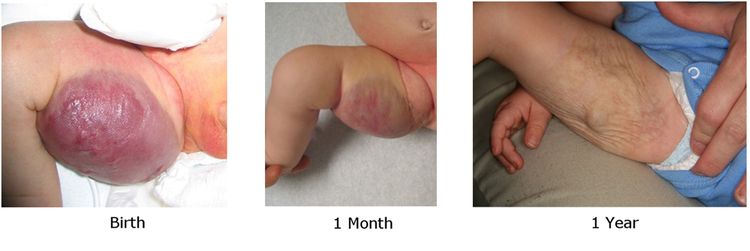
Congenital hemangioma is a benign vascular tumor that begins forming before birth and is fully formed when the baby is born. It is usually on the skin but can be in another organ. A congenital hemangioma may occur as a rash of purple spots. The skin around the spot may be lighter.

There are three types of congenital hemangiomas. The differences between the three types relate to how they shrink (involute) over time:

  • Rapidly involuting congenital hemangioma (RICH) goes away on its own 12 to 15 months after birth. It can form an ulcer, bleed, and cause temporary heart and blood clotting problems. The skin may look a little different even after the hemangioma goes away.
  • Partial involuting congenital hemangioma (PICH) may shrink on its own but does not go away completely.
  • Non-involuting congenital hemangioma (NICH) stays the same size and never goes away on its own.

If your child has symptoms that suggest a congenital hemangioma, the doctor will ask about your child’s personal health history and do a physical exam and ultrasound exam to make the diagnosis.

The types of treatment your child may receive depend on whether the congenital hemangioma will shrink on its own.

  • Treatment of rapidly involuting congenital hemangioma and partial involuting congenital hemangioma may be observation.
  • Treatment of non-involuting congenital hemangioma may be surgery to remove the tumor, depending on where it is and whether it is causing symptoms.

Learn more about these treatments in Types of treatment for childhood vascular tumors.

Benign vascular tumors of the liver

Benign vascular tumors of the liver may be:

  • a single lesion in one part of the liver (focal vascular lesion)
  • several lesions in one part of the liver (multiple liver lesions)
  • several lesions spread across different parts of the liver (diffuse liver lesions)

The liver has many functions, including filtering blood and making proteins that help with blood clotting. Sometimes, the tumor can block or slow the normal flow of blood through the liver. When this happens, blood is sent directly to the heart without going through the liver. This condition is known as a liver shunt. It can cause heart failure and problems with blood clotting.

If your child has symptoms that suggest a benign vascular tumor of the liver, the doctor will ask about your child’s personal health history and do a physical exam and ultrasound exam to make the diagnosis.

The treatment your child may receive depends on whether they have a focal vascular lesion, multiple liver lesions, or diffuse liver lesions.

A single lesion in one part of the liver (focal vascular lesion) is usually a rapidly involuting (shrinking) congenital hemangioma or a non-involuting congenital hemangioma. This lesion can be diagnosed before birth or shortly after the baby is born. Treatment of this type of lesion depends on whether symptoms occur and may include:

  • observation
  • embolization of the liver to treat symptoms
  • surgery, for lesions that do not respond to other treatments

Multiple and diffuse liver lesions are usually infantile hemangiomas. Diffuse liver lesions can cause serious effects, including problems with thyroid hormones and the heart. The liver can enlarge, press on other organs, and cause more symptoms.

Treatment of multiple liver lesions may include:

Treatment of diffuse liver lesions may include:

  • beta-blocker therapy (propranolol)
  • chemotherapy
  • steroid therapy
  • total hepatectomy and liver transplant, for lesions that do not respond to drug therapy or for diffuse liver lesions that are spreading and causing organ failure and there is no time to start treatment

Children with diffuse liver lesions may be diagnosed with hypothyroidism caused by the liver tumor using more of the thyroid hormone. These children may need thyroid hormone replacement therapy.

Learn more about these treatments in Types of treatment for childhood vascular tumors.

If a vascular liver lesion does not respond to treatment, a biopsy may be done to see if the tumor is cancer.

Spindle cell hemangioma

A spindle cell hemangioma contains cells called spindle cells. Under a microscope, spindle cells look long and slender. A spindle cell hemangioma is a painful red-brown or bluish lesion that usually appears on the arms or legs. It can begin as one lesion and develop into more lesions over the years. A spindle cell hemangioma can form in children and adults.

Some children may be at increased risk of developing a spindle cell hemangioma. A risk factor is anything that increases the chance of getting a disease. Not every child with a risk factor will develop a spindle cell hemangioma. And it can develop in some children who don’t have a known risk factor. Spindle cell hemangiomas are more likely to develop in children with the following syndromes:

Talk with your child’s doctor if you think your child may be at risk.

If your child has symptoms that suggest a spindle cell hemangioma, the doctor will ask about your child’s personal health history and do a physical exam to make the diagnosis. If needed, tests may be done. Learn more about Tests to diagnose childhood vascular tumors.

Although there is no standard treatment for spindle cell hemangioma, surgery may be used to remove the tumor. Spindle cell hemangioma may come back after surgery.

Epithelioid hemangioma

An epithelioid hemangioma most often forms on or in the skin, especially the head, but can occur in other areas, such as bone. An epithelioid hemangioma is sometimes caused by injury. It occurs in children and adults.

On the skin, an epithelioid hemangioma may appear as firm pink-to-red bumps and may be itchy. Epithelioid hemangioma of the bone may cause swelling, pain, and weakened bone in the affected area or symptoms of nerve injury. These symptoms may be caused by problems other than an epithelioid hemangioma. The only way to know is for your child to see a doctor.

If your child has symptoms that suggest an epithelioid hemangioma, the doctor will ask about your child’s personal health history and do a physical exam to make the diagnosis. An MRI, x-ray, or biopsy may also be done. Learn more about Tests to diagnose childhood vascular tumors.

There is no standard treatment for epithelioid hemangioma. Treatment may include:

Epithelioid hemangioma often comes back after treatment.

Learn more about these treatments in Types of treatment for childhood vascular tumors.

Pyogenic granuloma

Pyogenic granuloma is also called lobular capillary hemangioma. It is most common in older children and young adults but can occur at any age.

Pyogenic granuloma is sometimes caused by injury or from the use of certain medicines, including birth control pills and retinoids. It may also form for no known reason inside capillaries (the smallest blood vessels), arteries, veins, or other places on the body. Some lesions may be associated with capillary malformations.

Pyogenic granuloma is a raised, bright red lesion that may be small or large and smooth or bumpy. It grows quickly over weeks to months and may bleed a lot. The lesion is on the skin’s surface but may form in the tissues below the skin and look like other vascular lesions. Usually, there is only one lesion. Sometimes multiple lesions can occur in the same area or on different parts of the body. The only way to know if these symptoms are caused by a pyogenic granuloma is for your child to see a doctor.

If your child has symptoms that suggest a pyogenic granuloma, the doctor will ask about your child’s personal health history and do a physical exam to make the diagnosis. If needed, tests may be ordered. Learn more about Tests to diagnose childhood vascular tumors.

Pyogenic granuloma can go away without treatment. Sometimes a pyogenic granuloma needs treatment that may include:

Pyogenic granuloma often comes back after treatment.

Learn more about these treatments in Types of treatment for childhood vascular tumors.

Angiofibroma

Angiofibroma is rare and appears as red bumps on the face. It is a benign skin lesion that usually occurs with tuberous sclerosis, an inherited disorder that causes skin lesions, seizures, and mental disabilities. Talk to your child’s doctor if you think your child may have an angiofibroma.

If your child has symptoms that suggest an angiofibroma, the doctor will ask about your child’s personal health history and do a physical exam to make the diagnosis. If needed, tests may be ordered. Learn more about Tests to diagnose childhood vascular tumors.

Treatment of angiofibroma may include:

Learn more about these treatments in Types of treatment for childhood vascular tumors.

Juvenile nasopharyngeal angiofibroma

Juvenile nasopharyngeal angiofibroma is a tumor that is not cancer but can grow into nearby tissues. It is most common in males and may form around the time of puberty. Juvenile nasopharyngeal angiofibroma begins in the nasal cavity and may spread to the nasopharynx, the paranasal sinuses, the bone around the eyes, and sometimes to the brain.

If your child has symptoms that suggest juvenile nasopharyngeal angiofibroma, the doctor will ask about your child’s personal health history and do a physical exam to make the diagnosis. If needed, tests may be ordered. Learn more about Tests to diagnose childhood vascular tumors.

Treatment of juvenile nasopharyngeal angiofibroma may include:

This tumor may come back after treatment.

Learn more about these treatments in Types of treatment for childhood vascular tumors.

Intermediate tumors that may spread locally

Some intermediate tumors are likely to spread to the area around the tumor (locally), but not to other parts of the body.

Kaposiform hemangioendothelioma and tufted angioma

Kaposiform hemangioendothelioma and tufted angioma are blood vessel tumors that occur in infants or young children and affect males and females equally. These tumors can cause Kasabach-Merritt phenomenon, a condition in which the blood is not able to clot and serious bleeding may occur. This type of vascular tumor is not related to Kaposi sarcoma.

Symptoms of kaposiform hemangioendothelioma and tufted angioma

Kaposiform hemangioendothelioma and tufted angioma usually occur on the skin of the arms and legs, but may also form in deeper tissues, such as muscle or bone, or in the chest, abdomen, head, or neck.

Symptoms may include:

  • firm, warm, painful areas of skin that look bruised
  • purple or brownish-red areas of skin
  • pain with no visible lump
  • easy bruising
  • bleeding more than the usual amount from mucous membranes, wounds, and other tissues

People who have kaposiform hemangioendothelioma or tufted angioma may have anemia (weakness, feeling tired, or looking pale).

These symptoms may be caused by problems other than kaposiform hemangioendothelioma or tufted angioma. The only way to know is for your child to see a doctor.

Diagnosis of kaposiform hemangioendothelioma and tufted angioma

If a physical exam and MRI clearly show the tumor is a kaposiform hemangioendothelioma or a tufted angioma, a biopsy may not be needed. A biopsy is not always done because serious bleeding can occur. An ultrasound exam may also be used to diagnose a tufted angioma.

Learn more about Tests to diagnose childhood vascular tumors.

Treatment of kaposiform hemangioendothelioma and tufted angioma

Kaposiform hemangioendothelioma and tufted angioma are best treated by a vascular anomaly specialist. Treatment depends on the symptoms, size and location of the tumor, and the risk of bleeding. Infection, delay in treatment, and surgery can cause bleeding that is life-threatening.

Kaposiform hemangioendothelioma and tufted angioma may be called uncomplicated or complicated.

Uncomplicated tumors are in one area, smaller, cause few or no symptoms, and have a lower risk of bleeding. People with an uncomplicated tumor do not have Kasabach-Merritt phenomenon.

Treatment for uncomplicated kaposiform hemangioendothelioma and tufted angioma may include:

Complicated tumors are larger, may cause symptoms, and affect how the body functions. People with a complicated tumor may have Kasabach-Merritt phenomenon, a serious condition that can be life-threatening and requires treatment.

Treatment for complicated kaposiform hemangioendothelioma and tufted angioma may include:

  • chemotherapy, with or without steroid therapy
  • targeted therapy (sirolimus), with or without steroid therapy
  • surgery, with or without embolization

Even with treatment, these tumors do not fully go away and can come back. Pain and inflammation may get worse with age, often around puberty. Long-term effects include chronic pain, heart failure, bone problems, and lymphedema (the build up of lymph fluid in tissues).

Learn more about these treatments in Types of treatment for childhood vascular tumors.

Intermediate tumors that may spread to other parts of the body

Rarely, intermediate tumors spread to other parts of the body (metastasize).

Pseudomyogenic (epithelioid sarcoma–like) hemangioendothelioma

Pseudomyogenic hemangioendothelioma can occur in children, but is most common in men aged 20 to 50 years. This tumor is rare, and usually occurs on or under the skin or in bone. Pseudomyogenic hemangioendothelioma may appear as a lump in soft tissue or may cause pain in the affected area. It may spread to nearby tissue but usually does not spread to other parts of the body. In most cases, there are multiple tumors. Talk to your child’s doctor if you think your child may have pseudomyogenic hemangioendothelioma.

If your child has symptoms that suggest pseudomyogenic hemangioendothelioma, the doctor will ask about your child’s personal health history and do a physical exam to make the diagnosis. If needed, tests may be ordered. Learn about Tests to diagnose childhood vascular tumors.

Treatment of pseudomyogenic hemangioendothelioma may include:

Because pseudomyogenic hemangioendothelioma is so rare in children, treatment options are based on clinical trials in adults.

Learn more about these treatments in Types of treatment for childhood vascular tumors.

Retiform hemangioendothelioma

Retiform hemangioendothelioma is a slow-growing, flat tumor that occurs in young adults and sometimes children. This tumor usually occurs on or under the skin of the arms, legs, and trunk. It usually does not spread to other parts of the body. Talk to your child’s doctor if you think your child may have a retiform hemangioendothelioma.

If your child has symptoms that suggest retiform hemangioendothelioma, the doctor will ask about your child’s personal health history and do a physical exam to make the diagnosis. If needed, tests may be ordered. Learn about Tests to diagnose childhood vascular tumors.

Treatment of retiform hemangioendothelioma may include:

Retiform hemangioendothelioma may come back after treatment.

Learn more about these treatments in Types of treatment for childhood vascular tumors.

Papillary intralymphatic angioendothelioma

Papillary intralymphatic angioendothelioma is also called Dabska tumor. It occurs in children and adults.

Papillary intralymphatic angioendothelioma may appear as firm, raised, purplish bumps, which may be small or large. Papillary intralymphatic angioendothelioma forms in or under the skin anywhere on the body. Sometimes the lymph nodes are affected. Talk to your child’s doctor if you think your child may have papillary intralymphatic angioendothelioma.

If your child has symptoms that suggest papillary intralymphatic angioendothelioma, the doctor will ask about your child’s personal health history and do a physical exam to make the diagnosis. If needed, tests may be ordered. Learn about Tests to diagnose childhood vascular tumors.

Treatment of papillary intralymphatic angioendothelioma is surgery.

Learn more about this treatment in Types of treatment for childhood vascular tumors.

Composite hemangioendothelioma

Composite hemangioendothelioma has features of both benign and malignant vascular tumors. This tumor usually occurs on or under the skin of the arms or legs. It may also occur on the skin of the head, neck, or chest. Composite hemangioendothelioma is not likely to spread to nearby tissue or to other parts of the body, but it may come back in the same place. If the tumor spreads, it usually spreads to nearby lymph nodes. Composite hemangioendothelioma occurs in children and adults.

If your child has symptoms that suggest composite hemangioendothelioma, the doctor will ask about your child’s personal health history and do a physical exam to make the diagnosis. If needed, tests may be ordered. Learn about Tests to diagnose childhood vascular tumors.

Treatment of composite hemangioendothelioma may include:

Composite hemangioendothelioma may come back after treatment.

Learn more about these treatments in Types of treatment for childhood vascular tumors.

Kaposi sarcoma

Kaposi sarcoma is a cancer that causes lesions to grow in the skin; the mucous membranes lining the mouth, nose, and throat; lymph nodes; or other organs. It is caused by human herpesvirus 8. This cancer rarely occurs in children. In the United States, Kaposi sarcoma occurs most often in children who have a weak immune system caused by rare immune system disorders, HIV infection, or drugs used in organ transplants. In sub-Saharan Africa, Kaposi sarcoma is endemic and often occurs in children and young adults.

Kaposi sarcoma are lesions that form in the skin, mouth, or throat. The lesions are red, purple, or brown and change from flat, to raised, to scaly areas called plaques, to nodules. Sometimes Kaposi sarcoma causes swollen lymph nodes. These symptoms may be caused by problems other than Kaposi sarcoma. The only way to know is to see your child’s doctor.

If your child has symptoms that suggest Kaposi sarcoma, the doctor will ask about your child’s personal health history and do a physical exam to make the diagnosis. If needed, tests may be ordered. Learn about Tests to diagnose childhood vascular tumors.

Treatment of Kaposi sarcoma may include:

Learn more about these treatments in the Types of treatment for childhood vascular tumors.

Because Kaposi sarcoma is so rare in children, some treatment options are based on clinical trials in adults. Learn more at Kaposi Sarcoma Treatment.

Malignant tumors

Malignant tumors are cancer.

Epithelioid hemangioendothelioma

Epithelioid hemangioendothelioma can occur in children, but is most common in adults aged 30 to 50 years. It may occur in the liver, lung, bone, skin, or soft tissue. Epithelioid hemangioendothelioma may be fast growing or slow growing. In about a third of patients with a tumor in the soft tissue, the tumor spreads to other parts of the body very quickly.

Symptoms of epithelioid hemangioendothelioma

The symptoms of epithelioid hemangioendothelioma depend on where the tumor is in the body. It’s important to check with your child’s doctor if your child has:

  • red-brown patches on the skin that are raised and rounded or flat and feel warm
  • early symptoms of lesions in the lung, which may not occur in all children with lung lesions:
    • chest pain
    • spitting up blood
    • anemia (weakness, feeling tired, or looking pale)
    • trouble breathing (from scarred lung tissue)
  • broken bones
  • symptoms of lesions in the liver:
    • itching
    • yellowing of the skin or eye

These symptoms may be caused by problems other than an epithelioid hemangioendothelioma. The only way to know is for your child to see a doctor.

Diagnosis of epithelioid hemangioendothelioma

If your child has symptoms that suggest epithelioid hemangioendothelioma, the doctor will ask about your child’s personal health history and do a physical exam to make the diagnosis. The doctor may also order tests. Epithelioid hemangioendothelioma in the liver is found with an ultrasound exam, CT scan or MRI scan. X-rays of the chest or other areas of the body may also be done. Learn more about Tests to diagnose childhood vascular tumors.

Treatment of epithelioid hemangioendothelioma

Treatment of slow-growing epithelioid hemangioendothelioma may be observation. Surgery may be used when it is possible to remove the tumor.

Treatment of fast-growing epithelioid hemangioendothelioma may include:

Learn more about these treatments in the Types of treatment for childhood vascular tumors.

Angiosarcoma

Angiosarcoma is a fast-growing tumor that forms in blood vessels or lymph vessels in any part of the body, usually in the soft tissue. Most angiosarcoma is in the skin or in the soft tissue near the skin. Those in deeper soft tissue can form in the liver, spleen, and lung.

Angiosarcoma is very rare in children. Children sometimes have more than one tumor in the skin, liver, or both.

Causes and risk factors for angiosarcoma

A risk factor is anything that increases the chance of getting a disease. Not every child with a risk factor will develop angiosarcoma. And it can develop in some children who don’t have a known risk factor. Risk factors for angiosarcoma include:

Rarely, a benign vascular tumor, such as a hemangioma, may become an angiosarcoma.

Talk with your child’s doctor if you think your child may be at risk.

Symptoms of angiosarcoma

Symptoms of angiosarcoma depend on where the tumor is and may include:

  • red patches on the skin that bleed easily
  • purple tumors

These symptoms may be caused by problems other than an angiosarcoma. The only way to know is for your child to see a doctor.

Diagnosis of angiosarcoma

To make a diagnosis, the doctor will ask about your child’s personal health history and do a physical exam. If needed, tests may be ordered. Learn about Tests to diagnose childhood vascular tumors.

Treatment of angiosarcoma

Treatment of angiosarcoma may include:

Learn more about these treatments in Types of treatment for childhood vascular tumors.

Types of treatment for childhood vascular tumors

Who treats children with vascular tumors?

A pediatric oncologist, a doctor who specializes in treating children with cancer, oversees treatment for childhood vascular tumors. The pediatric oncologist works with other health care providers who are experts in treating children with cancer and also specialize in certain areas of medicine. Other specialists may include:

Treatment options

There are different types of treatment for children and adolescents with vascular tumors. You and your child’s care team will work together to decide treatment. Many factors will be considered, such as where the tumor is located, your child’s age and overall health, the type of vascular tumor, the risk of scarring, and the likelihood of completely treating the vascular tumor.

Your child’s treatment plan will include information about the tumor, the goals of treatment, treatment options, and the possible side effects. It will be helpful to talk with your child’s care team before treatment begins about what to expect. For help every step of the way, see our booklet, Children with Cancer: A Guide for Parents.

Types of treatment your child might have include:

Beta-blocker therapy

Beta-blockers are drugs commonly used to lower blood pressure and heart rate, but they can also shrink certain types of vascular tumors, such as infantile hemangiomas. Beta-blocker therapy may be injected into a vein, taken by mouth, or placed on the skin (topical). How beta-blocker therapy is given depends on the type of vascular tumor being treated and where it first formed.

The beta-blocker propranolol is usually the first treatment for hemangiomas. Infants younger than 4 weeks, who have an underlying condition, or who are treated with IV propranolol may need to have their treatment started in a hospital. Infantile hemangioma may also be treated with propranolol and steroid therapy or propranolol and topical beta-blocker therapy. Propranolol is also used to treat benign vascular tumors of liver.

Other beta-blockers used to treat vascular tumors include atenolol, nadolol, and timolol.

Surgery

The following types of surgery may be used to remove many types of vascular tumors:

  • Excision is surgery to remove the entire tumor and some of the healthy tissue around it.
  • Laser surgery uses a laser beam (a narrow beam of intense light) as a knife to make bloodless cuts in tissue or remove a skin lesion such as a tumor. Surgery with a pulsed dye laser may be used for some hemangiomas. This type of laser uses a beam of light that targets blood vessels in the skin. The light is changed into heat and the blood vessels are destroyed without damaging nearby skin.
  • Curettage uses a small, spoon-shaped instrument with a sharp edge called a curette to remove abnormal tissue.
  • Total hepatectomy and liver transplant removes the entire liver followed by a transplant of a healthy liver from a donor.
  • Amputation removes an arm or leg when there are multiple tumors in the bone.

The type of surgery used depends on the type of vascular tumor and where it formed in the body.

After the doctor removes all the cancer that can be seen at the time of the surgery, some people may be given chemotherapy or radiation therapy to kill any cancer cells that are left. Treatment given after the surgery to lower the risk that the cancer will come back is called adjuvant therapy.

Photocoagulation

Photocoagulation is the use of an intense beam of light, such as a laser, to seal off blood vessels or destroy tissue. It is used to treat pyogenic granuloma.

Cryotherapy

Cryotherapy uses an instrument to freeze and destroy abnormal tissue, such as abnormal blood vessels in pyogenic granuloma. This type of treatment is also called cryosurgery.

Learn more about Cryosurgery to Treat Cancer.

Embolization

Embolization uses particles, such as tiny gelatin sponges or beads, to block blood vessels in the liver. It may be used to block blood flow to some benign vascular tumors of the liver and kaposiform hemangioendothelioma.

Chemotherapy

Chemotherapy (also called chemo) uses drugs to stop the growth of tumor cells. Chemotherapy either kills the cancer cells or stops them from dividing. Chemotherapy may be given alone or with other types of treatment.

For some vascular tumors, chemotherapy is injected into a vein. When given this way, the drugs enter the bloodstream to reach tumor cells throughout the body.

Chemotherapy drugs that may be used alone or in combination to treat childhood vascular tumors include:

Other chemotherapy drugs not listed here may also be used.

Learn more about how chemotherapy works, how it is given, common side effects, and more at Chemotherapy to Treat Cancer.

Sclerotherapy

Sclerotherapy destroys the tumor and the blood vessels that lead to it. A liquid is injected into the blood vessels, causing them to scar and break down. Over time, the destroyed blood vessels are absorbed into normal tissue. The blood flows through nearby healthy veins instead. Sclerotherapy is used to treat epithelioid hemangioma.

Radiation therapy

Radiation therapy uses high-energy x-rays or other types of radiation to kill tumor cells or keep them from growing. External radiation therapy uses a machine outside the body to send radiation toward the area of the body with the tumor. It is used to treat some vascular tumors.

Learn more about External Beam Radiation Therapy for Cancer and Radiation Therapy Side Effects.

Targeted therapy

Targeted therapy uses drugs or other substances to block the action of specific enzymes, proteins, or other molecules involved in the growth and spread of cancer cells. Different types of targeted therapy are being used or studied to treat childhood vascular tumors:

Learn more about Targeted Therapy to Treat Cancer.

Immunotherapy

Immunotherapy helps a person’s immune system fight cancer. Interferon is a type of immunotherapy used to treat vascular tumors.

Learn more about Immunotherapy to Treat Cancer.

Other drug therapy

Other drugs used to treat childhood vascular tumors or manage their effects include:

  • Steroid therapy: Steroids are hormones made naturally in the body. They can also be made in a laboratory and used as drugs. Steroid drugs help shrink some vascular tumors. Corticosteroids, such as prednisone and methylprednisolone, are used to treat infantile hemangioma.
  • Immunosuppressant therapy: These drugs decrease the body’s immune responses. Immunosuppressant therapy has been used to help shrink vascular tumors. Topical tacrolimus is used to treat kaposiform hemangioendotheliomas and tufted angiomas.
  • Thyroid hormone replacement therapy: These drugs replace hormones made by the thyroid and are used to treat a rare form of hypothyroidism caused by some vascular tumors, such as liver hemangiomas.

Observation

Observation is closely monitoring a person’s condition without giving any treatment until symptoms appear or change.

Clinical trials

For some children, joining a clinical trial may be an option. There are different types of clinical trials for childhood cancer. For example, a treatment trial tests new treatments or new ways of using current treatments. Supportive care and palliative care trials look at ways to improve quality of life, especially for those who have side effects from cancer and its treatment.

You can use the clinical trial search to find NCI-supported cancer clinical trials accepting participants. The search allows you to filter trials based on the type of cancer, your child’s age, and where the trials are being done. Clinical trials supported by other organizations can be found on the ClinicalTrials.gov website.

Learn more about clinical trials, including how to find and join one, at Clinical Trials Information for Patients and Caregivers.

Side effects and late effects of treatment

Treatments for vascular tumors can cause side effects. Which side effects your child might have depends on the type of treatment they receive, the dose, and how their body reacts. Talk with your child’s treatment team about which side effects to look for and ways to manage them.

To learn more about side effects that begin during treatment for cancer, visit Side Effects.

Problems from cancer treatment that begin 6 months or later after treatment and continue for months or years are called late effects. Late effects of treatment may include:

  • physical problems
  • changes in mood, feelings, thinking, learning, or memory
  • second cancers (new types of cancer)

Some late effects may be treated or controlled. It is important to talk with your child’s doctors about the possible late effects caused by some treatments. Learn more about Late Effects of Treatment for Childhood Cancer.

Follow-up care

As your child goes through treatment, they will have follow-up tests or check-ups. Some of the tests that were done to diagnose the vascular tumor may be repeated to see how well the treatment is working. Decisions about whether to continue, change, or stop treatment may be based on the results of these tests.

Some of the tests will continue to be done from time to time after treatment has ended. The results of these tests can show if your child’s condition has changed or if the tumor has come back.

Learn more about follow-up tests in Tests to diagnose childhood vascular tumors.

Coping with your child's vascular tumor

When your child has a tumor, every member of the family needs support. Taking care of yourself during this difficult time is important. Reach out to your child’s treatment team and to people in your family and community for support. To learn more, visit Support for Families: Childhood Cancer and the booklet Children with Cancer: A Guide for Parents.

Related resources

About This PDQ Summary

About PDQ

Physician Data Query (PDQ) is the National Cancer Institute’s (NCI’s) comprehensive cancer information database. The PDQ database contains summaries of the latest published information on cancer prevention, detection, genetics, treatment, supportive care, and complementary and alternative medicine. Most summaries come in two versions. The health professional versions have detailed information written in technical language. The patient versions are written in easy-to-understand, nontechnical language. Both versions have cancer information that is accurate and up to date and most versions are also available in Spanish.

PDQ is a service of the NCI. The NCI is part of the National Institutes of Health (NIH). NIH is the federal government’s center of biomedical research. The PDQ summaries are based on an independent review of the medical literature. They are not policy statements of the NCI or the NIH.

Purpose of This Summary

This PDQ cancer information summary has current information about the treatment of childhood vascular tumors. It is meant to inform and help patients, families, and caregivers. It does not give formal guidelines or recommendations for making decisions about health care.

Reviewers and Updates

Editorial Boards write the PDQ cancer information summaries and keep them up to date. These Boards are made up of experts in cancer treatment and other specialties related to cancer. The summaries are reviewed regularly and changes are made when there is new information. The date on each summary (“Updated”) is the date of the most recent change.

The information in this patient summary was taken from the health professional version, which is reviewed regularly and updated as needed, by the PDQ Pediatric Treatment Editorial Board.

Clinical Trial Information

A clinical trial is a study to answer a scientific question, such as whether one treatment is better than another. Trials are based on past studies and what has been learned in the laboratory. Each trial answers certain scientific questions in order to find new and better ways to help cancer patients. During treatment clinical trials, information is collected about the effects of a new treatment and how well it works. If a clinical trial shows that a new treatment is better than one currently being used, the new treatment may become “standard.” Patients may want to think about taking part in a clinical trial. Some clinical trials are open only to patients who have not started treatment.

Clinical trials can be found online at NCI’s website. For more information, call the Cancer Information Service (CIS), NCI’s contact center, at 1-800-4-CANCER (1-800-422-6237).

Permission to Use This Summary

PDQ is a registered trademark. The content of PDQ documents can be used freely as text. It cannot be identified as an NCI PDQ cancer information summary unless the whole summary is shown and it is updated regularly. However, a user would be allowed to write a sentence such as “NCI’s PDQ cancer information summary about breast cancer prevention states the risks in the following way: [include excerpt from the summary].”

The best way to cite this PDQ summary is:

PDQ® Pediatric Treatment Editorial Board. PDQ Childhood Vascular Tumors. Bethesda, MD: National Cancer Institute. Updated <MM/DD/YYYY>. Available at: /types/soft-tissue-sarcoma/patient/child-vascular-tumors-treatment-pdq. Accessed <MM/DD/YYYY>. [PMID: 27253005]

Images in this summary are used with permission of the author(s), artist, and/or publisher for use in the PDQ summaries only. If you want to use an image from a PDQ summary and you are not using the whole summary, you must get permission from the owner. It cannot be given by the National Cancer Institute. Information about using the images in this summary, along with many other images related to cancer can be found in Visuals Online. Visuals Online is a collection of more than 3,000 scientific images.

Disclaimer

The information in these summaries should not be used to make decisions about insurance reimbursement. More information on insurance coverage is available on Cancer.gov on the Managing Cancer Care page.

Contact Us

More information about contacting us or receiving help with the Cancer.gov website can be found on our Contact Us for Help page. Questions can also be submitted to Cancer.gov through the website’s E-mail Us.

Childhood Vascular Tumors Treatment (PDQ®)–Health Professional Version

Childhood Vascular Tumors Treatment (PDQ®)–Health Professional Version

Vascular Anomalies

Vascular anomalies are a spectrum of rare diseases classified as either vascular tumors or vascular malformations. Generally, vascular tumors are proliferative, while vascular malformations enlarge through expansion of a developmental anomaly without underlying proliferation.

Although these anomalies are not oncological, it is important for oncologists to understand the biology and clinical management of common vascular malformations. This is because many vascular malformations are caused by targetable somatic variants, which means that pediatric oncologists will be asked to help manage these lesions. While information about vascular malformations is covered at the beginning of this summary, the remainder of this summary focuses on tumors, not malformations.

Vascular Malformations

Vascular malformations are distinguished from vascular tumors by their low cell turnover and lack of invasiveness.[1] They tend to grow in proportion to the child and are generally stable in adulthood. Nonetheless, endothelial cells isolated from vascular malformations have been found in vitro to have some tumor-like behaviors, such as increased growth, migration, and resistance to apoptosis.[2]

In the International Society for the Study of Vascular Anomalies (ISSVA) classification, vascular malformations are subdivided according to vessel type.[3] Fast-flow lesions include arterial-venous fistulas and arterial-venous malformations. These complicated lesions can cause bleeding, ulceration, and organ dysfunction.

Slow-flow lesions include venous, lymphatic, capillary, or combined lesions. Complications from slow-flow lesions include pain, infection, bleeding, thrombosis, and organ dysfunction.

Regular monitoring and assessment of changes or development of symptoms is warranted in patients with vascular malformations. Treatment requires an interdisciplinary approach to care and includes observation, surgery, endovascular intervention, and medical management. Only a low level of evidence supports the choice of treatment between these options. Recurrence rates of these lesions are relatively high.[4]

Vascular malformations are most commonly caused by variants in the MAP2K/PIK3CA pathway. Most are activating somatic variants but, rarely, germline variants are identified. Approximately one-third to one-half of venous malformations result from somatic or, rarely, germline variants in the TEK (or TIE2) gene.[5] Another one-third of venous malformations, and nearly all lymphatic malformations, are caused by somatic variants in PIK3CA.[6] In most cases, PIK3CA variants are identical to canonical cancer variants. Lesions harboring PIK3CA variants are frequently associated with overgrowth of adjacent tissues, as seen in patients with Klippel-Trénaunay syndrome and CLOVES syndrome (congenital lipomatous overgrowth, vascular malformations, epidermal nevis, spinal/skeletal anomalies/scoliosis).[7]

Sirolimus was initially used to target the PI3K pathway in slow-flow malformations, leading to symptomatic improvement in many patients. It is unclear whether treatment reduces the size of lesions because there is usually considerable fluctuation in size, and treatment generally begins when lesions are enlarged. The use of sirolimus in venous and lymphatic malformations is supported by level C evidence (case series, other observational study designs, phase II studies).[810] Both lesions with PIK3CA and TEK variants appear to respond equally to treatment with sirolimus. Phase III clinical trials are underway in Europe (e.g., NCT02638389, NCT03987152, and NCT04980872). A 2018 study reported promising level C evidence for the use of the PI3K inhibitor BYL719 (alpelisib) to treat patients who have lesions with a PIK3CA variant.[11] From these studies, preliminary FDA approval has been obtained. For information about ongoing studies, see the Treatment Options Under Clinical Evaluation section.

There is some support for targeted therapy in fast-flow malformations and complicated lymphatic anomalies that are caused by somatic and germline variants in the MAPK pathway, including gain of function variants in MAP2K1, KRAS, NRAS, and BRAF.[12] Limited data suggest that MEK pathway inhibition may soon have a role in treating patients with these aggressive, highly symptomatic, and sometimes fatal lesions.[1320] For information about ongoing studies, see the Treatment Options Under Clinical Evaluation section.

Treatment Options Under Clinical Evaluation

Information about National Cancer Institute (NCI)-supported clinical trials can be found on the NCI website. For information about clinical trials sponsored by other organizations, see the ClinicalTrials.gov website.

The following are examples of national and/or institutional clinical trials that are currently being conducted:

  • NCT04258046 (Trametinib in the Treatment of Complicated Extracranial Arterial Venous Malformation): This is a phase II study to assess the safety and efficacy of trametinib in the treatment of children and adults.
  • NCT05125471 (Cobimetinib in Extracranial Arteriovenous Malformations [COBI-AVM Study]): This is a phase II study to evaluate the safety and efficacy of cobimetinib in the treatment of children and adults.
  • NCT05948943 (Alpelisib in Pediatric and Adult Patients With Lymphatic Malformations Associated With a PIK3CA Variant): This is a phase II/III study of adults and children (aged 6–17 years) that will determine the dose of alpelisib in stage 1, followed by confirmation of efficacy and safety in stage 2 of the study.
References
  1. Mulliken JB, Glowacki J: Hemangiomas and vascular malformations in infants and children: a classification based on endothelial characteristics. Plast Reconstr Surg 69 (3): 412-22, 1982. [PUBMED Abstract]
  2. Lokmic Z, Mitchell GM, Koh Wee Chong N, et al.: Isolation of human lymphatic malformation endothelial cells, their in vitro characterization and in vivo survival in a mouse xenograft model. Angiogenesis 17 (1): 1-15, 2014. [PUBMED Abstract]
  3. Wassef M, Blei F, Adams D, et al.: Vascular Anomalies Classification: Recommendations From the International Society for the Study of Vascular Anomalies. Pediatrics 136 (1): e203-14, 2015. [PUBMED Abstract]
  4. van der Vleuten CJ, Kater A, Wijnen MH, et al.: Effectiveness of sclerotherapy, surgery, and laser therapy in patients with venous malformations: a systematic review. Cardiovasc Intervent Radiol 37 (4): 977-89, 2014. [PUBMED Abstract]
  5. Soblet J, Limaye N, Uebelhoer M, et al.: Variable Somatic TIE2 Mutations in Half of Sporadic Venous Malformations. Mol Syndromol 4 (4): 179-83, 2013. [PUBMED Abstract]
  6. Luks VL, Kamitaki N, Vivero MP, et al.: Lymphatic and other vascular malformative/overgrowth disorders are caused by somatic mutations in PIK3CA. J Pediatr 166 (4): 1048-54.e1-5, 2015. [PUBMED Abstract]
  7. Keppler-Noreuil KM, Rios JJ, Parker VE, et al.: PIK3CA-related overgrowth spectrum (PROS): diagnostic and testing eligibility criteria, differential diagnosis, and evaluation. Am J Med Genet A 167A (2): 287-95, 2015. [PUBMED Abstract]
  8. Adams DM, Trenor CC, Hammill AM, et al.: Efficacy and Safety of Sirolimus in the Treatment of Complicated Vascular Anomalies. Pediatrics 137 (2): e20153257, 2016. [PUBMED Abstract]
  9. Hammer J, Seront E, Duez S, et al.: Sirolimus is efficacious in treatment for extensive and/or complex slow-flow vascular malformations: a monocentric prospective phase II study. Orphanet J Rare Dis 13 (1): 191, 2018. [PUBMED Abstract]
  10. Maruani A, Tavernier E, Boccara O, et al.: Sirolimus (Rapamycin) for Slow-Flow Malformations in Children: The Observational-Phase Randomized Clinical PERFORMUS Trial. JAMA Dermatol 157 (11): 1289-1298, 2021. [PUBMED Abstract]
  11. Venot Q, Blanc T, Rabia SH, et al.: Targeted therapy in patients with PIK3CA-related overgrowth syndrome. Nature 558 (7711): 540-546, 2018. [PUBMED Abstract]
  12. Couto JA, Huang AY, Konczyk DJ, et al.: Somatic MAP2K1 Mutations Are Associated with Extracranial Arteriovenous Malformation. Am J Hum Genet 100 (3): 546-554, 2017. [PUBMED Abstract]
  13. Dori Y, Smith C, Pinto E, et al.: Severe Lymphatic Disorder Resolved With MEK Inhibition in a Patient With Noonan Syndrome and SOS1 Mutation. Pediatrics 146 (6): , 2020. [PUBMED Abstract]
  14. Nakano TA, Rankin AW, Annam A, et al.: Trametinib for Refractory Chylous Effusions and Systemic Complications in Children with Noonan Syndrome. J Pediatr 248: 81-88.e1, 2022. [PUBMED Abstract]
  15. Homayun-Sepehr N, McCarter AL, Helaers R, et al.: KRAS-driven model of Gorham-Stout disease effectively treated with trametinib. JCI Insight 6 (15): , 2021. [PUBMED Abstract]
  16. Foster JB, Li D, March ME, et al.: Kaposiform lymphangiomatosis effectively treated with MEK inhibition. EMBO Mol Med 12 (10): e12324, 2020. [PUBMED Abstract]
  17. Chowers G, Abebe-Campino G, Golan H, et al.: Treatment of severe Kaposiform lymphangiomatosis positive for NRAS mutation by MEK inhibition. Pediatr Res 94 (6): 1911-1915, 2023. [PUBMED Abstract]
  18. Lekwuttikarn R, Lim YH, Admani S, et al.: Genotype-Guided Medical Treatment of an Arteriovenous Malformation in a Child. JAMA Dermatol 155 (2): 256-257, 2019. [PUBMED Abstract]
  19. Nicholson CL, Flanagan S, Murati M, et al.: Successful management of an arteriovenous malformation with trametinib in a patient with capillary-malformation arteriovenous malformation syndrome and cardiac compromise. Pediatr Dermatol 39 (2): 316-319, 2022. [PUBMED Abstract]
  20. Cooke DL, Frieden IJ, Shimano KA: Angiographic evidence of response to trametinib therapy for a spinal cord arteriovenous malformation. J Vasc Anom (Phila) 2 (3): e018, 2021. Available online. Last accessed July 6, 2023..

Childhood Vascular Tumors

Vascular tumors are proliferative tumors that can be benign or malignant. Growth and/or expansion of vascular tumors can cause clinical problems such as disfigurement, chronic pain, coagulopathies, organ dysfunction, and death.

The quality of evidence regarding childhood vascular tumors is limited by retrospective data collection, small sample size, cohort selection and participation bias, and heterogeneity of the disorders. Lack of consistent criteria and medical terminology has led to unreliable conclusions from the historical medical literature.[13]

In the past, limited treatment options were available, and efficacy was not validated in prospective clinical trials. Historically, therapies consisted of interventional and surgical procedures used to palliate symptoms. Limited medical therapies were available. Newer therapy options with propranolol and sirolimus are now available for the treatment of patients with complex vascular tumors. The first prospective clinical trial using propranolol for infantile hemangioma has been published, as well as the first prospective clinical trial that studied the effectiveness of sirolimus for complicated vascular anomalies, including vascular tumors.[4,5]

With a prevalence of 4% to 5%, infantile hemangiomas are the most common benign tumors of infancy. Other vascular tumors are rare. The classification of these tumors has been difficult, especially in the pediatric population, because of their rarity, unusual morphologic appearance, diverse clinical behavior, and the lack of independent stratification for pediatric tumors. In 2020, the World Health Organization (WHO) updated the classification of soft tissue vascular tumors.[6]

The International Society for the Study of Vascular Anomalies (ISSVA) classification of tumors is based on the WHO classification, but it uses more precise terminology and phenotypes. The General Assembly of the ISSVA adopted an updated classification system in 2014, with further additions in 2018 (ISSVA).[7,8] For more information, see Tables 1 and 2.

Table 1. 2020 World Health Organization Classification of Vascular Tumorsa
Category Vascular Tumor Type
NOS = not otherwise specified.
aAdapted from the WHO Classification of Tumours Editorial Board.[6]
Benign Hemangioma NOS
Intramuscular hemangioma
Arteriovenous hemangioma
Venous hemangioma
Epithelioid hemangioma
Lymphangioma NOS
Cystic lymphangioma
Acquired tufted hemangioma
Intermediate (locally aggressive) Kaposiform hemangioendothelioma
Intermediate (rarely metastasizing) Retiform hemangioendothelioma
Papillary intralymphatic angioendothelioma
Composite hemangioendothelioma
Kaposi sarcoma
Pseudomyogenic (epithelioid sarcoma–like) hemangioendothelioma
Malignant Epithelioid hemangioendothelioma NOS
Angiosarcoma
Table 2. 2018 International Society for the Study of Vascular Anomalies (ISSVA) Classification of Vascular Tumorsa
Category Vascular Tumor Type (Causal Genes)
aAdapted from ISSVA Classification of Vascular Anomalies. ©2018 International Society for the Study of Vascular Anomalies. Available at “issva.org/classification.” Accessed June 2018.[7]
bSee the ISSVA classification 2018 for benign vascular tumors 2.[7]
cTufted angioma and kaposiform hemangioendothelioma are a spectrum of the same entity and will be discussed together.
Benign (type 1b) Infantile hemangioma/hemangioma of infancy
Congenital hemangioma (GNAQ, GNA11)
—Rapidly involuting (RICH)
—Non-involuting (NICH)
—Partially-involuting (PICH)
Tufted angiomac
Spindle cell hemangioma (IDH1, IDH2)
Epithelioid hemangioma (FOS)
Pyogenic granuloma (also known as lobular capillary hemangioma) (BRAF, RAS, GNA14)
Others
Locally aggressive or borderline Kaposiform hemangioendothelioma (KHE) (GNA14)
Retiform hemangioendothelioma
Papillary intralymphatic angioendothelioma (PILA), Dabska tumor
Composite hemangioendothelioma
Pseudomyogenic hemangioendothelioma (FOSB)
Polymorphous hemangioendothelioma
Hemangioendothelioma not otherwise specified
Kaposi sarcoma
Others
Malignant Angiosarcoma (MYC: postradiation therapy)
Epithelioid hemangioendothelioma (EHE) (CAMTA1, TFE3)
Others
References
  1. Liberale C, Rozell-Shannon L, Moneghini L, et al.: Stop Calling Me Cavernous Hemangioma! A Literature Review on Misdiagnosed Bony Vascular Anomalies. J Invest Surg 35 (1): 141-150, 2022. [PUBMED Abstract]
  2. Boulogeorgou K, Avramidou E, Koletsa T: Identifying erroneously used terms for vascular anomalies: A review of the English literature. Hippokratia 26 (4): 126-130, 2022. [PUBMED Abstract]
  3. Hassanein AH, Mulliken JB, Fishman SJ, et al.: Evaluation of terminology for vascular anomalies in current literature. Plast Reconstr Surg 127 (1): 347-351, 2011. [PUBMED Abstract]
  4. Léauté-Labrèze C, Hoeger P, Mazereeuw-Hautier J, et al.: A randomized, controlled trial of oral propranolol in infantile hemangioma. N Engl J Med 372 (8): 735-46, 2015. [PUBMED Abstract]
  5. Adams DM, Trenor CC, Hammill AM, et al.: Efficacy and Safety of Sirolimus in the Treatment of Complicated Vascular Anomalies. Pediatrics 137 (2): e20153257, 2016. [PUBMED Abstract]
  6. WHO Classification of Tumours Editorial Board: WHO Classification of Tumours. Volume 3: Soft Tissue and Bone Tumours. 5th ed., IARC Press, 2020.
  7. International Society for the Study of Vascular Anomalies: ISSVA Classification of Vascular Anomalies. Milwaukee, Wi: International Society for the Study of Vascular Anomalies, 2018. Available online. Last accessed June 7, 2022.
  8. Wassef M, Blei F, Adams D, et al.: Vascular Anomalies Classification: Recommendations From the International Society for the Study of Vascular Anomalies. Pediatrics 136 (1): e203-14, 2015. [PUBMED Abstract]

Special Considerations for the Treatment of Children With Cancer

Cancer in children and adolescents is rare, although the overall incidence has slowly increased since 1975.[1] Children and adolescents with cancer should be referred to medical centers that have a multidisciplinary team of cancer specialists with experience treating the cancers that occur during childhood and adolescence. This multidisciplinary team approach incorporates the skills of the following pediatric specialists and others to ensure that children receive treatment, supportive care, and rehabilitation to achieve optimal survival and quality of life:

  • Primary care physicians.
  • Pediatric surgeons.
  • Transplant surgeons.
  • Pathologists.
  • Pediatric radiation oncologists.
  • Pediatric medical oncologists and hematologists.
  • Ophthalmologists.
  • Rehabilitation specialists.
  • Pediatric oncology nurses.
  • Social workers.
  • Child-life professionals.
  • Psychologists.
  • Nutritionists.

For specific information about supportive care for children and adolescents with cancer, see the summaries on Supportive and Palliative Care.

The American Academy of Pediatrics has outlined guidelines for pediatric cancer centers and their role in the treatment of children and adolescents with cancer.[2] At these centers, clinical trials are available for most types of cancer that occur in children and adolescents, and the opportunity to participate is offered to most patients and their families. Clinical trials for children and adolescents diagnosed with cancer are generally designed to compare potentially better therapy with current standard therapy. Other types of clinical trials test novel therapies when there is no standard therapy for a cancer diagnosis. Most of the progress in identifying curative therapies for childhood cancers has been achieved through clinical trials. Information about ongoing clinical trials is available from the NCI website.

Dramatic improvements in survival have been achieved for children and adolescents with cancer.[35] Between 1975 and 2020, childhood cancer mortality decreased by more than 50%.[3,6,7] Childhood and adolescent cancer survivors require close monitoring because side effects of cancer therapy may persist or develop months or years after treatment. For specific information about the incidence, type, and monitoring of late effects in childhood and adolescent cancer survivors, see Late Effects of Treatment for Childhood Cancer.

References
  1. Smith MA, Seibel NL, Altekruse SF, et al.: Outcomes for children and adolescents with cancer: challenges for the twenty-first century. J Clin Oncol 28 (15): 2625-34, 2010. [PUBMED Abstract]
  2. American Academy of Pediatrics: Standards for pediatric cancer centers. Pediatrics 134 (2): 410-4, 2014. Also available online. Last accessed February 25, 2025.
  3. Smith MA, Altekruse SF, Adamson PC, et al.: Declining childhood and adolescent cancer mortality. Cancer 120 (16): 2497-506, 2014. [PUBMED Abstract]
  4. Childhood cancer. In: Howlader N, Noone AM, Krapcho M, et al., eds.: SEER Cancer Statistics Review, 1975-2010. National Cancer Institute, 2013, Section 28. Also available online. Last accessed August 21, 2023.
  5. Childhood cancer by the ICCC. In: Howlader N, Noone AM, Krapcho M, et al., eds.: SEER Cancer Statistics Review, 1975-2010. National Cancer Institute, 2013, Section 29. Also available online. Last accessed August 21, 2023.
  6. National Cancer Institute: NCCR*Explorer: An interactive website for NCCR cancer statistics. Bethesda, MD: National Cancer Institute. Available online. Last accessed February 25, 2025.
  7. Surveillance Research Program, National Cancer Institute: SEER*Explorer: An interactive website for SEER cancer statistics. Bethesda, MD: National Cancer Institute. Available online. Last accessed December 30, 2024.

Benign Tumors

Benign vascular tumors include the following:

Infantile Hemangioma

Incidence and epidemiology

Infantile hemangiomas (IH) are the most common benign vascular tumor of infancy, occurring in 4% to 5% of infants. The true incidence is unknown.[1] They are not usually present at birth and are diagnosed most commonly at age 3 to 6 weeks.[25] The lesion proliferates for an average of 5 months, stabilizes, and then involutes over several years.

Infantile hemangiomas are more common in females, non-Hispanic White patients, and premature infants. Multiple hemangiomas are more common in infants who are the product of multiple gestations or in vitro fertilization.[57] Infantile hemangiomas are associated with advanced maternal age, placenta previa, pre-eclampsia, and other placental anomalies.[5]

Clinical presentation

Most infantile hemangiomas are not present at birth, but precursor lesions such as telangiectasia or faint discoloration of the skin or hypopigmentation can often be seen. The lesion can be mistaken as a bruise from birth trauma or as a capillary malformation (port-wine stain) (see Figure 1).[8,9]

EnlargePhotos showing an infantile hemangioma premonitory mark; the photos on the left show a precursor lesion (faint color with halo). The photos on the right show a hemangioma after proliferation (slightly raised with a brighter central color).
Figure 1. The photos on the left depict the precursor lesion (faint color with halo). The photos on the right depict the hemangioma after proliferation (slightly raised with a brighter central color). Credit: Israel Fernandez-Pineda, M.D.

Infantile hemangiomas can be superficial in the dermis, deep in the subcutaneous tissue, combined, or in the viscera. Combined lesions are common and generally appear in the head and neck but can be anywhere on the body.

Infantile hemangiomas can be characterized as follows:

  • Local: Most lesions are localized and noted to be in a well-defined area without evidence of a geometric pattern.
  • Segmental: Most segmental hemangiomas occur in the head and neck region (PHACE syndrome) but can be seen in the genitourinary area, arm, chest, or legs (PELVIS/LUMBAR/SACRAL syndrome).
    • Diffuse hemangiomas of the face demonstrate defined cutaneous patterns. Several studies have evaluated the distributions of these hemangiomas and found the following four distinct patterns or segments:
      • Segment 1 involves the lateral forehead, anterior temporal scalp, and the lateral frontal scalp.
      • Segments 2 and 3 are located over the maxillary and mandibular area.
      • Segment 4 covers the medial frontal scalp, nose, and philtrum.

    Two papers have noted this observation and suggest the involvement of neural crest derivatives in facial hemangioma development.[10,11] Segmental hemangiomas commonly occur in females and are more likely associated with complications and other syndromes.[12,13]

    For information about PHACE syndrome or PELVIS/LUMBAR/SACRAL syndrome, see the Syndromes associated with infantile hemangioma section.

  • Multiple: More than one lesion but noted in the past as greater than five lesions, because of the increased risk of visceral involvement (mostly the liver).

The cutaneous appearance of infantile hemangiomas is usually red to crimson, firm, and warm in the proliferative phase. The lesion then lightens centrally and becomes less warm and softer; it then flattens and loses its color. The process of involution can take several years and once involution has occurred, regrowth is uncommon. In two patients treated with growth hormone, regrowth after involution was noted.[14] On further investigation, growth hormone receptors were found on the infantile hemangioma cells. Although preliminary, this may advance the research into the etiology of hemangioma growth.

Ulceration is the most common complication of infantile hemangiomas, occurring in 10% to 15% of patients. Ulceration typically occurs during the proliferative phase, and it can lead to bleeding and secondary infections.[15] Most other complications in the proliferative phase result from the impact of the mass on local structures (e.g., visual or auditory compromise, airway obstruction).[16]

Permanent sequelae, such as telangiectasia, anetodermal skin, redundant skin, and a persistent superficial component, can occur after hemangioma involution. Hemangiomas with a history of ulceration are more likely to cause scarring and potential local anatomical complications.[15] Rare instances of dysesthesias in sites of involuted infantile hemangiomas in the absence of ulceration have been described.[17][Level of evidence C1] In a retrospective cohort study of 184 hemangiomas, the overall incidence of significant sequelae was 54.9%. Sequelae were more common in combined hemangiomas, hemangiomas with a step or abrupt border, and cobblestone surface hemangiomas. Furthermore, this study revealed that the average age to hemangioma involution was 3.5 years.[18] Thus, prophylactic measures such as maintaining dermal integrity with moisturizing barrier agents are indicated for infantile hemangiomas and are important before and during the proliferative phase.[16] Once an ulceration has occurred, it is important to aggressively manage the ulceration to promote healing, prevent infection, and treat pain. In addition to pain control, management includes steroid ointments, antibiotic ointments or systemic antibiotics, laser therapy, or topical timolol.[16]

Biology and histopathology

Most infantile hemangiomas occur sporadically. However, they may rarely be caused by an abnormality of chromosome 5 and present in an autosomal dominant pattern.[19] In a study that evaluated inheritance patterns of infantile hemangiomas, 34% of patients had a family history of infantile hemangioma, most commonly in a first-degree relative.[19,20]

The exact mechanism that causes the initial proliferation of blood vessels followed by involution of the vascular component of hemangioma and replacement of fibrofatty tissue is unknown. Several cell types have been isolated from hemangiomas: progenitor/stem cells (HemSC), endothelial cells (HemEC), pericytes (HemPericytes), and mast cells.[21,22] These cells appear to play a role in the development of infantile hemangiomas.

HemSC represent a small percentage of proliferating hemangioma cells and have the ability for self renewal and multilineage differentiation. These cells differentiate into endothelial cells, adipocytes, and pericytes. When HemSC are implanted into immunodeficient mice, hemangioma-like lesions form and then spontaneously regress, similar to infantile hemangiomas.[23] This suggests that infantile hemangioma proliferation occurs during vasculogenesis (the formation of new blood vessels from angioblasts), as opposed to angiogenesis (the formation of new blood vessels from existing blood vessels).

HemEC are plump, metabolically active, and resemble fetal endothelial cells in the proliferative phase. Evaluation of infantile hemangioma endothelial cells suggest that they are clonal in nature.[2325]

HemPericytes surround the vasculature and are abundant in the proliferative phase. These cells express markers of pericytes and smooth muscle cells, such as neural-glial antigen 2 (NG2), platelet-derived growth factor receptor beta (PDGFR-beta), calponin, alpha smooth muscle actin (SMA), and NOTCH3. HemPericytes are proangiogenic, as they express increased vascular endothelial growth factor A (VEGF-A), decreased angiopoietin-1 (ANGPT1), increased proliferation, increased vessel formation in vivo, and decreased ability to suppress proliferation.[26] One study reported that proliferating infantile hemangiomas contained higher levels of messenger RNA, proteins for NOTCH1, 3, and 4 receptors and their ligands, and the downstream coactivator MAML1 than did normal skin, involuting infantile hemangiomas, and propranolol-treated infantile hemangiomas.[27]

Mast cells are found largely in the early involuting phase, but they are also found in small numbers in the proliferative phase and at the end of involution. Their function in infantile hemangiomas is unknown but they have been shown to play a role in other skin tumors such as basal cell carcinoma, squamous cell carcinoma, and melanoma.[22]

Provasculogenic factors are expressed during proliferation; these factors include VEGF, fibroblast growth factor (FGF), CD34, CD31, CD133, lymphatic vessel endothelial hyaluronan receptor 1 (LYVE1), and insulin-like growth factor 2 (IGF-2).[2831] During involution, infantile hemangiomas show increased apoptosis.[31] During this phase, there are also increased mast cells and levels of metalloproteinase, as well as upregulation of interferon and decreased basic FGF (bFGF).[3133] Throughout proliferation and involution, endothelial cells in infantile hemangioma express a particular phenotype showing positive staining for GLUT1 and placenta-associated antigens (Fc-gamma receptor II, merosin, and Lewis Y antigen). These markers are absent in normal capillaries and in other vascular tumors such as congenital hemangioma and vascular malformations. Placental chorionic villi share these same markers. However, no relationship between hemangiomas and placental chorionic villi has been found.[28]

Hypoxia appears to have a critical role in the pathogenesis of hemangiomas. There is an association between hemangiomas and placental hypoxia, which is increased in prematurity, multiple pregnancies, and placental anomalies.[2,5] Multiple targets of hypoxia [34,35] are demonstrated in proliferating hemangiomas, including VEGF-A, GLUT1, and IGF-2.[28,30,36] The hypothesis suggests that a proliferating hemangioma is an attempt to normalize hypoxic tissue that occurred in utero.

Diagnostic evaluation

Infantile hemangiomas are usually diagnosed by the history and clinical appearance. Biopsy is rarely needed and performed only if there is an atypical appearance and/or atypical history and presentation. Imaging is not usually necessary, but diagnostic ultrasonography is beneficial if there is a deeper lesion without a cutaneous component and reveals a well-circumscribed, hypoechoic, high-flow lesion with a typical Doppler wave characteristic.[37] Additionally, infants with five or more cutaneous hemangiomas should undergo ultrasonography of the liver to screen for hepatic hemangioma.[38]

Infantile hemangioma with minimal or arrested growth

Infantile hemangioma with minimal or arrested growth (IH-MAG) is a variant of hemangioma that can be confused with capillary malformation because of their unusual characteristics. These hemangiomas are mostly fully formed at birth and are characterized by telangiectasia and venules with light and dark areas of skin coloration (see Figure 2). They resolve spontaneously and are pathologically GLUT1 positive.[39] They are mainly located on the lower body but can be present in the head and neck area. If they are segmental, they can be associated with PHACE syndrome.[40] Associated soft tissue hypertrophy may persist through childhood.[41]

EnlargePhotographs showing (A) presentation and (B) resolution of an infantile hemangioma in patient 4 (upper left and right photos), and (C) presentation and (D) resolution of an infantile hemangioma in patient 5 (bottom left and right photos).
Figure 2. Patient 4 at (A) presentation and (B) resolution. Patient 5 at (C) presentation and (D) resolution. Ma, E. H., Robertson, S. J., Chow, C. W., and Bekhor, P. S. (2017), Infantile Hemangioma with Minimal or Arrested Growth: Further Observations on Clinical and Histopathologic Findings of this Unique but Underrecognized Entity. Pediatr Dermatol, 34: 64–71. doi:10.1111/pde.13022. Used with permission.

Airway infantile hemangioma

Airway infantile hemangiomas are usually associated with segmental hemangiomas in a bearded distribution, which may include all or some of the following—the preauricular skin, mandible, lower lip, chin, or anterior neck. It is important for an otolaryngologist to proactively assess lesions in this distribution before signs of stridor occur. Airway infantile hemangioma incidence increases with a larger area of bearded involvement.[42]

Airway infantile hemangiomas can occur without skin lesions. A retrospective study of the Vascular Anomaly Database at the Children’s Hospital of Pittsburgh analyzed 761 cases of infantile hemangioma. Thirteen patients (1.7%) had subglottic hemangiomas. Of those 13 patients, 4 (30%) had bearded distributions, 2 (15%) had cutaneous hemangiomas, and 7 (55%) had no cutaneous lesions.[43] For information about the treatment of airway infantile hemangiomas, see the Propranolol therapy section.

Ophthalmologic involvement of hemangiomas

Periorbital hemangiomas can cause visual compromise.[44] This usually occurs with hemangiomas of the upper medial eyelid but any hemangioma around the eye that is large enough can distort the cornea or obstruct the visual axis. Subcutaneous periocular hemangiomas can extend into the orbit, causing exophthalmos or globe displacement with only limited cutaneous manifestations. Issues with these lesions include astigmatism from direct pressure of the growing hemangioma, ptosis, proptosis, and strabismus. One of the leading causes of preventable blindness in children is stimulus-deprivation amblyopia caused by hemangioma obstruction. All periorbital hemangiomas or those with any possibility of potential visual impairment should have an ophthalmologic evaluation.

Two institutions in France and Canada performed a retrospective analysis of patients in a vascular anomalies practice. The investigators reviewed the records of all patients with a diagnosis of segmental facial or periorbital focal infantile hemangioma who had clinical photographs and brain magnetic resonance imaging (MRI) available.[45][Level of evidence C1] The study included 122 children (90 girls, 32 boys; mean age, 16.6 months). Forty-five children (36.9%) had a facial infantile hemangioma larger than 5 cm. Twenty-two patients (18.0%) had PHACES or possible PHACES syndrome. Cerebrovascular structural anomalies were seen in 14 of 22 patients with PHACES syndrome and no patients without PHACES syndrome. Brain anomalies were seen in 6 of 22 patients with PHACES syndrome and 1 patient without PHACES syndrome (P < .001). Cardiovascular anomalies were seen in six patients, and ocular anomalies were seen in eight patients. Of these 14 patients, 13 had PHACES syndrome. The authors concluded that clinical concern about associated extracutaneous anomalies is warranted for all children with facial segmental or periorbital focal infantile hemangiomas, including those with small hemangiomas.

Infantile hemangiomas can occur in the conjunctiva (see Figure 3). These hemangiomas can be associated with other ophthalmologic abnormalities and are treated with oral or topical beta-blockers.[46]

EnlargePhotographs showing different types of infantile hemangiomas involving the conjunctiva.
Figure 3. Proposed classification of infantile hemangiomas involving the conjunctiva. Theiler M, Baselga E, Gerth-Kahlert C, et al. Infantile hemangiomas with conjunctival involvement: An underreported occurrence. Pediatr Dermatol. 2017;34:681–685. https://doi.org/10.1111/pde.13305 Copyright © 2017 John Wiley & Sons, Inc.

Syndromes associated with infantile hemangioma

PHACE syndrome

Posterior fossa–brain malformations; Hemangiomas; Arterial, Cardiac, and Eye abnormalities (PHACE) syndrome:PHACE syndrome represents a spectrum of diseases and is defined by the presence of large segmental infantile hemangiomas, usually on the face or head, but can include the neck, chest, or arm, in association with one or more congenital malformations (see Figure 4).[47] PHACE syndrome is more common in girls and in full-term, normal birth weight and singleton infants.[13,4852] The syndrome is not rare among patients with infantile hemangiomas. A prospective study of 108 infants with large facial hemangiomas observed that 31% of patients had PHACE syndrome.[53] Rare cases of PHACE syndrome have been reported in infants with hemangiomas smaller than 5 cm.[45][Level of evidence C1]

EnlargePhotograph showing a large segmental hemangioma (plaque-like) in a bearded distribution on the right side of the face.
Figure 4. A large segmental infantile hemangioma (plaque-like) in a bearded distribution. This patient has an increased risk of PHACE syndrome, airway infantile hemangioma, and ulceration. A tracheostomy was placed secondary to a very diffuse airway hemangioma. Credit: Denise Adams, M.D. Garzon MC, Epstein LG, Heyer GL, et al.: PHACE Syndrome: Consensus-Derived Diagnosis and Care Recommendations. J Pediatr 178: 24-33.e2, 2016. PMID: 27659028

Consensus criteria for definite and possible PHACE syndrome were updated at an expert panel meeting, as follows:[47]

PHACE

  • Posterior fossa abnormalities. Posterior fossa malformations include Dandy-Walker complex, cerebellar hypoplasia, atrophy, and dysgenesis/agenesis of the vermis. Effects of these anomalies include developmental delays and pituitary dysfunction.[54]
  • Hemangiomas. [53,5558]
    • A large segmental hemangioma over the face and/or scalp with a surface area of 22 cm2 or greater (5 cm × 4.5 cm).
    • A large segmental hemangioma of the neck, trunk, or proximal upper extremity.

    Infants with two major criteria of PHACE (e.g., supraumbilical raphe and coarctation of the aorta) but lacking cutaneous infantile hemangiomas should undergo complete evaluation for PHACE.

  • Arterial abnormalities. Cerebrovascular anomalies can include carotid artery abnormalities (including tortuosity) and absence, dilation/aneurysm, or narrowing of cerebral vessels. These anomalies, especially related to the carotid arteries, can lead to progressive arterial occlusion and even stroke. The risk categories are as follows:[51,52,5961]
    • Low risk: This category includes patients with arterial anomalies frequently seen in a general screening population. It also includes findings that have either no or very minimal clinical impact on patient outcome, even if rarely seen in the general population. Examples are persistent embryonic arteries, anomalous arterial origin or course, and circle of Willis variants.
    • Intermediate risk: Includes patients with nonstenotic dysgenesis, including those with ectatic or segmentally enlarged arteries. It also includes patients with a narrowing or occlusion of arteries proximal to the circle of Willis, with no perceived hemodynamic risk. An evaluation of the patency of the circle of Willis is essential.
    • High risk: This category includes patients with one or more of the following:
      • Significant narrowing (>25%) or occlusion of principal cerebral vessels within or above the circle of Willis that results in an isolated circulation.
      • Tandem or multiple arterial stenoses associated with complex blood flow that may potentially result in diminished cerebral perfusion. Patients with cerebrovascular stenosis in the setting of coarctation of the aorta are likely at higher risk of transient and permanent neurological ischemic events.
      • Imaging findings in the brain parenchyma suggestive of chronic or silent ischemia, or progressive steno-occlusive disease. These parenchymal brain MRI findings include existing infarction, chronic or border zone ischemic changes, and presence of lenticulostriate collateral dilation or pial collaterals.
  • Cardiac abnormalities. Aortic arch anomalies observed in PHACE syndrome are unusually complex, with involvement of the transverse and descending aorta arch. The arch obstruction is most often long-segment. The obstruction is frequently characterized by areas of arch narrowing with adjacent segments of marked aneurysmal dilatation.
  • Eye abnormalities. Ophthalmologic anomalies can include microphthalmos, retinal vascular abnormalities, persistent fetal retinal vessels, exophthalmos, coloboma, and optic nerve atrophy. These abnormalities are rare and occur in 7% to 10% of patients.[62]

A retrospective review identified midline rhabdomyomatous mesenchymal hamartomas and chin hamartomas in a small number of children with PHACE or LUMBAR syndrome.[63] These are not currently included as minor criteria.

Diagnosis of PHACE syndrome requires clinical examination, cardiac evaluation with echocardiogram, ophthalmologic evaluation, and MRI/magnetic resonance angiogram (MRA) of the head and neck. All patients with intermediate-risk and high-risk central nervous system (CNS) findings should be monitored by a neurologist and/or neurosurgeon. Coarctation of the aorta requires immediate cardiology consultation, and a cardiac MRI/MRA may be warranted. A report of two patients with retro-orbital infantile hemangiomas and arteriopathy suggested a possible new presentation of PHACE syndrome.[58] For patients with proptosis, globe deviation, and strabismus, an MRI/MRA is recommended. Further workup for PHACE syndrome may be needed based on CNS findings.

Short- and long-term issues related to PHACE syndrome include the following:[6466]; [67][Level of evidence C1]

  • Headache.
    • May present at an early age.
    • Can be severe.
    • New-onset headaches should be evaluated for vasculopathy and/or cerebral ischemia.
    • Neurology referral is recommended.
    • Vasoconstrictive medications are contraindicated.
  • Hearing loss and speech-language delays.
    • Speech-language delays may be a consequence of hearing deficits, prolonged hospitalizations, or may occur because of other neurodevelopmental anomalies.
    • Sensorineural hearing loss is the most common type, and it is usually ipsilateral to the infantile hemangioma, which may involve the ipsilateral cranial nerve VIII.
    • Early detection is crucial.
    • All patients with PHACE syndrome should undergo hearing screening as a newborn and at least one follow-up if initial screening is normal.
  • Dysphagia, feeding disorders, speech disorders, and/or language delay.
    • Increased in patients with posterior fossa malformations, lip/oropharynx or airway hemangiomas, hearing loss, and those with a history of cardiac surgery.
    • Patients should undergo an initial speech language evaluation before age 24 months.
    • Patients with feeding difficulties should be referred for evaluation by a pediatric speech-language pathologist at any age.
    • Dysphagia may be secondary to the disease location (lip, oral cavity, and pharynx) or oral motor coordination.
  • Endocrine abnormalities.
    • Thyroid dysfunction and hypopituitarism resulting in growth hormone deficiency are the most frequently reported abnormalities.
    • Other manifestations of hypopituitarism, including hypogonadotropic hypogonadism and adrenal insufficiency, have been described.
    • Patients should undergo neonatal screening and repeat studies if symptomatic.
    • Growth hormone deficiency: Most reported cases are associated with hypopituitarism with empty or partially empty sella turcica noted on MRI, but it may also occur without evidence of central nervous system malformations.
    • Neonatal hypoglycemia can be a sign of hypopituitarism and should prompt additional endocrinologic evaluation.
    • Other consequences of pituitary dysfunction include hypogonadotropic hypogonadism, manifesting with delayed pubertal onset and late-onset adrenal insufficiency. These findings emphasize the importance of focused assessment of height, weight, and developmental milestones in the care of children with PHACE.
  • Dental abnormalities (enamel hypoplasia).
    • A study of 18 children with PHACE or possible PHACE syndrome revealed that 28% of patients had enamel hypoplasia. All of the affected children had intraoral hemangiomas. Five of 11 (45%) patients with intraoral hemangiomas had enamel defects. Children with enamel hypoplasia are at increased risk of developing caries.
    • Patients should be examined for the presence of intraoral hemangiomas. If they are present, patients should be referred to a pediatric dentist by age 1 year for early screening and management.
  • Long-term outcomes and quality of life.
    • An international group of experts published a report of a multicenter study that used cross-sectional interviews and chart review to examine long-term outcomes and quality of life for patients older than 10 years with PHACE syndrome.[68] Individuals were defined as having definite PHACE by previously reported guidelines.[47] This was the largest cohort of adolescents and adults with PHACE. Of 153 individuals who were contacted, 104 participated in the study (68%). The median age was 14 years (range, 10–77 years). This study found that PHACE syndrome was associated with long-term, mild-to-severe morbidities, including infantile hemangioma residua (94.1%), headaches/migraines (72.1%), learning differences (45.1%), and progressive arteriopathy (29.4%). Additional findings from the study are reported in Table 3.

      Most patients with hemangioma residua were satisfied or very satisfied with their appearance (89.5%). Those with surgery and/or ulceration were less likely to report a minimal impact on self-confidence. Of the 68 patients with arteriopathy and available follow-up imaging, 6 (8.8%) developed moyamoya vasculopathy or progressive stenoocclusion, leading to isolated circulation at or above the level of the circle of Willis. Despite this finding, the proportion of patients with ischemic stroke was low (2 of 104; 1.9%). Patient-Reported Outcomes Measurement Information System (PROMIS) global health scores were lower than population norms by at least 1 standard deviation. Given the overall prevalence of PHACE, it was not possible to obtain the proper power to accurately assess all outcomes. The authors of the study concluded that primary and specialty follow-up care is important for patients with PHACE into adulthood. Further study is needed to identify precise guidelines for long-term follow-up.[68]

      Table 3. Additional Findings Identified Among the PHACE Syndrome Cohorta
      Symptom Prevalence Symptom Prevalence
      ADHD = attention-deficit/hyperactivity syndrome; IH = infantile hemangioma.
      aAdapted from: Mitchell Braun, Ilona J. Frieden, Dawn H. Siegel, Elizabeth George, Christopher P. Hess, Christine K. Fox, Sarah L. Chamlin, Beth A. Drolet, Denise Metry, Elena Pope, Julie Powell, Kristen Holland, Caden Ulschmid, Marilyn G. Liang, Kelly K. Barry, Tina Ho, Chantal Cotter, Eulalia Baselga, David Bosquez, Surabhi Neerendranath Jain, Jordan K. Bui, Irene Lara-Corrales, Tracy Funk, Alison Small, Wenelia Baghoomian, Albert C. Yan, James R. Treat, Griffin Stockton Hogrogian, Charles Huang, Anita Haggstrom, Mary List, Catherine C. McCuaig, Victoria Barrio, Anthony J. Mancini, Leslie P. Lawley, Kerrie Grunnet-Satcher, Kimberly A. Horii, Brandon Newell, Amy Nopper, Maria C. Garzon, Margaret E. Scollan, Erin F. Mathes, Multicenter Study of Long-Term Outcomes and Quality of Life in PHACE Syndrome after Age 10, The Journal of Pediatrics, Volume 267, 2024, 113907, ISSN 0022-3476, https://doi.org/10.1016/j.jpeds.2024.113907. This is an open access article distributed under the terms of the Creative Commons CC-BY license, which permits unrestricted use, distribution, and reproduction in any medium, provided the original work is properly cited.
      bReport of ever having had a seizure.
      cIncluding Tourette syndrome, intention tremors, psychogenic movement disorder.
      IH late growth 13/104 (12.5%) Vision difficulty 56/104 (53.8%)
      Increased color 11/13 (84.6%) Unilateral legal blindness 5/104 (4.8%)
      Deep growth 2/13 (15.4%) Eye surgeries 26/104 (25%)
      Increased volume 6/13 (46.2%) Hearing loss 18/104 (17.3%)
      Additional neurological symptoms   Conductive 3/18 (16.7%)
      Seizuresb 15/104 (14.4%) Sensorineural 3/18 (16.7%)
      Speech difficulty 36/104 (34.6%) Mixed 6/18 (33.3%)
      Participated in speech therapy 30/104 (28.8%) Unknown 3/18 (16.7%)
      Balance problems 28/104 (26.9%) Use of hearing aids 12/104 (11.5%)
      Difficulty swallowing 11/104 (10.6%) Dental  
      Tic disordersc 6/104 (5.8%) Dental root problem 16/104 (15.4%)
      Learning diagnosis   Defects in enamel 31/104 (29.8%)
      ADHD 19/104 (18.3%)    
      Dyslexia 10/104 (9.6%)    
LUMBAR/PELVIS/SACRAL syndrome

Infantile hemangiomas located over the lumbar or sacral spine may be associated with genitourinary, anorectal anomalies, or neurological issues such as tethered cord.[6972] The following criteria have been used to describe segmental infantile hemangioma syndrome in the lumbar, pelvic, and sacral areas. This syndrome has been described in the literature using several acronyms.

LUMBAR

  • Lower-body hemangiomas and other cutaneous defects.
  • Urogenital anomalies or ulceration.
  • Myelopathy.
  • Bony deformities.
  • Anorectal malformations or arterial anomalies.
  • Renal anomalies.

PELVIS

  • Perineal hemangiomas.
  • External genital malformations.
  • Lipomyelomeningocele.
  • Vesicorenal abnormalities.
  • Imperforate anus.
  • Skin tag.

SACRAL

  • Spinal dysraphism.
  • Anogenital.
  • Cutaneous.
  • Renal and urologic anomalies Associated with an angioma of Lumbosacral localization.

Segmental lesions over the gluteal cleft and lumbar spine need to be evaluated with either ultrasonography or MRI, depending on the age of the patient. In several studies, ultrasonography evaluations have failed to identify some spinal abnormalities that were later found on MRI evaluation.[73,74]

Multiple hemangiomas

Infants with more than five hemangiomas need to be evaluated for visceral hemangiomas. The most common site of involvement is the liver, in which multiple or diffuse lesions can be noted.[7577] Often these lesions are asymptomatic, but in a minority of cases, symptoms such as heart failure secondary to large vessel shunts, compartment syndrome, or profound hypothyroidism can occur because of the expression of iodothyronine deiodinase by the hemangioma cells.[78] Multiple or diffuse liver hemangiomas can occur in the absence of skin lesions. Other rare potential complications of visceral hemangiomas depend on specific organ involvement and are caused by mass effects. These complications include gastrointestinal hemorrhage, obstructive jaundice, and CNS sequelae. For more information, see the Hepatic Vascular Tumors (HVT) section.

Treatment of infantile hemangioma

The decision to treat patients with hemangiomas is based on several factors, including the following:[79]

  • Size of the lesions.
  • Type of hemangioma.
  • Location of hemangioma.
  • Presence or risk of complications, including ulceration, possibility of scarring or disfigurement, the age of the patient, and the stage of growth of the hemangioma.

This decision is individualized among patients, and it is important to carefully consider the risks and benefits of treatment.

The American Academy of Pediatrics has published clinical practice guidelines on this topic. An early therapeutic intervention was noted to be critical for complex infantile hemangiomas to prevent medical complications and permanent disfigurement. The timing of interventions was noted to be best in the first 1 to 3 months of age. Photos were used to triage low-risk versus high-risk infantile hemangiomas,[80] and a scoring system was used for primary care physicians to encourage early referral to hemangioma specialists.[81] The guidelines indicated that hemangioma specialists are practitioners with expertise in the management and care of hemangiomas who have knowledge of risk stratification and treatment options. These providers consisted of experts in the fields of dermatology, hematology/oncology, pediatrics, plastic surgery, general surgery, otolaryngology, and ophthalmology.[82]

Treatment options for infantile hemangioma include the following:

  1. Propranolol therapy.
  2. Selective and other beta-blocker therapy.
  3. Corticosteroid therapy.
  4. Laser therapy.
    • Usually reserved for ulcerated infantile hemangiomas and residual lesions, such as telangiectasias after the proliferative period.[83] Pulsed dye laser therapy helps with pain from ulcerative infantile hemangiomas. The use of pulsed dye laser therapy as an up-front treatment for infantile hemangiomas is controversial.
    • A Russian pilot study employed multiline laser equipment using the Nd:YAP Q-Sw/KTP emitters combined with two wavelengths of 1079/540 nm to treat patients with infantile hemangiomas.[84] Laser treatment was performed on 109 patients with 119 hemangiomas. Evaluation of posttreatment samples revealed restoration of normal color, skin relief, and the absence of scars.
    • A retrospective study in China included 180 patients with superficial hemangiomas who were treated with a 595-nm pulsed dye laser. The study reported that younger children (aged <2 months) received fewer treatments, had shorter courses of disease, and experienced better effects with fewer adverse events when compared with older children.[85]
  5. Excisional surgery. With the advent of new medical treatments, the use of surgery is reserved for ulcerated lesions, residual lesions, large periocular lesions that interfere with vision, and facial lesions with aesthetic impact that do not respond to medical therapy.[86]
  6. Topical beta-blocker therapy.
  7. Combined therapy for complicated hemangiomas.
Propranolol therapy

Propranolol, a nonselective beta-blocker, is first-line therapy for infantile hemangiomas. Early studies suggested that propranolol might act through inducing vasoconstriction and/or by decreasing expression of VEGF and bFGF, leading to apoptosis.[87,88] Subsequent studies indicate that the activity of propranolol for infantile hemangiomas is not secondary to beta blockade resulting from action of the S(-) enantiomer of propranolol but rather resulting from the ability of the R(+) enantiomer of propranolol to inhibit SOX18, a transcription factor that acts as a master regulator of vasculogenesis.[8991] The R(+) enantiomer interferes with transcriptional activation by SOX18, disrupts SOX18-chromatin binding dynamics, and inhibits SOX18 dimer formation. These biochemical effects result in inhibition of hemangioma stem cell differentiation into endothelial cells and in inhibition of vasculogenesis.[91]

The use of propranolol was first noted in two infants treated for cardiac issues in Europe. A change in color, softening, and decrease in hemangioma size was noted. Since that time, the results of a randomized controlled trial have been reported.[92] In 2014, the U.S. Food and Drug Administration (FDA) approved Hemangeol, the pediatric formulation of propranolol hydrochloride, for the treatment of proliferating infantile hemangiomas. Generic propranolol remains in common use.

There are many other published reports about the efficacy and safety of propranolol.[9397] Lack of response to treatment is rare. Propranolol therapy is usually used during the proliferative phase but has been effective in patients older than 12 months with infantile hemangiomas.[98]; [99][Level of evidence C3]

Evidence (propranolol therapy):

  1. In a large industry-sponsored randomized trial, 456 infants aged 5 weeks to 5 months with proliferating infantile hemangiomas of at least 1.5 cm received either a placebo or propranolol (1 mg/kg per day or 3 mg/kg per day) for 3 or 6 months. After interim analysis of the first 188 patients who completed 24 weeks of trial treatment, the regimen of 3 mg/kg per day for 6 months was selected for the final efficacy analysis.[92][Level of evidence B3]
    • Of patients who received the selected regimen, 88% showed improvement by week 5, compared with 5% of patients who received the placebo.
    • Adverse events occurred infrequently.
  2. A retrospective study of 635 infants with infantile hemangiomas who were treated with propranolol (2 mg/kg per day) had the following results:[97][Level of evidence C3]
    • The overall response rate was 91%, with most patients demonstrating regression.
    • Two percent of patients had side effects, none of which were severe.
  3. A meta-analysis that evaluated 5,130 patients from 61 studies concluded that propranolol was more effective and safer than were other treatments for infantile hemangioma.[100]
  4. Airway infantile hemangioma lesions are rare. A meta-analysis of 61 patients reported the following results:[101]
    • There was a trend in decreased treatment failure with increased dosing strategies, which is consistent with the use of higher doses of propranolol in these patients (3 mg/kg per day).
    • The analysis also suggested that the concurrent use of steroids and propranolol may have reduced efficacy in patients with segmental airway hemangiomas, but prior treatment with steroids had no deleterious effect.
    • Additional prospective studies are needed to validate these findings.
    • Diffuse (segmental) hemangiomas of the airway are very rare, and their clinical behavior is different from that of isolated airway lesions.

Intralesional administration of propranolol has been used for periorbital lesions in a limited capacity and showed no advantages over oral administration.[102][Level of evidence B3]

Several expert consensus panel recommendations have been reported, including recommendations from the FDA and the European Medicines Agency after a randomized controlled trial of oral propranolol in infantile hemangioma patients led to FDA approval.[103105]

Considerations for the use of propranolol include the following:[103,105,106]

  • Initiation of treatment: Guidance from consensus panels suggested that treatment should be undertaken in consultation with a pediatric vascular anomaly specialist with expertise in the diagnosis and treatment of pediatric vascular tumors and in the use of propranolol in children. They suggested that hospitalization for initiation of oral propranolol be considered in the following circumstances:[103]
    • Infant aged 4 weeks or younger (corrected for gestational age).
    • Infant of any age with inadequate social support.
    • Infant of any age with comorbid conditions affecting the cardiovascular or respiratory system, including symptomatic airway infantile hemangiomas.
    • Infant of any age with conditions affecting blood glucose maintenance.

    The pretreatment evaluation (inpatient or outpatient) includes the following:

    • History, with focus on cardiovascular and respiratory abnormalities (e.g., poor feeding, dyspnea, tachypnea, diaphoresis, wheezing, heart murmur) and family history of heart block or arrhythmia.
    • Physical examination, including cardiac and pulmonary assessment and measurement of heart rate.
    • No need for echocardiogram or electrocardiogram for standard-risk patients. Two studies found no contraindication to beta-blocker therapy in 6.5% to 25% of patients who had electrocardiogram abnormalities.[106,107] Electrocardiogram should be considered in children with heart rate lower than normal for age and history of arrhythmia or arrhythmia detected during examination.
    • Family history of congenital heart disease or maternal history of connective tissue disease.
  • Dosing: According to the consensus panels, the dosing used is generally 1 mg/kg per day to 3 mg/kg per day divided into two or three doses. The starting dose varies depending on risk factors and location of initiation. Outpatients and inpatients are initially started at a dose of 0.5 mg/kg per day to 1 mg/kg per day and increased over time.[104106] A retrospective review of initial dosing indicates a starting dose of 2 mg/kg may also be well tolerated. This initial dosing could decrease the need for up-titration and more frequent clinic visits, although prospective studies are needed.[108] Initially, dosing of three times per day is recommended for infants younger than 5 weeks and for patients with PHACE syndrome.[47,103]
  • Monitoring: Monitoring varies depending on the institution. However, oral propranolol peaks at 1 to 3 hours after administration and most centers measure heart rate and blood pressure 1 and 2 hours after each dose with initiation and then when the dose is increased by at least 0.5 mg/kg per day. Parent and patient education includes when to withhold the medication, signs of hypoglycemia, feeding necessity through the night, and when to call the physician with issues, such as illness, that may interfere with oral intake or lead to dehydration or respiratory problems.

    A large retrospective multicenter study assessed the safety of outpatient administration of propranolol and evaluated the need for monitoring. In this study, 783 patients with 1,148 office visits were evaluated. No symptomatic bradycardia or hypotension was noted. Blood pressure evaluation was unreliable. The results suggested that outpatient evaluation may not be necessary for standard-risk patients with infantile hemangiomas.[109]

  • Contraindications: Propranolol treatment is contraindicated in infants and children with the following:[103105]
    • Sinus bradycardia.
    • Hypotension.
    • Heart block greater than first degree.
    • Heart failure.
    • Asthma.
    • Hypersensitivity.
    • PHACE syndrome. PHACE syndrome with CNS arterial disease and/or coarctation of the aorta may be a relative contraindication. A retrospective multi-institutional study that investigated the safety of propranolol therapy for patients with PHACE syndrome identified 76 infants, including 12 patients who were at high risk of having a stroke.[110] The incidence of adverse events in these patients was similar to the incidence in 726 infants who received oral propranolol therapy for hemangioma but did not meet the criteria for PHACE syndrome. A decision to treat should be made in consultation with neurology/neurosurgery and cardiology.
  • Adverse effects of propranolol include the following:[111]
    • Hypoglycemia.

      One study in Japan monitored hypoglycemia in infants with infantile hemangiomas who started treatment with propranolol.[112] After treatment with propranolol, the incidences of severe hypoglycemia and hypoglycemic convulsions were approximately 0.54% and 0.35%, respectively. The incidence of hypoglycemic convulsions appeared to be higher in Japan than in Western countries. Severe hypoglycemia was common in infants younger than 1 year when propranolol was used for 6 months or longer. Severe hypoglycemia often developed from 5:00 AM to 9:00 AM, and it was frequently associated with prolonged periods of fasting, poor feeding, or poor physical conditions.

    • Hypotension.
    • Bradycardia.
    • Sleep disturbance.
    • Diarrhea/constipation.
    • Cold extremities.

    These complications have been reported in several studies, and severe complications have been rare.[111,113] The risk of these complications is increased in patients with comorbidities and concomitant diseases, including diarrhea, vomiting, and respiratory infections. The need for close monitoring and possible periods of drug discontinuation should be considered during periods of illness.

    A retrospective review of 1,260 children with infantile hemangiomas who were treated with propranolol identified 26 patients (2.1%) with side effects that required discontinuation of propranolol.[114] Severe sleep disturbance was the most common reason for propranolol cessation, accounting for 65.4% of cases. In total, 23 patients received atenolol and 3 patients received prednisolone as second-line therapy. In the multivariate analysis, only younger age (95% confidence interval [CI], 1.201–2.793; P = .009) and lower body weight (95% CI, 1.036–1.972; P = .014) were associated with intolerable side effects.

  • Duration of treatment: There are no consensus guidelines for the treatment duration of propranolol. In a prospective, multi-institutional study that assessed efficacy and safety of propranolol in high-risk patients, the administration of propranolol for a minimum of 6 months, up to a maximum of age 12 months, increased treatment success; dosing of propranolol was 3 mg/kg per day. Treatment results were sustained for up to 3 months after discontinuation of therapy. Efficacy and safety of propranolol in this study were similar to those reported in other studies.[115]
  • Rebound growth after propranolol therapy: Rebound refers to the growth of infantile hemangiomas after propranolol cessation. A multi-institutional, retrospective review of 997 patients with infantile hemangiomas found a rebound rate of 25.3% in 912 patients with adequate data. On univariate analysis, the factors associated with rebound included discontinuation of treatment before age 9 months, female sex, location on the head or neck, segmental pattern, and deep or mixed skin involvement. On multivariate analysis, only deep infantile hemangiomas and female sex were significantly related.[116] A single-center retrospective review examined 198 patients with infantile hemangioma who underwent oral propranolol therapy. The study reported 35 patients (18%) with rebound growth 1 to 3 months after discontinuation of propranolol treatment. Of the 35 patients, 23 were re-treated with propranolol for up to 3 months. All patients had good responses.[117][Level of evidence C3]
  • Late growth of infantile hemangiomas: Hemangioma growth can occur in patients older than 3 years, and growth as late as age 8.5 years has been reported. Associated risk factors include segmental morphology, large hemangiomas, PHACE syndrome, and deep cutaneous and subcutaneous lesions in the head and neck.[118,119]
Selective and other beta-blocker therapy

Because of the nonselective and lipophilic nature of propranolol and its ability to cross the blood-brain barrier, other beta-blockers are being used for the treatment of infantile hemangiomas.

Evidence (beta-blocker therapy):

  1. In two small comparison studies, there was no difference in efficacy between propranolol and atenolol.[120,121]
  2. In support of a previous retrospective study, a prospective double-blind study compared nadolol with propranolol in 71 infants (aged 1–6 months).[122][Level of evidence A3]
    • The study demonstrated noninferiority with respect to efficacy and treatment.
  3. A prospective study of 76 infants treated with atenolol noted efficacy and safety similar to propranolol.[123][Level of evidence C3]

In one published report, nadolol was associated with the death of an infant (aged 17 weeks) after 10 days of no stool output.[124] There is limited information about the pharmacokinetics and safety of nadolol in infants. The drug has a narrow therapeutic index, and it is excreted and remains unchanged in the feces. If an infant is given nadolol, it is critical to monitor for regular stool output.

Additional studies are needed to assess differences between the toxicities of these agents and the toxicities of propranolol.

There is some suggestion that the more selective beta-blockers have fewer side effects.[125] A study has suggested that the R(+) enantiomer of propranolol, carried over in drug synthesis rather than the anti–beta-adrenergic L(-) enantiomer (commercially available drug is a racemic mixture), may carry the therapeutic anti-infantile hemangioma effect.[89,90]

Corticosteroid therapy

Before propranolol, corticosteroids were the first line of treatment for infantile hemangiomas. They were first used in the late 1950s but were never approved by the U.S. FDA for this indication. Corticosteroid therapy has become less popular because of the acute and long-term side effects of steroids (gastrointestinal irritability, immunosuppression, adrenocortical suppression, cushingoid features, and growth failure).

Corticosteroids (prednisone or methylprednisolone) are used at times when there is a contraindication to beta-blocker therapy or as initial treatment while a patient is started on beta-blocker therapy.[126]

Topical beta-blocker therapy

Topical beta-blockers are used mainly for the treatment of small, localized, superficial hemangiomas as an alternative to observation. They have also been used in combination with systemic therapy in complicated hemangiomas or to prevent rebound in hemangiomas being tapered off of systemic treatment.[127129] The same precautions (assessment of comorbidities and family history), as noted previously for propranolol, should be followed for topical beta-blockers. Systemic absorption (plasma and urine) of timolol is variable and prescreening for normal cardiac, pulmonary, and endocrine issues are essential. Recent medical histories and physical examinations are also important. Cautious administration is necessary for ulcerated and deep hemangiomas because higher plasma concentrations of timolol can be seen.[130,131]

The topical timolol that is used is the ophthalmic gel-forming solution 0.5%. One drop is applied to the hemangioma two times per day until stable response is achieved.

This treatment has limited side effects, but infants with a postmenstrual age of younger than 44 weeks and weight at treatment initiation of less than 2,500 grams may be at risk of adverse events, including bradycardia, hypotension, apnea, and hypothermia.[131,132] Close monitoring of temperature, blood pressure, and heart rate in premature and low birth weight infants with infantile hemangiomas at initiation of and during therapy with topical timolol is necessary.

Evidence (topical timolol therapy):

  1. A retrospective cohort study included 666 patients with infantile hemangioma who were treated with topical timolol for 12 months.[133]
    • Of these patients, 583 (87.5%) had visible reductions in the size of their lesions.
    • A total of 188 children (28.2%) had excellent responses (no remaining visible abnormality), 127 of whom had complete responses earlier than 12 months.
    • Of the remaining patients, 292 (43.8%) had good outcomes (i.e., the hemangioma was less than half its original size), while 103 (15.5%) had fair outcomes (i.e., a visibly smaller hemangioma but not less than half its original size), and 83 (12.5%) had poor outcomes (i.e., there was no change in hemangioma size or it was larger).
    • Patients aged 3 months and younger were more likely to have better outcomes than those older than 3 months (P < .001).
    • Patients with small infantile hemangiomas (maximum diameter, 1.5–≤5 cm) also had better outcomes than those with large infantile hemangiomas (5–≤10 cm) (P = .046).
  2. In a multicenter, retrospective, cohort study, 731 children with predominantly superficial hemangiomas were treated with topical timolol 0.5% twice daily.[129]
    • Ninety-two percent of patients showed significant improvement in hemangioma color.
    • Seventy-seven percent of patients showed improvement in hemangioma size, extent, and volume.
    • Topical timolol is generally well tolerated; however, data on its safety are limited.
  3. A Spanish consortium performed a prospective randomized trial to evaluate the efficacy and safety of topical timolol for the treatment of infantile hemangioma in the early proliferative stage.[134] This multicenter, randomized, double-blind, placebo-controlled, phase IIa pilot clinical trial included patients aged 10 to 60 days with focal or segmental hemangiomas (superficial, deep, mixed, or minimal/arrested growth). Patients were randomly assigned to treatment with either topical timolol maleate solution, 0.5%, or placebo, twice daily for 24 weeks.
    • At 24 weeks, there were no significant differences between the timolol treatment and the placebo for complete or nearly complete infantile hemangioma resolution (42% for timolol [n = 11] vs. 36% for placebo [n = 11]; P = .37).
Combined therapy for complicated hemangiomas

Combined therapy is considered either at initiation of treatment in complicated lesions in which there is functional impairment or organ compromise or used at the end of systemic therapy to prevent hemangioma rebound. Further investigation of efficacy and safety is needed for these regimens.

Evidence (combined therapy for complicated hemangiomas):

  1. A prospective randomized study that compared propranolol and 2 weeks of steroid therapy with propranolol alone revealed the following:[135]
    • Decreased sizes of hemangiomas at 2, 4, and 8 weeks in the combined-therapy group but no statistical difference in the sizes at 6 months.
  2. A prospective randomized study that compared timolol and propranolol with propranolol alone reported the following:[136]
    • Decreased color of the infantile hemangiomas in the timolol group but no difference in overall sizes of the infantile hemangiomas between the two treatment groups.
    • Other studies have supported this combination.[137,138][Level of evidence C3]

Treatment options under clinical evaluation for infantile hemangiomas

Information about National Cancer Institute (NCI)–supported clinical trials can be found on the NCI website. For information about clinical trials sponsored by other organizations, see the ClinicalTrials.gov website.

In response to the COVID-19 pandemic, the Hemangioma Investigator Group is studying the administration of propranolol for low-risk and standard-risk patients through virtual visits.[139]

Current Clinical Trials

Use our advanced clinical trial search to find NCI-supported cancer clinical trials that are now enrolling patients. The search can be narrowed by location of the trial, type of treatment, name of the drug, and other criteria. General information about clinical trials is also available.

Congenital Hemangiomas

Clinical features and diagnostic evaluation

Congenital hemangiomas can be difficult to diagnose, especially for clinicians who are unfamiliar with these lesions. Diagnostic criteria include a purpuric lesion fully formed at birth, frequently with a halo around the lesion, with high flow noted on ultrasound imaging. Essential to the diagnosis is serial observation for decrease or, at least stability, in size over time. These lesions do not enlarge unless there is hemorrhage into the tumor.

Congenital hemangiomas are divided into the following three forms:

  • Rapidly involuting congenital hemangiomas (RICH). These lesions are large high-flow lesions that are completely formed at birth but rapidly involute by age 12 to 15 months. They can ulcerate and bleed and can cause transient heart failure and mild coagulopathy. After involution, usually some residual changes in the skin are present (see Figure 5).[140143]

    In a retrospective case series of congenital hemangiomas, several high-risk ultrasound findings were noted for RICH. Venous lakes were associated with cardiac failure, and an increased risk of bleeding was noted with venous lakes and venous ectasia. Infants with RICH should be evaluated with ultrasonography and monitored closely if these high-risk features are noted.[144]

    EnlargePhotographs showing a cutaneous congenital hemangioma on the inner right thigh at birth (left panel), 1 month (middle panel), and 1 year (right panel).
    Figure 5. Typical appearance of a cutaneous congenital hemangioma at birth. Note the pedunculated mass. This RICH lesion involuted over time but some residual skin changes remained. Credit: Denise Adams, M.D.

  • Partial involuting congenital hemangiomas (PICH). These lesions are completely formed at birth and involute only partially.[145]
  • Non-involuting congenital hemangiomas (NICH). These lesions are formed at birth and never involute. Depending on the location of the lesions and whether they cause functional impairment, the lesions may need to be removed surgically.[146,147]

Histopathology and molecular features

Congenital hemangiomas are benign vascular tumors that proliferate in utero. Development of these lesions is complete at birth. Histologically, these lesions are GLUT1 negative, unlike infantile hemangiomas. They are usually cutaneous, but can be found in the viscera. Complications include hemorrhage, transient heart failure, and transient coagulopathy.[148]

Somatic activating variants of GNAQ and GNA11 have been found to be associated with congenital hemangiomas.[149] Additional research is necessary to assess the significance of these findings, as this may aid in diagnosis and pathophysiology.

Hepatic Vascular Tumors

With the development of the new WHO and ISSVA classifications, the terminology of pediatric hepatic vascular tumors (HVT) has changed.[150152] The historical Dehner classification of types 1, 2, and 3 liver hemangioendothelioma is no longer favored by pathologists.[153] The term hemangioendothelioma is not considered an isolated entity.

On MRI, vascular tumors of the liver are hyperintense on T2 imaging and hypointense on T1 imaging, with postcontrast imaging demonstrating early peripheral enhancement with eventual diffuse enhancement.[76] In practice, these tumors have been classified according to their clinical characteristics and radiological assessment.[76,154] In general, hepatic vascular tumors can be benign or malignant.

The differential diagnosis of vascular liver lesions always includes malignant liver tumors. Thus, alpha-fetoprotein (AFP) measurements should be included in the initial lab work. AFP is very high in all newborns but will rapidly fall to normal levels in several months. AFP levels should rapidly diminish, but failure to do so or a rising trend of AFP should elicit concern for a hepatoblastoma. There are no prospective studies investigating AFP elevation in patients with hemangiomas.[155,156] Some hypervascular hepatoblastomas in neonates with congestive heart failure have been mistaken for infantile hemangiomas. Other tumors in the differential diagnosis include angiosarcomas, metastatic neuroblastomas, and mesenchymal hamartomas. If there is any question about the diagnosis, a biopsy is recommended, although bleeding is a risk of the procedure.[157]

Benign hepatic vascular tumors

These lesions are usually divided into the following three categories:[76,154]

A more appropriate classification uses an interdisciplinary evaluation, including pathological classification with genomic assessment, radiological imaging evaluation, and clinical history and examination. This is based on the ISSVA and WHO classifications. A study analyzed clinicopathologic characteristics in 33 cases of pediatric hepatic vascular tumors diagnosed between 1970 and 2021.[158] Thirteen cases were identified as hepatic congenital hemangiomas. All were single lesions, and most of these were RICH. Ten patients had hepatic infantile hemangiomas. Three patients had hepatic angiosarcoma, and one patient had hepatic epithelioid hemangioendothelioma. Six patients were excluded from the study, five with vascular malformations and one with vascular dominant mesenchymal hamartoma. The study revealed the importance of an interdisciplinary team approach in the assessment of these tumors.

Congenital hemangiomas

Focal lesions of the liver are usually congenital hemangiomas (RICH or NICH, rarely PICH) (see Figure 6). RICH can present with symptoms of heart failure and mild to moderate coagulopathy but are typically detected by antenatal ultrasonography or as an asymptomatic mass in the newborn period.

Treatment options for focal vascular lesions of the liver include the following:

  1. Supportive management. Most lesions are asymptomatic and can be monitored through involution using ultrasonography.
  2. Embolization. This procedure is considered for severe symptomatic shunting that is unresponsive to treatment for congestive heart failure. These procedures need to be performed by interventional radiologists with expertise in vascular anomalies.[159]
  3. Surgery. Patients with massive focal symptomatic hepatic congenital hemangioma unresponsive to supportive management or radiological intervention may be candidates for surgical resection. This is a rare circumstance and needs to be evaluated by an interdisciplinary vascular anomaly team. Indication for surgical removal includes rupture, bleeding, and nonresolving coagulopathy. Two patients were reported to require surgical resection after the development of clinically significant ascites as their RICH involuted.[160,161]

No medication has proven to be an effective treatment for these lesions, and infants need to be supported during the initial period until involution begins.[76,154] These lesions may be diagnosed prenatally. In rare situations, maternal treatment with medications such as steroids appeared to be effective but, more likely, natural involution may have been responsible.[162]

EnlargeMRI image of a single liver lesion (intrahepatic congenital hemangioma).
Figure 6. Single liver lesion (intrahepatic congenital hemangioma). MRI image of a congenital hemangioma. Note the central enhancement, which is typical for an intrahepatic congenital hemangioma. Credit: Denise Adams, M.D.

Infantile hemangiomas

Multifocal hepatic lesions are infantile hemangiomas. Multifocal lesions may not need to be treated if the patient is asymptomatic. These lesions typically follow the same proliferative and involution course as cutaneous hemangiomas.[76,154] These lesions are monitored closely and if there is growth, propranolol therapy should be considered. If propranolol is needed, doses of up to 2 mg/kg per day are effective.

Diffuse hepatic infantile hemangiomas

Diffuse liver lesions are very serious (see Figure 7). Complications include hypothyroidism caused by the expression of iodothyronine deiodinase, high-output or congestive heart failure, and abdominal compartment syndrome.[75,76,163,164]

EnlargeCT image of diffuse liver lesions.
Figure 7. Diffuse liver lesions with classical imaging on CT. Note the peripheral enhancement in early contrast phase. Credit: Denise Adams, M.D.

Treatment options for diffuse liver lesions may include the following:

  1. Propranolol: Beta-blockers are the most common treatment for diffuse and some multifocal infantile hemangiomas of the liver. Treatment doses of 2 to 3 mg/kg per day are indicated.[92]
  2. Thyroid hormone replacement: Thyroid hormone replacement therapy must be aggressive if hypothyroidism is diagnosed. Treatment with higher doses of hormones may be needed because the deficiency is caused by the aggressive consumption of the hormone by the tumor.[78]
  3. Chemotherapy: Steroids, cyclophosphamide, and vincristine have been used to treat diffuse liver infantile hemangioma.[76,165,166]
  4. Liver transplant: If a patient does not respond to medical management, a transplant may be indicated.[167] Transplant is considered only for patients with severe diffuse lesions who have multisystem organ failure and there is insufficient time for effective pharmacologic therapy.

Malignant hepatic vascular tumors

There have been isolated reports of malignancy in patients with diffuse hepatic infantile hemangiomas.[168,169] It is not clear that all cases were caused by the transformation of a benign lesion to a malignant phenotype. However, if the lesion does not respond to standard therapy, biopsy should be considered. Further evaluation and consensus is needed to assess whether these patients need to be monitored over a longer period of time with liver ultrasonography. For more information, see the Angiosarcoma section.

Hepatic angiosarcoma

Hepatic angiosarcoma (HA) in children is extremely rare, and there are approximately 80 cases reported in the medical literature. There is a female predilection, and the median age at diagnosis is 40 months. Hepatic angiosarcomas present rapidly and are diagnosed by histopathology (see Table 4). These malignant tumors are treated with chemotherapy, embolization, antiangiogenic agents, and liver transplant (for those without metastases). Patients have a poor prognosis.[170] For more information, see the Angiosarcoma section.

Hepatic epithelioid hemangioendothelioma

Hepatic epithelioid hemangioendothelioma is a very rare tumor in the liver, especially in children. There is also a female predilection, and it occurs most frequently in young adults. This tumor may often involve extrahepatic disease, reportedly in over one-third of patients. It presents similarly to other hepatic tumors, with hepatomegaly and abdominal pain. However, hepatic epithelioid hemangioendothelioma behaves more moderately than hepatic angiosarcoma, and patients with hepatic epithelioid hemangioendothelioma have better outcomes.

Imaging is very helpful in diagnosing hepatic epithelioid hemangioendothelioma. In addition to a hypoechoic lesion on ultrasonography, ultrasonography with contrast or MRI with contrast show a typical target sign, due to concentric filling of the tumor. This is thought to be due to concentric areas of necrosis and alternating areas of dense, active tumor cells. In addition, hepatic epithelioid hemangioendothelioma is known for avid glucose uptake. The use of fluorine F 18-fludeoxyglucose (18F-FDG) positron emission tomography (PET), with computed tomography (CT) or MRI (PET-CT/PET-MRI), is helpful in confirming the diagnosis and determining organ involvement since these tumors often have extrahepatic disease. This imaging modality is also helpful in monitoring patients for disease recurrence after intervention. Ultimately, histopathological diagnosis is the preferred method, and these tumors exhibit both epithelioid and histiocytic/dendritic cells and a characteristic immunohistochemistry pattern in tumor samples (see Table 4).[171]

Treatment of hepatic epithelioid hemangioendothelioma is primarily surgical resection, either hepatectomy or liver transplant. Interventional procedures can also be used. Studies are evaluating whether incorporating chemotherapy into the treatment plan improves outcomes.[171]

Table 4. Distinguishing Features of Pediatric Hepatic Vascular Tumorsa
Features Hepatic Congenital Hemangioma (HCH) Hepatic Infantile Hemangioma (HIH) Hepatic Angiosarcoma (HA) Hepatic Epithelioid Hemangioendothelioma (HEHE)
CHF = congestive heart failure; NICH = noninvoluting congenital hemangioma; PICH = partially involuting congenital hemangioma; RICH = rapidly involuting congenital hemangioma.
aAdapted from Berklite et al.[158]
Clinical Presentation Noted at birth or prenatally; CHF; transient coagulopathy; single lesions; RICH, PICH, rarely NICH Noted postnatally, usually associated with skin lesions; diffuse lesions with significant hypothyroidism and CHF Rare in pediatrics, has been seen in neonates and toddlers; very aggressive Very rare; associated with other lesions (bone, lung); variable course
Imaging Solid lesion Multiple or diffuse lesions Large infiltrative, can be multiple or diffuse, can be seen with IH, or rarely transformation can occur to HA Solid or multiple lesions
Histology Involutional changes (calcification, necrosis), dilated, fibrotic stroma capillary vessels Anastomosing sinusoidal vasculature, dense normal appearing endothelial cells Marked cytological atypia, infiltrative, epithelioid to spindle tumor cells, marked mitotic activity Epithelioid endothelial cells in a background of myxohyaline stroma
GLUT1 Negative Positive Positive in 20% of tumors Negative
Somatic Variants or Gene Fusions GNAQ, GNA11 None KRAS, KDR, PTPRB, FLT4, PLCG1, PIK3CA, TP53, TIE1, AKT1, CIC YAP1::TFE3, WWTR1::CAMTA1

Spindle Cell Hemangioma

Clinical presentation, molecular features, and histopathology

Spindle cell hemangiomas, initially called spindle cell hemangioendotheliomas, often occur as superficial (skin and subcutis), painful lesions involving distal extremities in children and adults.[172,173] The tumors appear as red-brown or bluish lesions that can begin as a single nodule and develop into multifocal painful lesions over years. The hemangiomas are well circumscribed, occasionally contain phleboliths, and consist of cavernous blood spaces alternating with areas of nodular spindle cell proliferation. A significant percentage of spindle cell hemangiomas are completely intravascular. The vein containing the tumor is abnormal, as are blood vessels apart from the tumor mass.[174,175]

Spindle cell hemangiomas can be seen in patients with Maffucci syndrome (cutaneous spindle cell hemangiomas occurring with cartilaginous tumors, enchondromas) and Klippel-Trénaunay syndrome (capillary/lymphatic/venous malformations), generalized lymphatic anomalies, lymphedema, and organized thrombus.[174,175] In Maffucci syndrome, spindle cell hemangiomas are associated with IDH1 or IDH2 variants.[176]

Treatment of spindle cell hemangioma

There is no standard treatment for spindle cell hemangioma because it has not been studied in clinical trials. Surgical removal is usually curative, although there is a risk of recurrence.[174,175]

Epithelioid Hemangioma

Clinical presentation and histopathology

Epithelioid hemangiomas (EH) are benign lesions that usually occur in the skin and subcutis but can occur in other areas such as the bone, with focal and multifocal lesions.[174,177] Epithelioid hemangiomas may be a reactive process, as they can be associated with local trauma and can develop in pregnancy. Patients usually present with local swelling and pain at the involved site. In the bone, they present as well-defined lytic lesions that involve the metaphysis and diaphysis of long bones.[174,178] They can have a mixed lytic and sclerotic pattern of bone destruction.

On pathological evaluation, epithelioid hemangiomas have small caliber capillaries with eosinophilic, vacuolated cytoplasm and large oval, grooved, and lobulated nuclei. The endothelial cells are plump and are mature, well-formed vessels surrounded by multiple epithelioid endothelial cells within abundant cytoplasm. They lack cellular atypia and mitotic activity.[174,177179]

In a study of 58 cases of epithelioid hemangiomas, 29% were found to have FOS gene rearrangements. FOS gene rearrangements were noted more often in cellular epithelioid hemangiomas and intraosseous lesions compared with lesions in the skin, soft tissue, and head and neck. This genetic abnormality can be helpful in distinguishing epithelioid hemangiomas from other malignant epithelioid vascular tumors.[179]

A single-institution report reviewed 11 patients with epithelioid hemangiomas (median age, 14.4 years) who were diagnosed between 1999 and 2017. Lesions occurred in the lower extremities (five patients), skull (three patients), pelvis (two patients), and spine (one patient). Five patients had multifocal disease. Patients presented with localized pain and neurological symptoms, including cranial nerve injury. No significant cytological atypia was noted, and the endothelial cells were positive for CD31 and ERG, and negative for cytokeratin and CAMPTA1. Median follow-up was 1.5 years. Various modalities of treatments were used, including surgery, endovascular embolization, cryoablation, and medical management. One patient received sirolimus, and another patient received interferon; the lesions of both patients shrank within the first year of follow-up. The youngest patient, aged 2.5 years, had multifocal skull lesions that partially regressed 1 year later without treatment.[180]

Treatment of epithelioid hemangioma

There is no standard treatment for epithelioid hemangioma because it has not been studied in clinical trials. Treatment usually consists of curettage, sclerotherapy, or resection. In rare cases, radiation therapy may be used.[174,178]

Pyogenic Granuloma (Lobular Capillary Hemangioma)

Clinical presentation, histopathology, and molecular features

Pyogenic granulomas (PG), known as lobular capillary hemangiomas, are benign reactive lesions. Pyogenic granulomas can present at any age—including at birth (congenitally), during the neonatal period, during infancy, or during pregnancy—although they are most common in older children and young adults. These lesions can arise spontaneously, in sites of trauma, or within capillary and arteriovenous malformations. Pyogenic granulomas have also been associated with medications including oral contraceptives and retinoids.

Pyogenic granulomas occur as solitary growths, but multiple (grouped) or rarely disseminated lesions have been described.[181] These lesions appear as small or large, smooth or lobulated vascular nodules that can grow rapidly, sometimes over weeks to months and have a tendency to bleed profusely. These lesions are usually cutaneous, but deep-seated/subcutaneous pyogenic granulomas have been reported and mimic other vascular lesions.[182] Histologically, these lesions are composed of capillaries and venules with plump endothelial cells separated into lobules by fibromyxoid stroma. Some untreated lesions eventually atrophy, become fibromatous, and slowly regress. A retrospective review of a series of eight children with disseminated congenital or neonatal pyogenic granulomas reported the occurrence of hemorrhagic central nervous system lesions in seven patients, five of whom developed neurological sequelae. Four of the eight patients had transient coagulopathy.[183][Level of evidence C2]

The pathogenesis of pyogenic granulomas associated with capillary malformations and those that are sporadic are unknown. A study investigated ten patients with pyogenic granulomas arising from a capillary malformation and found eight with BRAF c.1799T>A variants, one with an NRAS c.182A>G variant, and one with a GNAQ c.548G>A variant. This GNAQ variant was also found in the underlying capillary malformation. In 25 patients with pyogenic granulomas and no capillary malformation, 3 patients had BRAF c.1799T>A variants and 1 patient had a KRAS c.37G>C variant. These genetic findings will help with future treatment modalities for this benign vascular tumor.[184]

Treatment of pyogenic granuloma

Full-thickness excision is the treatment with the lowest recurrence rate (around 3%),[185] but curettage, laser photocoagulation, or cryotherapy can also be used.[186] Topical timolol and propranolol have also been used.

Evidence (topical beta-blockers):

  1. In a single-arm series of patients with acquired ocular pyogenic granulomas, a small number of pediatric patients were treated for 21 days to 6 weeks with twice-daily topical timolol, 0.5%.[187,188][Level of evidence C3]
    • Complete or near-complete responses without subsequent recurrence or progression were noted in 75% to 100% of the patients (all ages).
  2. A study of 22 patients with cutaneous pyogenic granulomas who were treated with topical 1% propranolol ointment with occlusion had the following results:[189]
    • Fifty-nine percent of patients achieved complete responses (mean, 66 days), 18% of patients had stable disease, and 22% of patients did not respond to the treatment.
    • In this study, only skin toxicity was assessed.
    • The authors did not comment on the penetrance of the propranolol formulation or include a safety evaluation of the side effects such as hypoglycemia and the effects on heart rate or blood pressure.

Angiofibroma

Clinical presentation

Angiofibromas are rare, benign neoplasms in the pediatric population. Typically, they are cutaneous lesions associated with tuberous sclerosis, appearing as red papules on the face.

Treatment of angiofibroma

Excision of the tumor, laser treatments, and topical treatments, such as sirolimus, have been used.[190192]

Evidence (topical sirolimus):

  1. A prospective, randomized, placebo-controlled trial of nine cancer centers that included 62 patients who received sirolimus gel demonstrated the following results:[193]
    • Sixty percent of patients who received sirolimus showed significant improvement in the size and color of the lesion, which was assessed at week 12.
  2. In another prospective, multicenter, randomized, double-blind, vehicle-controlled study that included six monthly clinic visits, 179 patients with tuberous sclerosis complex–related facial angiofibromas were treated with topical rapamycin (sirolimus) 0.3 g per 30 g (1%).[194]
    • According to the Angiofibroma Grading Scale, patients who were treated with topical rapamycin showed a statistically and clinically significant improvement in facial angiofibromas.

Juvenile Nasopharyngeal Angiofibroma

Clinical presentation and histopathology

Juvenile nasopharyngeal angiofibromas (JNA) account for 0.5% of all head and neck tumors.[195] They typically occur in peri-pubertal males. While juvenile nasopharyngeal angiofibromas have not classically been included among vascular tumors, histologically, these tumors appear to be vascular tumors, with cells expressing vascular endothelial marker CD31, VEGFA, and VEGFR1.

Despite their benign-appearing histology, juvenile nasopharyngeal angiofibromas can be locally destructive, spreading from the nasal cavity to the nasopharynx, paranasal sinuses, and orbit skull base, with intracranial extension. Some publications have suggested a hormonal influence on juvenile nasopharyngeal angiofibromas, with emphasis on the molecular mechanisms involved.[196,197] Nineteen patients with clinico-radiologically diagnosed primary juvenile nasopharyngeal angiofibromas underwent gallium Ga 68-[DOTA, 1-Nal3]-octreotide (68Ga-DOTANOC) PET-CT scans.[198] The rationale for using this scan was the high expression of somatostatin receptors (SSTRs) in these tumors. DOTANOC expression was noted in all 19 cases of primary juvenile nasopharyngeal tumors (100%). The mean DOTANOC maximum standardized uptake value ratio of tumor and background was 6.9 (±1.4) (range, 3.8–9.5). Intracranial extension in 13 of 19 patients was prominently visualized because of the absence of DOTANOC uptake in the brain. The authors suggested that these findings open possibilities for physiological diagnostic imaging, with a promise of greater specificity and sensitivity. This scan may be applicable in ambivalent diagnostic situations, such as the detection of recurrence.

Treatment of juvenile nasopharyngeal angiofibroma

Surgical excision is the treatment of choice, but this can be challenging because of the extent of the lesion. A single-institution retrospective review of juvenile nasopharyngeal angiofibromas identified 37 patients with lateral extension.[199] Anterior lateral extension to the pterygopalatine fossa occurred in 36 patients (97%) and further to the infratemporal fossa in 20 patients (54%). In 16 patients (43%), posterior lateral spread was observed (posterior to the pterygoid process and/or between its plates). The recurrence rate was 29.7% (11 of 37 patients). The recurrence rate in patients with anterior and/or posterior lateral extension was significantly higher than in patients with anterior lateral extension only.

Juvenile nasopharyngeal angiofibromas have also been treated with radiation therapy, chemotherapy, alpha-interferon therapy, and sirolimus.[200204]

References
  1. Kilcline C, Frieden IJ: Infantile hemangiomas: how common are they? A systematic review of the medical literature. Pediatr Dermatol 25 (2): 168-73, 2008 Mar-Apr. [PUBMED Abstract]
  2. Munden A, Butschek R, Tom WL, et al.: Prospective study of infantile haemangiomas: incidence, clinical characteristics and association with placental anomalies. Br J Dermatol 170 (4): 907-13, 2014. [PUBMED Abstract]
  3. Darrow DH, Greene AK, Mancini AJ, et al.: Diagnosis and Management of Infantile Hemangioma. Pediatrics 136 (4): e1060-104, 2015. [PUBMED Abstract]
  4. Darrow DH, Greene AK, Mancini AJ, et al.: Diagnosis and Management of Infantile Hemangioma: Executive Summary. Pediatrics 136 (4): 786-91, 2015. [PUBMED Abstract]
  5. Haggstrom AN, Drolet BA, Baselga E, et al.: Prospective study of infantile hemangiomas: demographic, prenatal, and perinatal characteristics. J Pediatr 150 (3): 291-4, 2007. [PUBMED Abstract]
  6. Dickison P, Christou E, Wargon O: A prospective study of infantile hemangiomas with a focus on incidence and risk factors. Pediatr Dermatol 28 (6): 663-669, 2011. [PUBMED Abstract]
  7. Chen XD, Ma G, Chen H, et al.: Maternal and perinatal risk factors for infantile hemangioma: a case-control study. Pediatr Dermatol 30 (4): 457-61, 2013. [PUBMED Abstract]
  8. Chang LC, Haggstrom AN, Drolet BA, et al.: Growth characteristics of infantile hemangiomas: implications for management. Pediatrics 122 (2): 360-7, 2008. [PUBMED Abstract]
  9. Tollefson MM, Frieden IJ: Early growth of infantile hemangiomas: what parents’ photographs tell us. Pediatrics 130 (2): e314-20, 2012. [PUBMED Abstract]
  10. Haggstrom AN, Lammer EJ, Schneider RA, et al.: Patterns of infantile hemangiomas: new clues to hemangioma pathogenesis and embryonic facial development. Pediatrics 117 (3): 698-703, 2006. [PUBMED Abstract]
  11. Waner M, North PE, Scherer KA, et al.: The nonrandom distribution of facial hemangiomas. Arch Dermatol 139 (7): 869-75, 2003. [PUBMED Abstract]
  12. Chiller KG, Passaro D, Frieden IJ: Hemangiomas of infancy: clinical characteristics, morphologic subtypes, and their relationship to race, ethnicity, and sex. Arch Dermatol 138 (12): 1567-76, 2002. [PUBMED Abstract]
  13. Metry DW, Garzon MC, Drolet BA, et al.: PHACE syndrome: current knowledge, future directions. Pediatr Dermatol 26 (4): 381-98, 2009 Jul-Aug. [PUBMED Abstract]
  14. Munabi NC, Tan QK, Garzon MC, et al.: Growth Hormone Induces Recurrence of Infantile Hemangiomas After Apparent Involution: Evidence of Growth Hormone Receptors in Infantile Hemangioma. Pediatr Dermatol 32 (4): 539-43, 2015 Jul-Aug. [PUBMED Abstract]
  15. Olsen GM, Nackers A, Drolet BA: Infantile and congenital hemangiomas. Semin Pediatr Surg 29 (5): 150969, 2020. [PUBMED Abstract]
  16. Haggstrom AN, Drolet BA, Baselga E, et al.: Prospective study of infantile hemangiomas: clinical characteristics predicting complications and treatment. Pediatrics 118 (3): 882-7, 2006. [PUBMED Abstract]
  17. Braun M, Metry D, Frieden IJ, et al.: Persistent dysesthesias in involuted infantile hemangiomas: An uncommon complication in a common condition. Pediatr Dermatol 38 (5): 1061-1065, 2021. [PUBMED Abstract]
  18. Baselga E, Roe E, Coulie J, et al.: Risk Factors for Degree and Type of Sequelae After Involution of Untreated Hemangiomas of Infancy. JAMA Dermatol 152 (11): 1239-1243, 2016. [PUBMED Abstract]
  19. Blei F, Walter J, Orlow SJ, et al.: Familial segregation of hemangiomas and vascular malformations as an autosomal dominant trait. Arch Dermatol 134 (6): 718-22, 1998. [PUBMED Abstract]
  20. Castrén E, Salminen P, Vikkula M, et al.: Inheritance Patterns of Infantile Hemangioma. Pediatrics 138 (5): , 2016. [PUBMED Abstract]
  21. Greenberger S, Bischoff J: Pathogenesis of infantile haemangioma. Br J Dermatol 169 (1): 12-9, 2013. [PUBMED Abstract]
  22. Biswas A, Richards JE, Massaro J, et al.: Mast cells in cutaneous tumors: innocent bystander or maestro conductor? Int J Dermatol 53 (7): 806-11, 2014. [PUBMED Abstract]
  23. Khan ZA, Boscolo E, Picard A, et al.: Multipotential stem cells recapitulate human infantile hemangioma in immunodeficient mice. J Clin Invest 118 (7): 2592-9, 2008. [PUBMED Abstract]
  24. Boye E, Yu Y, Paranya G, et al.: Clonality and altered behavior of endothelial cells from hemangiomas. J Clin Invest 107 (6): 745-52, 2001. [PUBMED Abstract]
  25. Yu Y, Flint AF, Mulliken JB, et al.: Endothelial progenitor cells in infantile hemangioma. Blood 103 (4): 1373-5, 2004. [PUBMED Abstract]
  26. Boscolo E, Mulliken JB, Bischoff J: Pericytes from infantile hemangioma display proangiogenic properties and dysregulated angiopoietin-1. Arterioscler Thromb Vasc Biol 33 (3): 501-9, 2013. [PUBMED Abstract]
  27. Zhang H, Wei T, Johnson A, et al.: NOTCH pathway activation in infantile hemangiomas. J Vasc Surg Venous Lymphat Disord 9 (2): 489-496, 2021. [PUBMED Abstract]
  28. Barnés CM, Huang S, Kaipainen A, et al.: Evidence by molecular profiling for a placental origin of infantile hemangioma. Proc Natl Acad Sci U S A 102 (52): 19097-102, 2005. [PUBMED Abstract]
  29. Walter JW, North PE, Waner M, et al.: Somatic mutation of vascular endothelial growth factor receptors in juvenile hemangioma. Genes Chromosomes Cancer 33 (3): 295-303, 2002. [PUBMED Abstract]
  30. Ritter MR, Dorrell MI, Edmonds J, et al.: Insulin-like growth factor 2 and potential regulators of hemangioma growth and involution identified by large-scale expression analysis. Proc Natl Acad Sci U S A 99 (11): 7455-60, 2002. [PUBMED Abstract]
  31. Takahashi K, Mulliken JB, Kozakewich HP, et al.: Cellular markers that distinguish the phases of hemangioma during infancy and childhood. J Clin Invest 93 (6): 2357-64, 1994. [PUBMED Abstract]
  32. Ritter MR, Reinisch J, Friedlander SF, et al.: Myeloid cells in infantile hemangioma. Am J Pathol 168 (2): 621-8, 2006. [PUBMED Abstract]
  33. Bielenberg DR, Bucana CD, Sanchez R, et al.: Progressive growth of infantile cutaneous hemangiomas is directly correlated with hyperplasia and angiogenesis of adjacent epidermis and inversely correlated with expression of the endogenous angiogenesis inhibitor, IFN-beta. Int J Oncol 14 (3): 401-8, 1999. [PUBMED Abstract]
  34. Colonna V, Resta L, Napoli A, et al.: Placental hypoxia and neonatal haemangioma: clinical and histological observations. Br J Dermatol 162 (1): 208-9, 2010. [PUBMED Abstract]
  35. de Jong S, Itinteang T, Withers AH, et al.: Does hypoxia play a role in infantile hemangioma? Arch Dermatol Res 308 (4): 219-27, 2016. [PUBMED Abstract]
  36. North PE, Waner M, Mizeracki A, et al.: A unique microvascular phenotype shared by juvenile hemangiomas and human placenta. Arch Dermatol 137 (5): 559-70, 2001. [PUBMED Abstract]
  37. Dubois J, Patriquin HB, Garel L, et al.: Soft-tissue hemangiomas in infants and children: diagnosis using Doppler sonography. AJR Am J Roentgenol 171 (1): 247-52, 1998. [PUBMED Abstract]
  38. Horii KA, Drolet BA, Frieden IJ, et al.: Prospective study of the frequency of hepatic hemangiomas in infants with multiple cutaneous infantile hemangiomas. Pediatr Dermatol 28 (3): 245-53, 2011 May-Jun. [PUBMED Abstract]
  39. Ma EH, Robertson SJ, Chow CW, et al.: Infantile Hemangioma with Minimal or Arrested Growth: Further Observations on Clinical and Histopathologic Findings of this Unique but Underrecognized Entity. Pediatr Dermatol 34 (1): 64-71, 2017. [PUBMED Abstract]
  40. Valdivielso-Ramos M, Torrelo A, Martin-Santiago A, et al.: Infantile hemangioma with minimal or arrested growth as the skin manifestation of PHACE syndrome. Pediatr Dermatol 35 (5): 622-627, 2018. [PUBMED Abstract]
  41. Planas-Ciudad S, Roé Crespo E, Sánchez-Carpintero I, et al.: Infantile hemangiomas with minimal or arrested growth associated with soft tissue hypertrophy: a case series of 10 patients. J Eur Acad Dermatol Venereol 31 (11): 1924-1929, 2017. [PUBMED Abstract]
  42. Elluru RG, Friess MR, Richter GT, et al.: Multicenter Evaluation of the Effectiveness of Systemic Propranolol in the Treatment of Airway Hemangiomas. Otolaryngol Head Neck Surg 153 (3): 452-60, 2015. [PUBMED Abstract]
  43. McCormick AA, Tarchichi T, Azbell C, et al.: Subglottic hemangioma: Understanding the association with facial segmental hemangioma in a beard distribution. Int J Pediatr Otorhinolaryngol 113: 34-37, 2018. [PUBMED Abstract]
  44. Xue L, Sun C, Xu DP, et al.: Clinical Outcomes of Infants With Periorbital Hemangiomas Treated With Oral Propranolol. J Oral Maxillofac Surg 74 (11): 2193-2199, 2016. [PUBMED Abstract]
  45. Proisy M, Powell J, McCuaig C, et al.: PHACES Syndrome and Associated Anomalies: Risk Associated With Small and Large Facial Hemangiomas. AJR Am J Roentgenol 217 (2): 507-514, 2021. [PUBMED Abstract]
  46. Theiler M, Baselga E, Gerth-Kahlert C, et al.: Infantile hemangiomas with conjunctival involvement: An underreported occurrence. Pediatr Dermatol 34 (6): 681-685, 2017. [PUBMED Abstract]
  47. Garzon MC, Epstein LG, Heyer GL, et al.: PHACE Syndrome: Consensus-Derived Diagnosis and Care Recommendations. J Pediatr 178: 24-33.e2, 2016. [PUBMED Abstract]
  48. Frieden IJ, Reese V, Cohen D: PHACE syndrome. The association of posterior fossa brain malformations, hemangiomas, arterial anomalies, coarctation of the aorta and cardiac defects, and eye abnormalities. Arch Dermatol 132 (3): 307-11, 1996. [PUBMED Abstract]
  49. Metry D, Heyer G, Hess C, et al.: Consensus Statement on Diagnostic Criteria for PHACE Syndrome. Pediatrics 124 (5): 1447-56, 2009. [PUBMED Abstract]
  50. Metry DW, Haggstrom AN, Drolet BA, et al.: A prospective study of PHACE syndrome in infantile hemangiomas: demographic features, clinical findings, and complications. Am J Med Genet A 140 (9): 975-86, 2006. [PUBMED Abstract]
  51. Drolet BA, Dohil M, Golomb MR, et al.: Early stroke and cerebral vasculopathy in children with facial hemangiomas and PHACE association. Pediatrics 117 (3): 959-64, 2006. [PUBMED Abstract]
  52. Heyer GL, Dowling MM, Licht DJ, et al.: The cerebral vasculopathy of PHACES syndrome. Stroke 39 (2): 308-16, 2008. [PUBMED Abstract]
  53. Haggstrom AN, Garzon MC, Baselga E, et al.: Risk for PHACE syndrome in infants with large facial hemangiomas. Pediatrics 126 (2): e418-26, 2010. [PUBMED Abstract]
  54. Poindexter G, Metry DW, Barkovich AJ, et al.: PHACE syndrome with intracerebral hemangiomas, heterotopia, and endocrine dysfunction. Pediatr Neurol 36 (6): 402-6, 2007. [PUBMED Abstract]
  55. Brandon K, Burrows P, Hess C, et al.: Arteriovenous malformation: a rare manifestation of PHACE syndrome. Pediatr Dermatol 28 (2): 180-4, 2011 Mar-Apr. [PUBMED Abstract]
  56. Chan YC, Eichenfield LF, Malchiodi J, et al.: Small facial haemangioma and supraumbilical raphe–a forme fruste of PHACES syndrome? Br J Dermatol 153 (5): 1053-7, 2005. [PUBMED Abstract]
  57. Nabatian AS, Milgraum SS, Hess CP, et al.: PHACE without face? Infantile hemangiomas of the upper body region with minimal or absent facial hemangiomas and associated structural malformations. Pediatr Dermatol 28 (3): 235-41, 2011 May-Jun. [PUBMED Abstract]
  58. Antonov NK, Spence-Shishido A, Marathe KS, et al.: Orbital Hemangioma with Intracranial Vascular Anomalies and Hemangiomas: A New Presentation of PHACE Syndrome? Pediatr Dermatol 32 (6): e267-72, 2015 Nov-Dec. [PUBMED Abstract]
  59. Burrows PE, Robertson RL, Mulliken JB, et al.: Cerebral vasculopathy and neurologic sequelae in infants with cervicofacial hemangioma: report of eight patients. Radiology 207 (3): 601-7, 1998. [PUBMED Abstract]
  60. Hess CP, Fullerton HJ, Metry DW, et al.: Cervical and intracranial arterial anomalies in 70 patients with PHACE syndrome. AJNR Am J Neuroradiol 31 (10): 1980-6, 2010. [PUBMED Abstract]
  61. Yu J, Siegel DH, Drolet BA, et al.: Prevalence and Clinical Characteristics of Headaches in PHACE Syndrome. J Child Neurol 31 (4): 468-73, 2016. [PUBMED Abstract]
  62. Samuelov L, Kinori M, Mancini AJ, et al.: Ocular Complications in PHACE Syndrome: A True Association or a Coincidence? J Pediatr 204: 214-218.e2, 2019. [PUBMED Abstract]
  63. Stefanko NS, Davies OMT, Beato MJ, et al.: Hamartomas and midline anomalies in association with infantile hemangiomas, PHACE, and LUMBAR syndromes. Pediatr Dermatol 37 (1): 78-85, 2020. [PUBMED Abstract]
  64. Martin KL, Arvedson JC, Bayer ML, et al.: Risk of dysphagia and speech and language delay in PHACE syndrome. Pediatr Dermatol 32 (1): 64-9, 2015 Jan-Feb. [PUBMED Abstract]
  65. Chiu YE, Siegel DH, Drolet BA, et al.: Tooth enamel hypoplasia in PHACE syndrome. Pediatr Dermatol 31 (4): 455-8, 2014 Jul-Aug. [PUBMED Abstract]
  66. Duffy KJ, Runge-Samuelson C, Bayer ML, et al.: Association of hearing loss with PHACE syndrome. Arch Dermatol 146 (12): 1391-6, 2010. [PUBMED Abstract]
  67. Letertre O, Boccara O, Prey S, et al.: Segmental facial infantile haemangiomas in the era of propranolol: evaluation at 6 years of age. J Eur Acad Dermatol Venereol 36 (4): 610-614, 2022. [PUBMED Abstract]
  68. Braun M, Frieden IJ, Siegel DH, et al.: Multicenter Study of Long-Term Outcomes and Quality of Life in PHACE Syndrome after Age 10. J Pediatr 267: 113907, 2024. [PUBMED Abstract]
  69. Iacobas I, Burrows PE, Frieden IJ, et al.: LUMBAR: association between cutaneous infantile hemangiomas of the lower body and regional congenital anomalies. J Pediatr 157 (5): 795-801.e1-7, 2010. [PUBMED Abstract]
  70. Girard C, Bigorre M, Guillot B, et al.: PELVIS Syndrome. Arch Dermatol 142 (7): 884-8, 2006. [PUBMED Abstract]
  71. Stockman A, Boralevi F, Taïeb A, et al.: SACRAL syndrome: spinal dysraphism, anogenital, cutaneous, renal and urologic anomalies, associated with an angioma of lumbosacral localization. Dermatology 214 (1): 40-5, 2007. [PUBMED Abstract]
  72. Anwar T, Malm-Buatsi E: Propranolol as an effective therapy for infantile haemangioma of the urinary bladder. BMJ Case Rep 12 (2): , 2019. [PUBMED Abstract]
  73. Drolet BA, Chamlin SL, Garzon MC, et al.: Prospective study of spinal anomalies in children with infantile hemangiomas of the lumbosacral skin. J Pediatr 157 (5): 789-94, 2010. [PUBMED Abstract]
  74. Sardana K, Gupta R, Garg VK, et al.: A prospective study of cutaneous manifestations of spinal dysraphism from India. Pediatr Dermatol 26 (6): 688-95, 2009 Nov-Dec. [PUBMED Abstract]
  75. Rialon KL, Murillo R, Fevurly RD, et al.: Risk factors for mortality in patients with multifocal and diffuse hepatic hemangiomas. J Pediatr Surg 50 (5): 837-41, 2015. [PUBMED Abstract]
  76. Hsi Dickie B, Fishman SJ, Azizkhan RG: Hepatic vascular tumors. Semin Pediatr Surg 23 (4): 168-72, 2014. [PUBMED Abstract]
  77. Ji Y, Chen S, Yang K, et al.: Screening for infantile hepatic hemangioma in patients with cutaneous infantile hemangioma: A multicenter prospective study. J Am Acad Dermatol 84 (5): 1378-1384, 2021. [PUBMED Abstract]
  78. Huang SA, Tu HM, Harney JW, et al.: Severe hypothyroidism caused by type 3 iodothyronine deiodinase in infantile hemangiomas. N Engl J Med 343 (3): 185-9, 2000. [PUBMED Abstract]
  79. Fernández Faith E, Shah S, Witman PM, et al.: Clinical Features, Prognostic Factors, and Treatment Interventions for Ulceration in Patients With Infantile Hemangioma. JAMA Dermatol 157 (5): 566-572, 2021. [PUBMED Abstract]
  80. de Graaf M, Knol MJ, Totté JE, et al.: E-learning enables parents to assess an infantile hemangioma. J Am Acad Dermatol 70 (5): 893-8, 2014. [PUBMED Abstract]
  81. Léauté-Labrèze C, Baselga Torres E, Weibel L, et al.: The Infantile Hemangioma Referral Score: A Validated Tool for Physicians. Pediatrics 145 (4): , 2020. [PUBMED Abstract]
  82. Krowchuk DP, Frieden IJ, Mancini AJ, et al.: Clinical Practice Guideline for the Management of Infantile Hemangiomas. Pediatrics 143 (1): , 2019. [PUBMED Abstract]
  83. Kessels JP, Hamers ET, Ostertag JU: Superficial hemangioma: pulsed dye laser versus wait-and-see. Dermatol Surg 39 (3 Pt 1): 414-21, 2013. [PUBMED Abstract]
  84. Trapeznikova TV, Pisklakova TP, Khomchenko VV, et al.: New technology for coagulation of dilated vessels using the combined effects of several modes of generation and wavelengths in one laser pulse for the treatment of pediatric hemangiomas: Open prospective study. Dermatol Ther 33 (3): e13341, 2020. [PUBMED Abstract]
  85. He HY, Shi WK, Jiang JC, et al.: An exploration of optimal time and safety of 595-nm pulsed dye laser for the treatment of early superficial infantile hemangioma. Dermatol Ther 35 (5): e15406, 2022. [PUBMED Abstract]
  86. Keller RG, Patel KG: Evidence-Based Medicine in the Treatment of Infantile Hemangiomas. Facial Plast Surg Clin North Am 23 (3): 373-92, 2015. [PUBMED Abstract]
  87. Sharifpanah F, Saliu F, Bekhite MM, et al.: β-Adrenergic receptor antagonists inhibit vasculogenesis of embryonic stem cells by downregulation of nitric oxide generation and interference with VEGF signalling. Cell Tissue Res 358 (2): 443-52, 2014. [PUBMED Abstract]
  88. Ma X, Zhao T, Ouyang T, et al.: Propranolol enhanced adipogenesis instead of induction of apoptosis of hemangiomas stem cells. Int J Clin Exp Pathol 7 (7): 3809-17, 2014. [PUBMED Abstract]
  89. Sasaki M, North PE, Elsey J, et al.: Propranolol exhibits activity against hemangiomas independent of beta blockade. NPJ Precis Oncol 3: 27, 2019. [PUBMED Abstract]
  90. Overman J, Fontaine F, Wylie-Sears J, et al.: R-propranolol is a small molecule inhibitor of the SOX18 transcription factor in a rare vascular syndrome and hemangioma. Elife 8: , 2019. [PUBMED Abstract]
  91. Seebauer CT, Graus MS, Huang L, et al.: Non-beta blocker enantiomers of propranolol and atenolol inhibit vasculogenesis in infantile hemangioma. J Clin Invest 132 (3): , 2022. [PUBMED Abstract]
  92. Léauté-Labrèze C, Hoeger P, Mazereeuw-Hautier J, et al.: A randomized, controlled trial of oral propranolol in infantile hemangioma. N Engl J Med 372 (8): 735-46, 2015. [PUBMED Abstract]
  93. Bauman NM: Propanolol effectively treats significant infantile hemangiomas. J Pediatr 167 (1): 210, 2015. [PUBMED Abstract]
  94. Chang L, Ye X, Qiu Y, et al.: Is Propranolol Safe and Effective for Outpatient Use for Infantile Hemangioma? A Prospective Study of 679 Cases From One Center in China. Ann Plast Surg 76 (5): 559-63, 2016. [PUBMED Abstract]
  95. Ames JA, Sykes JM: Current trends in medical management of infantile hemangioma. Curr Opin Otolaryngol Head Neck Surg 23 (4): 286-91, 2015. [PUBMED Abstract]
  96. Lou Y, Peng WJ, Cao Y, et al.: The effectiveness of propranolol in treating infantile haemangiomas: a meta-analysis including 35 studies. Br J Clin Pharmacol 78 (1): 44-57, 2014. [PUBMED Abstract]
  97. Luo Y, Zeng Y, Zhou B, et al.: A retrospective study of propranolol therapy in 635 infants with infantile hemangioma. Pediatr Dermatol 32 (1): 151-2, 2015 Jan-Feb. [PUBMED Abstract]
  98. Vivas-Colmenares GV, Bernabeu-Wittel J, Alonso-Arroyo V, et al.: Effectiveness of propranolol in the treatment of infantile hemangioma beyond the proliferation phase. Pediatr Dermatol 32 (3): 348-52, 2015 May-Jun. [PUBMED Abstract]
  99. Kridin K, Pam N, Bergman R, et al.: Oral propranolol administration is effective for infantile hemangioma in late infancy: A retrospective cohort study. Dermatol Ther 33 (3): e13331, 2020. [PUBMED Abstract]
  100. Liu X, Qu X, Zheng J, et al.: Effectiveness and Safety of Oral Propranolol versus Other Treatments for Infantile Hemangiomas: A Meta-Analysis. PLoS One 10 (9): e0138100, 2015. [PUBMED Abstract]
  101. Hardison S, Wan W, Dodson KM: The use of propranolol in the treatment of subglottic hemangiomas: A literature review and meta-analysis. Int J Pediatr Otorhinolaryngol 90: 175-180, 2016. [PUBMED Abstract]
  102. Mehta A, Bajaj MS, Pushker N, et al.: To compare intralesional and oral propranolol for treating periorbital and eyelid capillary hemangiomas. Indian J Ophthalmol 67 (12): 1974-1980, 2019. [PUBMED Abstract]
  103. Drolet BA, Frommelt PC, Chamlin SL, et al.: Initiation and use of propranolol for infantile hemangioma: report of a consensus conference. Pediatrics 131 (1): 128-40, 2013. [PUBMED Abstract]
  104. Solman L, Glover M, Beattie PE, et al.: Oral propranolol in the treatment of proliferating infantile haemangiomas: British Society for Paediatric Dermatology consensus guidelines. Br J Dermatol 179 (3): 582-589, 2018. [PUBMED Abstract]
  105. Hoeger PH, Harper JI, Baselga E, et al.: Treatment of infantile haemangiomas: recommendations of a European expert group. Eur J Pediatr 174 (7): 855-65, 2015. [PUBMED Abstract]
  106. Raphael MF, Breugem CC, Vlasveld FA, et al.: Is cardiovascular evaluation necessary prior to and during beta-blocker therapy for infantile hemangiomas?: A cohort study. J Am Acad Dermatol 72 (3): 465-72, 2015. [PUBMED Abstract]
  107. Streicher JL, Riley EB, Castelo-Soccio LA: Reevaluating the Need for Electrocardiograms Prior to Initiation of Treatment With Propranolol for Infantile Hemangiomas. JAMA Pediatr 170 (9): 906-7, 2016. [PUBMED Abstract]
  108. Huang CY, Perman MJ, Yan AC: Regimen for accelerated propranolol initial dosing (RAPID). Pediatr Dermatol 41 (4): 621-627, 2024. [PUBMED Abstract]
  109. Püttgen KB, Hansen LM, Lauren C, et al.: Limited utility of repeated vital sign monitoring during initiation of oral propranolol for complicated infantile hemangioma. J Am Acad Dermatol 85 (2): 345-352, 2021. [PUBMED Abstract]
  110. Olsen GM, Hansen LM, Stefanko NS, et al.: Evaluating the Safety of Oral Propranolol Therapy in Patients With PHACE Syndrome. JAMA Dermatol 156 (2): 186-190, 2020. [PUBMED Abstract]
  111. Prey S, Voisard JJ, Delarue A, et al.: Safety of Propranolol Therapy for Severe Infantile Hemangioma. JAMA 315 (4): 413-5, 2016. [PUBMED Abstract]
  112. Morimoto A, Ozeki M, Sasaki S, et al.: Severe hypoglycemia in propranolol treatment for infantile hemangiomas. Pediatr Int 64 (1): e15278, 2022. [PUBMED Abstract]
  113. Wedgeworth E, Glover M, Irvine AD, et al.: Propranolol in the treatment of infantile haemangiomas: lessons from the European Propranolol In the Treatment of Complicated Haemangiomas (PITCH) Taskforce survey. Br J Dermatol 174 (3): 594-601, 2016. [PUBMED Abstract]
  114. Ji Y, Chen S, Wang Q, et al.: Intolerable side effects during propranolol therapy for infantile hemangioma: frequency, risk factors and management. Sci Rep 8 (1): 4264, 2018. [PUBMED Abstract]
  115. Baselga E, Dembowska-Baginska B, Przewratil P, et al.: Efficacy of Propranolol Between 6 and 12 Months of Age in High-Risk Infantile Hemangioma. Pediatrics 142 (3): , 2018. [PUBMED Abstract]
  116. Shah SD, Baselga E, McCuaig C, et al.: Rebound Growth of Infantile Hemangiomas After Propranolol Therapy. Pediatrics 137 (4): , 2016. [PUBMED Abstract]
  117. Frongia G, Byeon JO, Mehrabi A, et al.: Recurrence rate of infantile hemangioma after oral propranolol therapy. Eur J Pediatr 180 (2): 585-590, 2021. [PUBMED Abstract]
  118. O’Brien KF, Shah SD, Pope E, et al.: Late growth of infantile hemangiomas in children >3 years of age: A retrospective study. J Am Acad Dermatol 80 (2): 493-499, 2019. [PUBMED Abstract]
  119. Phillips RJ, Crock CM, Penington AJ, et al.: Prolonged tumour growth after treatment of infantile haemangioma with propranolol. Med J Aust 206 (3): 131, 2017. [PUBMED Abstract]
  120. Ábarzúa-Araya A, Navarrete-Dechent CP, Heusser F, et al.: Atenolol versus propranolol for the treatment of infantile hemangiomas: a randomized controlled study. J Am Acad Dermatol 70 (6): 1045-9, 2014. [PUBMED Abstract]
  121. Bayart CB, Tamburro JE, Vidimos AT, et al.: Atenolol Versus Propranolol for Treatment of Infantile Hemangiomas During the Proliferative Phase: A Retrospective Noninferiority Study. Pediatr Dermatol 34 (4): 413-421, 2017. [PUBMED Abstract]
  122. Pope E, Lara-Corrales I, Sibbald C, et al.: Noninferiority and Safety of Nadolol vs Propranolol in Infants With Infantile Hemangioma: A Randomized Clinical Trial. JAMA Pediatr 176 (1): 34-41, 2022. [PUBMED Abstract]
  123. Ji Y, Wang Q, Chen S, et al.: Oral atenolol therapy for proliferating infantile hemangioma: A prospective study. Medicine (Baltimore) 95 (24): e3908, 2016. [PUBMED Abstract]
  124. McGillis E, Baumann T, LeRoy J: Death Associated With Nadolol for Infantile Hemangioma: A Case for Improving Safety. Pediatrics 145 (1): , 2020. [PUBMED Abstract]
  125. Bernabeu-Wittel J, Narváez-Moreno B, de la Torre-García JM, et al.: Oral Nadolol for Children with Infantile Hemangiomas and Sleep Disturbances with Oral Propranolol. Pediatr Dermatol 32 (6): 853-7, 2015 Nov-Dec. [PUBMED Abstract]
  126. Chinnadurai S, Fonnesbeck C, Snyder KM, et al.: Pharmacologic Interventions for Infantile Hemangioma: A Meta-analysis. Pediatrics 137 (2): e20153896, 2016. [PUBMED Abstract]
  127. Xu DP, Cao RY, Tong S, et al.: Topical timolol maleate for superficial infantile hemangiomas: an observational study. J Oral Maxillofac Surg 73 (6): 1089-94, 2015. [PUBMED Abstract]
  128. Tawfik AA, Alsharnoubi J: Topical timolol solution versus laser in treatment of infantile hemangioma: a comparative study. Pediatr Dermatol 32 (3): 369-76, 2015 May-Jun. [PUBMED Abstract]
  129. Püttgen K, Lucky A, Adams D, et al.: Topical Timolol Maleate Treatment of Infantile Hemangiomas. Pediatrics 138 (3): , 2016. [PUBMED Abstract]
  130. Drolet BA, Boakye-Agyeman F, Harper B, et al.: Systemic timolol exposure following topical application to infantile hemangiomas. J Am Acad Dermatol 82 (3): 733-736, 2020. [PUBMED Abstract]
  131. Weibel L, Barysch MJ, Scheer HS, et al.: Topical Timolol for Infantile Hemangiomas: Evidence for Efficacy and Degree of Systemic Absorption. Pediatr Dermatol 33 (2): 184-90, 2016 Mar-Apr. [PUBMED Abstract]
  132. Frommelt P, Juern A, Siegel D, et al.: Adverse Events in Young and Preterm Infants Receiving Topical Timolol for Infantile Hemangioma. Pediatr Dermatol 33 (4): 405-14, 2016. [PUBMED Abstract]
  133. Xia M, Ding K, Ji Y, et al.: The timing and safety of topical timolol treatment for superficial infantile hemangioma: a retrospective cohort study. Eur J Pediatr 184 (2): 151, 2025. [PUBMED Abstract]
  134. Muñoz-Garza FZ, Ríos M, Roé-Crespo E, et al.: Efficacy and Safety of Topical Timolol for the Treatment of Infantile Hemangioma in the Early Proliferative Stage: A Randomized Clinical Trial. JAMA Dermatol 157 (5): 583-587, 2021. [PUBMED Abstract]
  135. Aly MM, Hamza AF, Abdel Kader HM, et al.: Therapeutic superiority of combined propranolol with short steroids course over propranolol monotherapy in infantile hemangioma. Eur J Pediatr 174 (11): 1503-9, 2015. [PUBMED Abstract]
  136. Li G, Xu DP, Tong S, et al.: Oral Propranolol With Topical Timolol Maleate Therapy for Mixed Infantile Hemangiomas in Oral and Maxillofacial Regions. J Craniofac Surg 27 (1): 56-60, 2016. [PUBMED Abstract]
  137. Tong S, Xu DP, Liu ZM, et al.: Evaluation of the efficacy and safety of topical timolol maleate combined with oral propranolol treatment for parotid mixed infantile hemangiomas. Oncol Lett 12 (3): 1806-1810, 2016. [PUBMED Abstract]
  138. Ge J, Zheng J, Zhang L, et al.: Oral propranolol combined with topical timolol for compound infantile hemangiomas: a retrospective study. Sci Rep 6: 19765, 2016. [PUBMED Abstract]
  139. Frieden IJ, Püttgen KB, Drolet BA, et al.: Management of infantile hemangiomas during the COVID pandemic. Pediatr Dermatol 37 (3): 412-418, 2020. [PUBMED Abstract]
  140. Maguiness S, Uihlein LC, Liang MG, et al.: Rapidly involuting congenital hemangioma with fetal involution. Pediatr Dermatol 32 (3): 321-6, 2015 May-Jun. [PUBMED Abstract]
  141. Scalise R, Bolton J, Gibbs NF: Rapidly involuting congenital hemangioma (RICH): a brief case report. Dermatol Online J 20 (11): , 2014. [PUBMED Abstract]
  142. Kumarasamy MT, Castrisios G, Sharma BK: Rapidly involuting congenital haemangioma in a term neonate. BMJ Case Rep 2014: , 2014. [PUBMED Abstract]
  143. Hughes R, McAleer M, Watson R, et al.: Rapidly involuting congenital hemangioma with pustules: two cases. Pediatr Dermatol 31 (3): 398-400, 2014 May-Jun. [PUBMED Abstract]
  144. Waelti SL, Rypens F, Damphousse A, et al.: Ultrasound findings in rapidly involuting congenital hemangioma (RICH) – beware of venous ectasia and venous lakes. Pediatr Radiol 48 (4): 586-593, 2018. [PUBMED Abstract]
  145. Nasseri E, Piram M, McCuaig CC, et al.: Partially involuting congenital hemangiomas: a report of 8 cases and review of the literature. J Am Acad Dermatol 70 (1): 75-9, 2014. [PUBMED Abstract]
  146. Lee PW, Frieden IJ, Streicher JL, et al.: Characteristics of noninvoluting congenital hemangioma: a retrospective review. J Am Acad Dermatol 70 (5): 899-903, 2014. [PUBMED Abstract]
  147. Enjolras O, Mulliken JB, Boon LM, et al.: Noninvoluting congenital hemangioma: a rare cutaneous vascular anomaly. Plast Reconstr Surg 107 (7): 1647-54, 2001. [PUBMED Abstract]
  148. Vildy S, Macher J, Abasq-Thomas C, et al.: Life-threatening hemorrhaging in neonatal ulcerated congenital hemangioma: two case reports. JAMA Dermatol 151 (4): 422-5, 2015. [PUBMED Abstract]
  149. Ayturk UM, Couto JA, Hann S, et al.: Somatic Activating Mutations in GNAQ and GNA11 Are Associated with Congenital Hemangioma. Am J Hum Genet 98 (4): 789-95, 2016. [PUBMED Abstract]
  150. WHO Classification of Tumours Editorial Board: WHO Classification of Tumours. Volume 3: Soft Tissue and Bone Tumours. 5th ed., IARC Press, 2020.
  151. International Society for the Study of Vascular Anomalies: ISSVA Classification of Vascular Anomalies. Milwaukee, Wi: International Society for the Study of Vascular Anomalies, 2018. Available online. Last accessed June 7, 2022.
  152. Wassef M, Blei F, Adams D, et al.: Vascular Anomalies Classification: Recommendations From the International Society for the Study of Vascular Anomalies. Pediatrics 136 (1): e203-14, 2015. [PUBMED Abstract]
  153. Dehner LP, Ishak KG: Vascular tumors of the liver in infants and children. A study of 30 cases and review of the literature. Arch Pathol 92 (2): 101-11, 1971. [PUBMED Abstract]
  154. Kulungowski AM, Alomari AI, Chawla A, et al.: Lessons from a liver hemangioma registry: subtype classification. J Pediatr Surg 47 (1): 165-70, 2012. [PUBMED Abstract]
  155. Sari N, Yalçin B, Akyüz C, et al.: Infantile hepatic hemangioendothelioma with elevated serum alpha-fetoprotein. Pediatr Hematol Oncol 23 (8): 639-47, 2006. [PUBMED Abstract]
  156. Seo IS, Min KW, Mirkin LD: Hepatic hemangioendothelioma of infancy associated with elevated alpha fetoprotein and catecholamine by-products. Pediatr Pathol 8 (6): 625-31, 1988. [PUBMED Abstract]
  157. Langham MR, Furman WL, Fernandez-Pineda I: Current Management of Neonatal Liver Tumors. Curr Pediatr Rev 11 (3): 195-204, 2015. [PUBMED Abstract]
  158. Berklite L, Malik F, Ranganathan S, et al.: Pediatric hepatic vascular tumors: clinicopathologic characteristics of 33 cases and proposed updates to current classification schemes. Hum Pathol 141: 78-89, 2023. [PUBMED Abstract]
  159. Kayaalp C, Sabuncuoglu MZ: Embolization of Liver Hemangiomas. Hepat Mon 15 (8): e30334, 2015. [PUBMED Abstract]
  160. Klein M, Chang AK, Vasudevan SA, et al.: Clinically significant ascites as an indication for resection of rapidly involuting congenital hepatic hemangiomas. Pediatr Blood Cancer 65 (8): e27222, 2018. [PUBMED Abstract]
  161. Gourgiotis S, Moustafellos P, Zavos A, et al.: Surgical treatment of hepatic haemangiomas: a 15-year experience. ANZ J Surg 76 (9): 792-5, 2006. [PUBMED Abstract]
  162. Schmitz R, Heinig J, Klockenbusch W, et al.: Antenatal diagnosis of a giant fetal hepatic hemangioma and treatment with maternal corticosteroid. Ultraschall Med 30 (3): 223-6, 2009. [PUBMED Abstract]
  163. Rialon KL, Murillo R, Fevurly RD, et al.: Impact of Screening for Hepatic Hemangiomas in Patients with Multiple Cutaneous Infantile Hemangiomas. Pediatr Dermatol 32 (6): 808-12, 2015 Nov-Dec. [PUBMED Abstract]
  164. Yeh I, Bruckner AL, Sanchez R, et al.: Diffuse infantile hepatic hemangiomas: a report of four cases successfully managed with medical therapy. Pediatr Dermatol 28 (3): 267-75, 2011 May-Jun. [PUBMED Abstract]
  165. Wasserman JD, Mahant S, Carcao M, et al.: Vincristine for successful treatment of steroid-dependent infantile hemangiomas. Pediatrics 135 (6): e1501-5, 2015. [PUBMED Abstract]
  166. Vlahovic A, Simic R, Djokic D, et al.: Diffuse neonatal hemangiomatosis treatment with cyclophosphamide: a case report. J Pediatr Hematol Oncol 31 (11): 858-60, 2009. [PUBMED Abstract]
  167. Sundar Alagusundaramoorthy S, Vilchez V, Zanni A, et al.: Role of transplantation in the treatment of benign solid tumors of the liver: a review of the United Network of Organ Sharing data set. JAMA Surg 150 (4): 337-42, 2015. [PUBMED Abstract]
  168. Jeng MR, Fuh B, Blatt J, et al.: Malignant transformation of infantile hemangioma to angiosarcoma: response to chemotherapy with bevacizumab. Pediatr Blood Cancer 61 (11): 2115-7, 2014. [PUBMED Abstract]
  169. Rutten C, Ackermann O, Lambert V, et al.: Pediatric hepatic hemangiomas: spectrum and prognostic significance of initial ultrasound findings. Pediatr Radiol 53 (12): 2446-2457, 2023. [PUBMED Abstract]
  170. Lucas B, Ravishankar S, Pateva I: Pediatric Primary Hepatic Tumors: Diagnostic Considerations. Diagnostics (Basel) 11 (2): , 2021. [PUBMED Abstract]
  171. Kou K, Chen YG, Zhou JP, et al.: Hepatic epithelioid hemangioendothelioma: Update on diagnosis and therapy. World J Clin Cases 8 (18): 3978-3987, 2020. [PUBMED Abstract]
  172. Perkins P, Weiss SW: Spindle cell hemangioendothelioma. An analysis of 78 cases with reassessment of its pathogenesis and biologic behavior. Am J Surg Pathol 20 (10): 1196-204, 1996. [PUBMED Abstract]
  173. Fletcher CD, Beham A, Schmid C: Spindle cell haemangioendothelioma: a clinicopathological and immunohistochemical study indicative of a non-neoplastic lesion. Histopathology 18 (4): 291-301, 1991. [PUBMED Abstract]
  174. Enjolras O, Mulliken JB, Kozakewich HPW: Vascular tumors and tumor-like lesions. In: Mulliken JB, Burrows PE, Fishman SJ, eds.: Mulliken & Young’s Vascular Anomalies: Hemangiomas and Malformations. 2nd ed. Oxford University Press, 2013, pp 259-324.
  175. Hoeger PH, Colmenero I: Vascular tumours in infants. Part I: benign vascular tumours other than infantile haemangioma. Br J Dermatol 171 (3): 466-73, 2014. [PUBMED Abstract]
  176. Pansuriya TC, van Eijk R, d’Adamo P, et al.: Somatic mosaic IDH1 and IDH2 mutations are associated with enchondroma and spindle cell hemangioma in Ollier disease and Maffucci syndrome. Nat Genet 43 (12): 1256-61, 2011. [PUBMED Abstract]
  177. Guo R, Gavino AC: Angiolymphoid hyperplasia with eosinophilia. Arch Pathol Lab Med 139 (5): 683-6, 2015. [PUBMED Abstract]
  178. O’Connell JX, Nielsen GP, Rosenberg AE: Epithelioid vascular tumors of bone: a review and proposal of a classification scheme. Adv Anat Pathol 8 (2): 74-82, 2001. [PUBMED Abstract]
  179. Huang SC, Zhang L, Sung YS, et al.: Frequent FOS Gene Rearrangements in Epithelioid Hemangioma: A Molecular Study of 58 Cases With Morphologic Reappraisal. Am J Surg Pathol 39 (10): 1313-21, 2015. [PUBMED Abstract]
  180. Liu KX, Duggan EM, Al-Ibraheemi A, et al.: Characterization of long-term outcomes for pediatric patients with epithelioid hemangioma. Pediatr Blood Cancer 66 (1): e27451, 2019. [PUBMED Abstract]
  181. Browning JC, Eldin KW, Kozakewich HP, et al.: Congenital disseminated pyogenic granuloma. Pediatr Dermatol 26 (3): 323-7, 2009 May-Jun. [PUBMED Abstract]
  182. Putra J, Rymeski B, Merrow AC, et al.: Four cases of pediatric deep-seated/subcutaneous pyogenic granuloma: Review of literature and differential diagnosis. J Cutan Pathol 44 (6): 516-522, 2017. [PUBMED Abstract]
  183. Alomari MH, Kozakewich HPW, Kerr CL, et al.: Congenital Disseminated Pyogenic Granuloma: Characterization of an Aggressive Multisystemic Disorder. J Pediatr 226: 157-166, 2020. [PUBMED Abstract]
  184. Groesser L, Peterhof E, Evert M, et al.: BRAF and RAS Mutations in Sporadic and Secondary Pyogenic Granuloma. J Invest Dermatol 136 (2): 481-6, 2016. [PUBMED Abstract]
  185. Lee J, Sinno H, Tahiri Y, et al.: Treatment options for cutaneous pyogenic granulomas: a review. J Plast Reconstr Aesthet Surg 64 (9): 1216-20, 2011. [PUBMED Abstract]
  186. Patrizi A, Gurioli C, Dika E: Pyogenic granulomas in childhood: New treatment modalities. Dermatol Ther 28 (5): 332, 2015 Sep-Oct. [PUBMED Abstract]
  187. Oke I, Alkharashi M, Petersen RA, et al.: Treatment of Ocular Pyogenic Granuloma With Topical Timolol. JAMA Ophthalmol 135 (4): 383-385, 2017. [PUBMED Abstract]
  188. Jaiswal H, Patidar N, Shah C, et al.: Topical timolol 0.5% as the primary treatment of ophthalmic pyogenic granuloma: A prospective, single-arm study. Indian J Ophthalmol 69 (5): 1155-1160, 2021. [PUBMED Abstract]
  189. Neri I, Baraldi C, Balestri R, et al.: Topical 1% propranolol ointment with occlusion in treatment of pyogenic granulomas: An open-label study in 22 children. Pediatr Dermatol 35 (1): 117-120, 2018. [PUBMED Abstract]
  190. Haemel AK, O’Brian AL, Teng JM: Topical rapamycin: a novel approach to facial angiofibromas in tuberous sclerosis. Arch Dermatol 146 (7): 715-8, 2010. [PUBMED Abstract]
  191. Pignatti M, Spaggiari A, Sala P, et al.: Laser treatment of angiofibromas in tuberous sclerosis. Minerva Pediatr 66 (6): 585-6, 2014. [PUBMED Abstract]
  192. Lee YI, Lee JH, Kim DY, et al.: Comparative Effects of Topical 0.2% Sirolimus for Angiofibromas in Adults and Pediatric Patients with Tuberous Sclerosis Complex. Dermatology 234 (1-2): 13-22, 2018. [PUBMED Abstract]
  193. Wataya-Kaneda M, Ohno Y, Fujita Y, et al.: Sirolimus Gel Treatment vs Placebo for Facial Angiofibromas in Patients With Tuberous Sclerosis Complex: A Randomized Clinical Trial. JAMA Dermatol 154 (7): 781-788, 2018. [PUBMED Abstract]
  194. Koenig MK, Bell CS, Hebert AA, et al.: Efficacy and Safety of Topical Rapamycin in Patients With Facial Angiofibromas Secondary to Tuberous Sclerosis Complex: The TREATMENT Randomized Clinical Trial. JAMA Dermatol 154 (7): 773-780, 2018. [PUBMED Abstract]
  195. Coutinho-Camillo CM, Brentani MM, Nagai MA: Genetic alterations in juvenile nasopharyngeal angiofibromas. Head Neck 30 (3): 390-400, 2008. [PUBMED Abstract]
  196. Riggs S, Orlandi RR: Juvenile nasopharyngeal angiofibroma recurrence associated with exogenous testosterone therapy. Head Neck 32 (6): 812-5, 2010. [PUBMED Abstract]
  197. Liu Z, Wang J, Wang H, et al.: Hormonal receptors and vascular endothelial growth factor in juvenile nasopharyngeal angiofibroma: immunohistochemical and tissue microarray analysis. Acta Otolaryngol 135 (1): 51-7, 2015. [PUBMED Abstract]
  198. Sakthivel P, Kumar R, Arunraj ST, et al.: 68 Ga DOTANOC PET/CT Scan in Primary Juvenile Nasopharyngeal Angiofibroma – A Pilot Study. Laryngoscope 131 (7): 1509-1515, 2021. [PUBMED Abstract]
  199. Szymańska A, Szymański M, Czekajska-Chehab E, et al.: Two types of lateral extension in juvenile nasopharyngeal angiofibroma: diagnostic and therapeutic management. Eur Arch Otorhinolaryngol 272 (1): 159-66, 2015. [PUBMED Abstract]
  200. Samanta D: Topical mTOR (mechanistic target of rapamycin) inhibitor therapy in facial angiofibroma. Indian J Dermatol Venereol Leprol 81 (5): 540-1, 2015 Sep-Oct. [PUBMED Abstract]
  201. Krakowski AC, Nguyen TA: Inhibition of Angiofibromas in a Tuberous Sclerosis Patient Using Topical Timolol 0.5% Gel. Pediatrics 136 (3): e709-13, 2015. [PUBMED Abstract]
  202. Mallick S, Benson R, Bhasker S, et al.: Long-term treatment outcomes of juvenile nasopharyngeal angiofibroma treated with radiotherapy. Acta Otorhinolaryngol Ital 35 (2): 75-9, 2015. [PUBMED Abstract]
  203. Peters T, Traboulsi D, Tibbles LA, et al.: Sirolimus: a therapeutic advance for dermatologic disease. Skin Therapy Lett 19 (4): 1-4, 2014 Jul-Aug. [PUBMED Abstract]
  204. Fernández KS, de Alarcon A, Adams DM, et al.: Sirolimus for the treatment of juvenile nasopharyngeal angiofibroma. Pediatr Blood Cancer 67 (4): e28162, 2020. [PUBMED Abstract]

Intermediate Tumors (Locally Aggressive)

Kaposiform Hemangioendothelioma and Tufted Angioma

Kaposiform hemangioendothelioma (KHE) and tufted angioma are rare vascular tumors that typically occur during infancy or early childhood but have been reported in adults. Both tumors are thought to be a spectrum of the same disease, because both can be locally aggressive and cause Kasabach-Merritt phenomenon, a serious life-threatening coagulopathy characterized by profound thrombocytopenia and hypofibrinogenemia. They are discussed here as a single entity, kaposiform hemangioendothelioma.

Incidence

The exact incidence of kaposiform hemangioendothelioma is unknown but is estimated to be 0.07 cases per 100,000 children per year.[13] This lesion affects both sexes equally, with most developing in the neonatal period, one-half presenting at birth, and others presenting during childhood or adulthood.[4]

Clinical presentation

Kaposiform hemangioendothelioma most frequently involves the extremities and less frequently involves the trunk and head and neck area.[3] Most lesions involve the skin (see Figure 8). Deeper lesions (retroperitoneum, thoracic cavity, and muscle) can appear as a bluish-purpuric hue on the skin, whereas superficial lesions can be firm, purpuric or ecchymotic, and painful. Primary bone lesions may cause pain or other nonspecific findings, even without an obvious mass on physical examination.[5][Level of evidence C2] Lesions are usually unifocal and growth is expansive and contiguous. Local lymph nodes may be involved, but there are no reports of distant metastasis. Rare multifocal presentations have been reported, mostly in the bone.[13]

EnlargePhotograph showing a Kaposiform hemangioendothelioma lesion on the right side of the face and neck.
Figure 8. Kaposiform hemangioendothelioma with Kasabach-Merritt phenomenon. The lesion is indurated, firm, and warm with petechiae and purpura. Credit: Denise Adams, M.D.

Fifty to seventy percent of patients with kaposiform hemangioendothelioma develop Kasabach-Merritt phenomenon (KMP), which is a life-threatening complication. The risk of developing Kasabach-Merritt phenomenon is highest in patients with congenital lesions, lesions larger than 6 to 8 cm, deeper lesions, and when kaposiform hemangioendothelioma arises in the retroperitoneum or mediastinum.[3,6,7] This condition is characterized by profound thrombocytopenia (range, 3,000/µL–60,000/µL) and hypofibrinogenemia (<1 g/L). D-dimer and fibrin degradation products are elevated. Severe anemia can occur secondary to tumor sequestration. Severe hemorrhage is rare; however, trauma (biopsy, surgical procedure), ulceration, infection, or delay in initiating treatment may induce progression to disseminated intravascular coagulation, serious bleeding, and even death. Aggressive replacement of blood products, especially platelets, can increase the size of the lesion, causing significant pain and should only be considered with active bleeding and under the direction of a vascular anomalies specialist.[3] The mortality rate is unclear but it has been reported to be as high as 30%.[3,6]

Histopathology

Kaposiform hemangioendothelioma is characterized by sheets of spindle cells with an infiltrative pattern in the dermis, subcutaneous fat, and muscle. There are often areas of fibrosis, with dilated thin-walled vessels infiltrated around the areas of spindle cells. Mixed within these areas are nests of rounded epithelioid cells of vascular origin and aggregates of capillaries with round or irregularly shaped lumens containing platelet-rich fibrin thrombi. There are usually abnormal lymphatic spaces, either within or at the periphery of the lesion. The rate of mitosis is usually low but can be variable. Tufted angioma is characterized by multiple, discrete lobules of tightly packed capillaries (tufts) scattered in the dermis and sometimes in the subcutis, a so-called cannonball pattern.[8] Mitoses are rare.

The pathogenesis is poorly understood. There is some evidence that kaposiform hemangioendothelioma may be derived from lymphatic endothelium, as the spindle cell expresses the vascular markers CD31 and CD34, the vascular endothelial growth factor receptor-3 (VEGFR-3) (a receptor required for lymphangiogenesis), and the lymphatic markers D2-40 and PROX1.[810] There is no evidence of association with human herpesvirus 8 infection as is present in Kaposi sarcoma.[10]

Genomic data are limited. There have been reports of a small number of patients with GNA14 variants but not in all cases.[11,12]

High serum levels of angiopoietin-2 (Ang-2) have been found in high-risk patients with kaposiform hemangioendothelioma and kaposiform lymphangiomatosis. The Ang-2 levels have also been noted to decrease in response to therapy with sirolimus, which raises the possibility of an effect on the endothelial cells of the kaposiform hemangioendothelioma tumor.[13] Ang-2 is produced and stored in the endothelial cells and acts as a TEK tyrosine kinase antagonist. Ang-2 can promote neovascularization in conjunction with VEGF, and in humans, Ang-2 is greatly increased in vascular remodeling that occurs with sepsis, inflammation, and lymphangiogenesis.[14] These levels have been used for the diagnosis of vascular tumors and assessment of response to therapy.

Diagnostic evaluation

The diagnosis is based on the combination of clinical, histological, and imaging features. Laboratory evaluation is essential for the diagnosis of Kasabach-Merritt phenomenon. Whenever possible, histological confirmation should be obtained, because prolonged therapy is often needed. However, if clinical and imaging findings are highly suggestive of the diagnosis, deferring biopsy may be an option, but this decision should be reached via an interdisciplinary discussion and approach.

Magnetic resonance imaging (MRI) is the preferred imaging modality, especially for kaposiform hemangioendothelioma with Kasabach-Merritt phenomenon and large lesions. T1-weighted sequences typically show a poorly circumscribed soft tissue mass with soft tissue and dermal thickening and diffuse enhancement with gadolinium. T2-weighted sequences show a diffuse increased signal, with stranding in the subcutaneous fat. Gradient sequences show mildly dilated vessels in and around the soft-tissue mass.[3]

For small and superficial lesions, ultrasonography can be useful for diagnosis and can distinguish tufted angioma from kaposiform hemangioendothelioma. Tufted angiomas are more superficial, with well-defined borders and are hyperechoic. Kaposiform hemangioendothelioma has a more infiltrative pattern, with ill-defined borders and mixed echogenicity. Kaposiform hemangioendotheliomas also have an increased vascular density than do tufted angiomas.[15]

Treatment of kaposiform hemangioendothelioma and tufted angioma

Treatment of uncomplicated kaposiform hemangioendothelioma and tufted angioma

There is no evidence-based standard of care for kaposiform hemangioendotheliomas and tufted angiomas. Treatment varies according to size, location, presence of symptoms, and severity of coagulopathy.

Treatment options for uncomplicated kaposiform hemangioendotheliomas and tufted angiomas include the following:

  1. Observation.
  2. Surgical excision.
  3. Pulse-dye laser.
  4. Topical agents.
  5. Propranolol.
  6. Sirolimus with or without steroid therapy.

Observation is an option for patients with low-risk tumors (i.e., no Kasabach-Merritt phenomenon, small tumor size, asymptomatic). Spontaneous regression and/or stability has been noted.[16]

Kaposiform hemangioendotheliomas and tufted angiomas that are uncomplicated and localized can be treated with surgical excision, pulse-dye laser, or topical agents (steroids, sirolimus, or tacrolimus).[1618]

Propranolol therapy has been reported as a treatment option for patients with kaposiform hemangioendotheliomas based on positive results of propranolol use for other more benign vascular tumors. Results have been mixed, with a report of improved effectiveness using higher doses of propranolol.[19,20] Preliminary results indicate that propranolol should be reserved for patients with kaposiform hemangioendotheliomas without Kasabach-Merritt phenomenon and with smaller, less complicated lesions.

Treatment of complicated kaposiform hemangioendothelioma and tufted angioma

Patients who have Kasabach-Merritt phenomenon and/or functional compromise and are symptomatic need aggressive therapy. An American and Canadian multidisciplinary expert panel published guidelines for the management of complicated kaposiform hemangioendotheliomas.[21] A number of treatment therapies have been reported but none have been uniformly effective.[22,23]

Treatment options for complicated kaposiform hemangioendotheliomas and Kasabach-Merritt phenomenon include the following:

Vincristine with or without steroid therapy

The most common treatment option for complicated kaposiform hemangioendotheliomas with or without Kasabach-Merritt phenomenon has traditionally been steroid therapy with or without vincristine or other agents.[2126] However, many institutions are now using the mTOR inhibitor sirolimus, with or without steroid therapy, as primary treatment for high-risk patients.[2731] Steroid therapy has not been effective as a single agent for complicated kaposiform hemangioendotheliomas, even at high doses. Patients treated with steroid therapy have a response rate of 10% to 20% and a significant number of side effects.[21]

Vincristine was shown to have a hematologic response and reduction in tumor volume in patients with high-risk kaposiform hemangioendotheliomas.[22] Furthermore, in a retrospective review of 37 children with kaposiform hemangioendotheliomas whose lesions did not respond to steroids, 26 of the lesions achieved complete remission, with platelet counts reaching normal levels within 7.6 (± 5.2) weeks after vincristine treatment.[24][Level of evidence C3] Vincristine monotherapy in other studies has not been shown to be effective.[27,31] Successful management of patients with kaposiform hemangioendotheliomas who were treated with vincristine and ticlopidine has also been reported.[32]

In 2013, consensus guidelines for the management of complicated kaposiform hemangioendotheliomas proposed the use of vincristine with or without steroids as first-line therapy. This recommendation was based on available evidence.[21]

Sirolimus with or without steroid therapy

Secondary to promising case reports, case series, and a prospective clinical trial, sirolimus may be considered an alternative first-line therapy for patients with kaposiform hemangioendotheliomas.[28,29,33] There are limited studies investigating the effect of sirolimus on kaposiform hemangioendotheliomas/tufted angiomas without Kasabach-Merritt phenomenon.

Evidence (sirolimus therapy):

  1. In a prospective study that assessed the efficacy and safety of sirolimus for the treatment of complicated vascular anomalies, 13 patients with kaposiform hemangioendotheliomas were treated with sirolimus.[31]
    • In patients with kaposiform hemangioendotheliomas and Kasabach-Merritt phenomenon, ten of ten patients had partial responses, with normalization of their platelet count and fibrinogen at the end of 6 and 12 courses.
    • Of the three patients with kaposiform hemangioendotheliomas without Kasabach-Merritt phenomenon, two patients experienced partial responses by the end of course 12, and the third patient with multifocal bony disease had disease progression.
    • Side effects were minimal in this group of young patients, and no patient with a kaposiform hemangioendothelioma required a dose adjustment or was removed from the study because of toxicity of sirolimus.
  2. A retrospective study of sirolimus therapy in patients who had nearly all received previous other treatments reported a complete response rate of 73%. All patients assessed as having Kasabach-Merritt phenomenon had recovery of platelet counts between 1 day and 3 weeks (mean, 1.3 weeks).[34]
    • One death occurred in this study from a respiratory infection in a child with a pleural effusion and multifocal disease involving the thoracic cavity. Biopsy was not routinely used in this study to confirm diagnosis, raising the possibility that this child had an unrecognized complex lymphatic anomaly.
  3. A multicenter, retrospective cohort study analyzed 52 Chinese patients with progressive kaposiform hemangioendotheliomas. Thirty-seven patients (71%) had Kasabach-Merritt phenomenon. Those without Kasabach-Merritt phenomenon received sirolimus alone, and 21 of the patients with Kasabach-Merritt phenomenon received a combination of sirolimus and prednisone.[30]
    • Overall, 96% of patients at 6 months and 98% of patients at 12 months demonstrated improvement in notable symptoms and/or had improved complications.
  4. A single case report of a child with a kaposiform hemangioendothelioma who developed recurrence of pain and fibrosis years after initial therapy and was treated with sirolimus for 26 months observed the following:[33]
    • The patient’s contracture and range of motion improved, the lesion shrank, and the child was well 2 years later.
  5. A prospective randomized study of patients with kaposiform hemangioendothelioma associated with Kasabach-Merritt syndrome compared sirolimus monotherapy with sirolimus in combination with prednisolone. The primary outcome was defined as achievement of a durable platelet response (platelet count >100 × 109/L) at week 4.[35][Level of evidence A3]
    • At week 4, a durable platelet response was achieved in 35 of 37 patients who received sirolimus and prednisolone, compared with 24 of 36 patients who received sirolimus monotherapy (difference, 27.9%; 95% CI, 10.0%–44.7%).
    • Compared with the sirolimus monotherapy group, the combination treatment group showed improvements in measures of durable platelet responses at all points during the initial 3-week treatment period. The durable platelet responses included median platelet counts during weeks 1 to 4, an increased number of patients achieving fibrinogen stabilization at week 4, and objective lesion responses at 12 months.
    • The frequencies of total and serious adverse events were similar in both groups.
    • Patients who received combination therapy had lower total disease sequelae and fewer blood transfusions.

Most high-risk patients (kaposiform hemangioendothelioma with Kasabach-Merritt phenomenon) are treated with sirolimus to achieve serum blood levels of 8 to 15 ng/mL.[30,31,36,37]

Supportive care and close monitoring of infants on sirolimus

A case report described two children with kaposiform hemangioendotheliomas and Kasabach-Merritt syndrome who died of pulmonary infections after treatment with sirolimus.[38] Another child who received sirolimus and prednisolone developed Pneumocystis jirovecii pneumonia.[39] P. jirovecii pneumonia prophylaxis and close monitoring of patients on sirolimus (especially infants) is encouraged.

Surgical excision

Surgical excision may be possible for lesions that did not respond to medical management or are life threatening. Embolization may be performed in conjunction with surgery or medical therapy; usually, it is a temporizing measure.[40]

Long-term outcomes

Even with therapy, these lesions do not fully regress and can recur. Worsened symptomatology (pain, inflammation) can occur with age, especially around the time of puberty.[41]

Long-term effects include chronic pain, lymphedema, heart failure, and orthopedic issues.[40,41] These lesions prove to be a difficult dilemma for the practitioner because they have a varied clinical spectrum and response to therapy.

Treatment options under clinical evaluation for kaposiform hemangioendothelioma

Information about National Cancer Institute (NCI)–supported clinical trials can be found on the NCI website. For information about clinical trials sponsored by other organizations, see the ClinicalTrials.gov website.

References
  1. Rodriguez V, Lee A, Witman PM, et al.: Kasabach-merritt phenomenon: case series and retrospective review of the mayo clinic experience. J Pediatr Hematol Oncol 31 (7): 522-6, 2009. [PUBMED Abstract]
  2. Ryan C, Price V, John P, et al.: Kasabach-Merritt phenomenon: a single centre experience. Eur J Haematol 84 (2): 97-104, 2010. [PUBMED Abstract]
  3. Croteau SE, Liang MG, Kozakewich HP, et al.: Kaposiform hemangioendothelioma: atypical features and risks of Kasabach-Merritt phenomenon in 107 referrals. J Pediatr 162 (1): 142-7, 2013. [PUBMED Abstract]
  4. Lee B, Chiu M, Soriano T, et al.: Adult-onset tufted angioma: a case report and review of the literature. Cutis 78 (5): 341-5, 2006. [PUBMED Abstract]
  5. Kuo C, Warren M, Malvar J, et al.: Kaposiform hemangioendothelioma of the bone in children and adolescents. Pediatr Blood Cancer 69 (1): e29392, 2022. [PUBMED Abstract]
  6. Ji Y, Yang K, Peng S, et al.: Kaposiform haemangioendothelioma: clinical features, complications and risk factors for Kasabach-Merritt phenomenon. Br J Dermatol 179 (2): 457-463, 2018. [PUBMED Abstract]
  7. Chen C, Yan H, Yao W, et al.: Analysis of Risk Factors for Kasabach Merritt Phenomenom in Children With Kaposiform Hemangioendothelioma. J Pediatr Surg 60 (2): 161932, 2025. [PUBMED Abstract]
  8. Enjolras O, Soupre V, Picard A: Uncommon benign infantile vascular tumors. Adv Dermatol 24: 105-24, 2008. [PUBMED Abstract]
  9. Zukerberg LR, Nickoloff BJ, Weiss SW: Kaposiform hemangioendothelioma of infancy and childhood. An aggressive neoplasm associated with Kasabach-Merritt syndrome and lymphangiomatosis. Am J Surg Pathol 17 (4): 321-8, 1993. [PUBMED Abstract]
  10. Arai E, Kuramochi A, Tsuchida T, et al.: Usefulness of D2-40 immunohistochemistry for differentiation between kaposiform hemangioendothelioma and tufted angioma. J Cutan Pathol 33 (7): 492-7, 2006. [PUBMED Abstract]
  11. Lim YH, Bacchiocchi A, Qiu J, et al.: GNA14 Somatic Mutation Causes Congenital and Sporadic Vascular Tumors by MAPK Activation. Am J Hum Genet 99 (2): 443-50, 2016. [PUBMED Abstract]
  12. Lim YH, Fraile C, Antaya RJ, et al.: Tufted angioma with associated Kasabach-Merritt phenomenon caused by somatic mutation in GNA14. Pediatr Dermatol 36 (6): 963-964, 2019. [PUBMED Abstract]
  13. Le Cras TD, Mobberley-Schuman PS, Broering M, et al.: Angiopoietins as serum biomarkers for lymphatic anomalies. Angiogenesis 20 (1): 163-173, 2017. [PUBMED Abstract]
  14. Saharinen P, Eklund L, Alitalo K: Therapeutic targeting of the angiopoietin-TIE pathway. Nat Rev Drug Discov 16 (9): 635-661, 2017. [PUBMED Abstract]
  15. Gong X, Ying H, Zhang Z, et al.: Ultrasonography and magnetic resonance imaging features of kaposiform hemangioendothelioma and tufted angioma. J Dermatol 46 (10): 835-842, 2019. [PUBMED Abstract]
  16. Osio A, Fraitag S, Hadj-Rabia S, et al.: Clinical spectrum of tufted angiomas in childhood: a report of 13 cases and a review of the literature. Arch Dermatol 146 (7): 758-63, 2010. [PUBMED Abstract]
  17. Burleigh A, Kanigsberg N, Lam JM: Topical rapamycin (sirolimus) for the treatment of uncomplicated tufted angiomas in two children and review of the literature. Pediatr Dermatol 35 (5): e286-e290, 2018. [PUBMED Abstract]
  18. Zhang X, Yang K, Chen S, et al.: Tacrolimus ointment for the treatment of superficial kaposiform hemangioendothelioma and tufted angioma. J Dermatol 46 (10): 898-901, 2019. [PUBMED Abstract]
  19. Filippi L, Tamburini A, Berti E, et al.: Successful Propranolol Treatment of a Kaposiform Hemangioendothelioma Apparently Resistant to Propranolol. Pediatr Blood Cancer 63 (7): 1290-2, 2016. [PUBMED Abstract]
  20. Wang Z, Li K, Dong K, et al.: Variable response to propranolol treatment of kaposiform hemangioendothelioma, tufted angioma, and Kasabach-Merritt phenomenon. Pediatr Blood Cancer 61 (8): 1518-9, 2014. [PUBMED Abstract]
  21. Drolet BA, Trenor CC, Brandão LR, et al.: Consensus-derived practice standards plan for complicated Kaposiform hemangioendothelioma. J Pediatr 163 (1): 285-91, 2013. [PUBMED Abstract]
  22. Haisley-Royster C, Enjolras O, Frieden IJ, et al.: Kasabach-merritt phenomenon: a retrospective study of treatment with vincristine. J Pediatr Hematol Oncol 24 (6): 459-62, 2002 Aug-Sep. [PUBMED Abstract]
  23. Hauer J, Graubner U, Konstantopoulos N, et al.: Effective treatment of kaposiform hemangioendotheliomas associated with Kasabach-Merritt phenomenon using four-drug regimen. Pediatr Blood Cancer 49 (6): 852-4, 2007. [PUBMED Abstract]
  24. Wang Z, Li K, Yao W, et al.: Steroid-resistant kaposiform hemangioendothelioma: a retrospective study of 37 patients treated with vincristine and long-term follow-up. Pediatr Blood Cancer 62 (4): 577-80, 2015. [PUBMED Abstract]
  25. Fernandez-Pineda I, Lopez-Gutierrez JC, Ramirez G, et al.: Vincristine-ticlopidine-aspirin: an effective therapy in children with Kasabach-Merritt phenomenon associated with vascular tumors. Pediatr Hematol Oncol 27 (8): 641-5, 2010. [PUBMED Abstract]
  26. Fernandez-Pineda I, Lopez-Gutierrez JC, Chocarro G, et al.: Long-term outcome of vincristine-aspirin-ticlopidine (VAT) therapy for vascular tumors associated with Kasabach-Merritt phenomenon. Pediatr Blood Cancer 60 (9): 1478-81, 2013. [PUBMED Abstract]
  27. Kai L, Wang Z, Yao W, et al.: Sirolimus, a promising treatment for refractory Kaposiform hemangioendothelioma. J Cancer Res Clin Oncol 140 (3): 471-6, 2014. [PUBMED Abstract]
  28. Hammill AM, Wentzel M, Gupta A, et al.: Sirolimus for the treatment of complicated vascular anomalies in children. Pediatr Blood Cancer 57 (6): 1018-24, 2011. [PUBMED Abstract]
  29. Blatt J, Stavas J, Moats-Staats B, et al.: Treatment of childhood kaposiform hemangioendothelioma with sirolimus. Pediatr Blood Cancer 55 (7): 1396-8, 2010. [PUBMED Abstract]
  30. Ji Y, Chen S, Xiang B, et al.: Sirolimus for the treatment of progressive kaposiform hemangioendothelioma: A multicenter retrospective study. Int J Cancer 141 (4): 848-855, 2017. [PUBMED Abstract]
  31. Adams DM, Trenor CC, Hammill AM, et al.: Efficacy and Safety of Sirolimus in the Treatment of Complicated Vascular Anomalies. Pediatrics 137 (2): e20153257, 2016. [PUBMED Abstract]
  32. López V, Martí N, Pereda C, et al.: Successful management of Kaposiform hemangioendothelioma with Kasabach-Merritt phenomenon using vincristine and ticlopidine. Pediatr Dermatol 26 (3): 365-6, 2009 May-Jun. [PUBMED Abstract]
  33. Oza VS, Mamlouk MD, Hess CP, et al.: Role of Sirolimus in Advanced Kaposiform Hemangioendothelioma. Pediatr Dermatol 33 (2): e88-92, 2016 Mar-Apr. [PUBMED Abstract]
  34. Wang Z, Yao W, Sun H, et al.: Sirolimus therapy for kaposiform hemangioendothelioma with long-term follow-up. J Dermatol 46 (11): 956-961, 2019. [PUBMED Abstract]
  35. Ji Y, Chen S, Zhou J, et al.: Sirolimus plus prednisolone vs sirolimus monotherapy for kaposiform hemangioendothelioma: a randomized clinical trial. Blood 139 (11): 1619-1630, 2022. [PUBMED Abstract]
  36. Zhang G, Chen H, Gao Y, et al.: Sirolimus for treatment of Kaposiform haemangioendothelioma with Kasabach-Merritt phenomenon: a retrospective cohort study. Br J Dermatol 178 (5): 1213-1214, 2018. [PUBMED Abstract]
  37. Mariani LG, Schmitt IR, Garcia CD, et al.: Low dose sirolimus treatment for refractory tufted angioma and congenital kaposiform hemangioendothelioma, both with Kasabach-Merritt phenomenon. Pediatr Blood Cancer 66 (8): e27810, 2019. [PUBMED Abstract]
  38. Ying H, Qiao C, Yang X, et al.: A Case Report of 2 Sirolimus-Related Deaths Among Infants With Kaposiform Hemangioendotheliomas. Pediatrics 141 (Suppl 5): S425-S429, 2018. [PUBMED Abstract]
  39. Russell TB, Rinker EK, Dillingham CS, et al.: Pneumocystis Jirovecii Pneumonia During Sirolimus Therapy for Kaposiform Hemangioendothelioma. Pediatrics 141 (Suppl 5): S421-S424, 2018. [PUBMED Abstract]
  40. Ji Y, Chen S, Yang K, et al.: Kaposiform hemangioendothelioma: current knowledge and future perspectives. Orphanet J Rare Dis 15 (1): 39, 2020. [PUBMED Abstract]
  41. Schaefer BA, Wang D, Merrow AC, et al.: Long-term outcome for kaposiform hemangioendothelioma: A report of two cases. Pediatr Blood Cancer 64 (2): 284-286, 2017. [PUBMED Abstract]

Intermediate Tumors (Rarely Metastasizing)

Intermediate vascular tumors (rarely metastasizing) include the following:

Pseudomyogenic (Epithelioid Sarcoma–Like) Hemangioendothelioma

Clinical presentation

Pseudomyogenic hemangioendotheliomas usually present in young men aged 20 to 50 years.[1,2] Multifocal disease occurs in 70% of patients and sites of involvement include the dermis, subcutis, and bones. Patients usually present with pain or a soft tissue mass.[1,3]

Histopathology and molecular features

Pseudomyogenic hemangioendotheliomas are rare, newly designated, distinct vascular tumors. They are characterized as intermediate-grade tumors with moderately aggressive local spread and rare distant metastatic disease. The etiology for this tumor is unclear, although a balanced translocation t(7;19) resulting in the SERPINE1::FOSB fusion gene has been reported.[4]

Pseudomyogenic hemangioendotheliomas are characterized by loose fascicles of plump spindle and epithelioid cells with abundant eosinophils, cytoplasm, and coexpression of keratins and endothelial markers.[1,2,5]

Treatment of pseudomyogenic hemangioendothelioma

Most patients with pseudomyogenic hemangioendotheliomas are treated with surgery, including amputation for multifocal bony disease.[1] In reported cases, chemotherapy has produced responses.[6,7] Recently, the mammalian target of rapamycin (mTOR) inhibitors have been considered as treatment options.[7,8] An additional case report noted efficacy of sirolimus with the addition of zoledronic acid in a patient with multifocal bony disease.[9] Tyrosine kinase inhibitors (pazopanib and telatinib) have also been used to successfully treat pseudomyogenic hemangioendothelioma.[10,11]

Retiform Hemangioendothelioma

Clinical presentation

Retiform hemangioendotheliomas are slow growing, exophytic, flat tumors found in young adults and occasionally children.[12] They are usually located in the limbs and trunk. Local recurrences are common, but distinct metastases are extremely rare.[13]

Histopathology

Histologically, retiform hemangioendotheliomas are located in the dermis and subcutaneous tissue. Vessels exhibit a pattern resembling the rete testis and are lined by protruding endothelial cells. They do not express lymphatic markers but stain positive for endothelial markers.[13]

Treatment of retiform hemangioendothelioma

Treatment for patients with retiform hemangioendotheliomas includes surgical excision with adequate tumor margins and monitoring for local recurrence. There are case reports describing the use of radiation therapy and chemotherapy for inoperable and recurrent tumors.[1417]

Papillary Intralymphatic Angioendothelioma

Clinical presentation

Papillary intralymphatic angioendotheliomas, also known as Dabska tumors, can occur in the adult and pediatric population.[18] The lesions occur in the dermis and subcutis on all body parts and there have been some reports of lymph node involvement. They can be large or small raised purplish firm nodules.

Histopathology

Pathologically, papillary intralymphatic angioendothelioma lesions reveal intravascular growth of well-differentiated endothelial cells in a columnar configuration. They have thickened hyaline walls with hobnailed endothelium. Vascular endothelial growth factor receptor type 3, a marker for lymphatic endothelium, is positive in most cases. There is minimal cytological atypia.[19] Some lesions are associated with vascular malformations.

Treatment of papillary intralymphatic angioendothelioma

Surgical excision is the treatment of choice for patients with papillary intralymphatic angioendotheliomas.[20]

Composite Hemangioendothelioma

Clinical presentation

Composite hemangioendotheliomas usually occur in the dermis and subcutis of the distal extremities but has been found in other areas such as the head, neck, and mediastinum.[21] They have been reported in all age groups.[21]

Composite hemangioendotheliomas recur locally and rarely metastasize.[21,22] Regional lymph nodes are the most likely site of metastasis and require imaging evaluation for surveillance.[21]

Histopathology

Composite hemangioendotheliomas are very rare vascular tumors classified as intermediate because of the combined benign and malignant vascular components. Usually, combined epithelioid and retiform variants are noted but some tumors have three components (epithelioid, retiform, and spindle cell).[21] Angiosarcoma foci have been noted. Pathology reveals positivity for CD31, factor VIII, and vimentin.[21,22] Rarely, D-240 is positive with a Ki-67 index of approximately 20%.[21]

Treatment of composite hemangioendothelioma

Surgical removal is the treatment of choice for patients with composite hemangioendotheliomas, although radiation therapy and chemotherapy have been used for metastatic disease.[23,24]

Kaposi Sarcoma

Clinical presentation

Kaposi sarcoma (KS) is a rare malignant vascular tumor associated with a viral etiology (human herpesvirus 8).[25] The skin lesions were first described in 1872 by Moritz Kaposi. The incidence has increased worldwide because of the HIV-AIDS epidemic. It is an extremely rare diagnosis in children. Epidemic and iatrogenic forms of Kaposi sarcoma in children result from profound acquired T-cell deficiency that is caused by HIV infections, rare immune disorders, or solid organ transplants.

A retrospective study has investigated the presentation of Kaposi sarcoma in children in endemic areas of Africa. Children usually present with cutaneous lesions, lymphadenopathy, and intrathoracic and oral lesions. Cutaneous lesions initially appear as red, purple, or brown macules, later developing into plaques and then nodules.[2628]

Treatment of Kaposi sarcoma

Children with Kaposi sarcoma have responded to treatment with chemotherapy regimens, including bleomycin, vincristine, and taxanes, although there are no prospective clinical trials. Because Kaposi sarcoma is rare in the pediatric population, there are few evidence-based studies.

Evidence (chemotherapy):

  1. Fifty-six Malawian children aged 3 to 12 years with Kaposi sarcoma were treated with six courses of vincristine, bleomycin, and oral etoposide. This was a high-risk population because 48 of the patients (86%) were HIV positive, 36 of whom (77%) were on antiretroviral therapy (ART).[29][Level of evidence C1]
    • Eighteen patients (32%) had a complete remission.
    • At 12 months, the overall survival (OS) rate was 71%, and the event-free survival rate was 50%.
    • Quality of life improved in 45 patients (80%).

In one retrospective series, 207 children and adolescents with endemic or HIV-related Kaposi sarcoma were treated with unspecified protocols and ART between 2006 and 2015. The study reported a 7-year OS rate of 37% (76 patients). Of these patients, 62% had complete responses, and 8% had stable partial responses. Four of the patients with complete responses had been treated with ART without chemotherapy.[30][Level of evidence C1]

Even in adults, the evidence and quality of studies are poor, and it is difficult to recommend particular treatment regimens. Other treatment options have been based on adult studies (refer directly below).

In a systematic review of treatment for classic Kaposi sarcoma, 26 articles published from 1980 to 2010 were reviewed. Articles describing populations at high risk secondary to previous transplant and endemic and epidemic Kaposi sarcoma were excluded.[31] All articles had a minimum of five patients per intervention. A greater than 50% decrease in the size of the lesions or lymphedema was considered a response. The quality of the articles was considered poor, primarily because of lack of uniform staging criteria and variable means of assessing response. The following response rates for systemic treatments were noted:

  • Pegylated doxorubicin: 71% to 100%.
  • Vinca alkaloids: 58% to 90%.
  • Etoposide: 74% to 76%.
  • Taxanes: 93% to 100%.
  • Gemcitabine: 100%.
  • Vinblastine and bleomycin: 97%.
  • Interferon alfa-2: 71% to 100%.

For local therapies, the following response rates were reported:

  • Intralesional vincristine: 62%.
  • Intralesional interferon alfa-2: 50% to 90%.
  • Imiquimod: 56%.
  • Radiation therapy: 63% to 93%.[3234]

For more information about the treatment of Kaposi sarcoma in adults, see Kaposi Sarcoma Treatment.

References
  1. Hornick JL, Fletcher CD: Pseudomyogenic hemangioendothelioma: a distinctive, often multicentric tumor with indolent behavior. Am J Surg Pathol 35 (2): 190-201, 2011. [PUBMED Abstract]
  2. Billings SD, Folpe AL, Weiss SW: Epithelioid sarcoma-like hemangioendothelioma. Am J Surg Pathol 27 (1): 48-57, 2003. [PUBMED Abstract]
  3. Amary MF, O’Donnell P, Berisha F, et al.: Pseudomyogenic (epithelioid sarcoma-like) hemangioendothelioma: characterization of five cases. Skeletal Radiol 42 (7): 947-57, 2013. [PUBMED Abstract]
  4. Walther C, Tayebwa J, Lilljebjörn H, et al.: A novel SERPINE1-FOSB fusion gene results in transcriptional up-regulation of FOSB in pseudomyogenic haemangioendothelioma. J Pathol 232 (5): 534-40, 2014. [PUBMED Abstract]
  5. Mirra JM, Kessler S, Bhuta S, et al.: The fibroma-like variant of epithelioid sarcoma. A fibrohistiocytic/myoid cell lesion often confused with benign and malignant spindle cell tumors. Cancer 69 (6): 1382-95, 1992. [PUBMED Abstract]
  6. Pranteda G, Magri F, Muscianese M, et al.: The management of pseudomyogenic hemangioendothelioma of the foot: A case report and review of the literature. Dermatol Ther 31 (6): e12725, 2018. [PUBMED Abstract]
  7. Joseph J, Wang WL, Patnana M, et al.: Cytotoxic and targeted therapy for treatment of pseudomyogenic hemangioendothelioma. Clin Sarcoma Res 5: 22, 2015. [PUBMED Abstract]
  8. Ozeki M, Nozawa A, Kanda K, et al.: Everolimus for Treatment of Pseudomyogenic Hemangioendothelioma. J Pediatr Hematol Oncol 39 (6): e328-e331, 2017. [PUBMED Abstract]
  9. Danforth OM, Tamulonis K, Vavra K, et al.: Effective Use of Sirolimus and Zoledronic Acid for Multiosteotic Pseudomyogenic Hemangioendothelioma of the Bone in a Child: Case Report and Review of Literature. J Pediatr Hematol Oncol 41 (5): 382-387, 2019. [PUBMED Abstract]
  10. Alhanash A, Aseafan M, Atallah J: Pazopanib as Treatment Option for Pseudomyogenic Hemangioendothelioma: A Case Report. Cureus 14 (5): e25250, 2022. [PUBMED Abstract]
  11. van IJzendoorn DGP, Sleijfer S, Gelderblom H, et al.: Telatinib Is an Effective Targeted Therapy for Pseudomyogenic Hemangioendothelioma. Clin Cancer Res 24 (11): 2678-2687, 2018. [PUBMED Abstract]
  12. El Darouti M, Marzouk SA, Sobhi RM, et al.: Retiform hemangioendothelioma. Int J Dermatol 39 (5): 365-8, 2000. [PUBMED Abstract]
  13. Colmenero I, Hoeger PH: Vascular tumours in infants. Part II: vascular tumours of intermediate malignancy [corrected] and malignant tumours. Br J Dermatol 171 (3): 474-84, 2014. [PUBMED Abstract]
  14. Keiler SA, Honda K, Bordeaux JS: Retiform hemangioendothelioma treated with Mohs micrographic surgery. J Am Acad Dermatol 65 (1): 233-5, 2011. [PUBMED Abstract]
  15. Hirsh AZ, Yan W, Wei L, et al.: Unresectable retiform hemangioendothelioma treated with external beam radiation therapy and chemotherapy: a case report and review of the literature. Sarcoma 2010: , 2010. [PUBMED Abstract]
  16. Enjolras O, Mulliken JB, Kozakewich HPW: Vascular tumors and tumor-like lesions. In: Mulliken JB, Burrows PE, Fishman SJ, eds.: Mulliken & Young’s Vascular Anomalies: Hemangiomas and Malformations. 2nd ed. Oxford University Press, 2013, pp 259-324.
  17. Tamhankar AS, Vaidya A, Pai P: Retiform hemangioendothelioma over forehead: A rare tumor treated with chemoradiation and a review of literature. J Cancer Res Ther 11 (3): 657, 2015 Jul-Sep. [PUBMED Abstract]
  18. Dabska M: Malignant endovascular papillary angioendothelioma of the skin in childhood. Clinicopathologic study of 6 cases. Cancer 24 (3): 503-10, 1969. [PUBMED Abstract]
  19. Fanburr-Smith JC: Papillary intralymphatic angioendothelioma. In: Fletcher CDM, Bridge JA, Hogendoorn P, et al., eds.: WHO Classification of Tumours of Soft Tissue and Bone. 4th ed. IARC Press, 2013, pp 148.
  20. Neves RI, Stevenson J, Hancey MJ, et al.: Endovascular papillary angioendothelioma (Dabska tumor): underrecognized malignant tumor in childhood. J Pediatr Surg 46 (1): e25-8, 2011. [PUBMED Abstract]
  21. Shang Leen SL, Fisher C, Thway K: Composite hemangioendothelioma: clinical and histologic features of an enigmatic entity. Adv Anat Pathol 22 (4): 254-9, 2015. [PUBMED Abstract]
  22. Mahmoudizad R, Samrao A, Bentow JJ, et al.: Composite hemangioendothelioma: An unusual presentation of a rare vascular tumor. Am J Clin Pathol 141 (5): 732-6, 2014. [PUBMED Abstract]
  23. Tateishi J, Saeki H, Ito K, et al.: Cutaneous composite hemangioendothelioma on the nose treated with electron beam. Int J Dermatol 52 (12): 1618-9, 2013. [PUBMED Abstract]
  24. Soldado F, Fontecha CG, Haddad S, et al.: Composite vascularized fibular epiphyseo-osteo-periosteal transfer for hip reconstruction after proximal femoral tumoral resection in a 4-year-old child. Microsurgery 32 (6): 489-92, 2012. [PUBMED Abstract]
  25. Jackson CC, Dickson MA, Sadjadi M, et al.: Kaposi Sarcoma of Childhood: Inborn or Acquired Immunodeficiency to Oncogenic HHV-8. Pediatr Blood Cancer 63 (3): 392-7, 2016. [PUBMED Abstract]
  26. Dow DE, Cunningham CK, Buchanan AM: A Review of Human Herpesvirus 8, the Kaposi’s Sarcoma-Associated Herpesvirus, in the Pediatric Population. J Pediatric Infect Dis Soc 3 (1): 66-76, 2014. [PUBMED Abstract]
  27. El-Mallawany NK, Kamiyango W, Slone JS, et al.: Clinical Factors Associated with Long-Term Complete Remission versus Poor Response to Chemotherapy in HIV-Infected Children and Adolescents with Kaposi Sarcoma Receiving Bleomycin and Vincristine: A Retrospective Observational Study. PLoS One 11 (4): e0153335, 2016. [PUBMED Abstract]
  28. Rees CA, Keating EM, Lukolyo H, et al.: Mapping the Epidemiology of Kaposi Sarcoma and Non-Hodgkin Lymphoma Among Children in Sub-Saharan Africa: A Review. Pediatr Blood Cancer 63 (8): 1325-31, 2016. [PUBMED Abstract]
  29. Macken M, Dale H, Moyo D, et al.: Triple therapy of vincristine, bleomycin and etoposide for children with Kaposi sarcoma: Results of a study in Malawian children. Pediatr Blood Cancer 65 (2): , 2018. [PUBMED Abstract]
  30. Silverstein A, Kamiyango W, Villiera J, et al.: Long-term outcomes for children and adolescents with Kaposi sarcoma. HIV Med 23 (2): 197-203, 2022. [PUBMED Abstract]
  31. Régnier-Rosencher E, Guillot B, Dupin N: Treatments for classic Kaposi sarcoma: a systematic review of the literature. J Am Acad Dermatol 68 (2): 313-31, 2013. [PUBMED Abstract]
  32. Tsao MN, Sinclair E, Assaad D, et al.: Radiation therapy for the treatment of skin Kaposi sarcoma. Ann Palliat Med 5 (4): 298-302, 2016. [PUBMED Abstract]
  33. Singh NB, Lakier RH, Donde B: Hypofractionated radiation therapy in the treatment of epidemic Kaposi sarcoma–a prospective randomized trial. Radiother Oncol 88 (2): 211-6, 2008. [PUBMED Abstract]
  34. Lebbe C, Garbe C, Stratigos AJ, et al.: Diagnosis and treatment of Kaposi’s sarcoma: European consensus-based interdisciplinary guideline (EDF/EADO/EORTC). Eur J Cancer 114: 117-127, 2019. [PUBMED Abstract]

Malignant Tumors

Malignant vascular tumors include the following:

Epithelioid Hemangioendothelioma

Incidence and outcome

Epithelioid hemangioendothelioma was first described in soft tissue by Weiss and Enzinger in 1982. These tumors can occur in younger patients, but the peak incidence is in the fourth and fifth decades of life. The number of pediatric patients reported in the literature is limited.

Epithelioid hemangioendotheliomas can have an indolent or very aggressive course, with an overall survival rate of 73% at 5 years. There are case reports of patients with untreated multiple lesions who have a very benign course. However, other patients have a very aggressive course. Some pathologists have tried to stratify patients to evaluate risks and adjust treatment, but more research is needed.[17]

A multi-institutional case series reported on 24 patients aged 2 to 26 years with epithelioid hemangioendotheliomas.[8][Level of evidence C2] Most patients presented with multiorgan disease. Progression was seen in 63% of patients, with a mean time to progression of 18.4 months (range, 0–72 months).

The presence of effusions, tumor size larger than 3 cm, and a high mitotic index (>3 mitoses/50 high-power fields) have been associated with unfavorable outcomes.[3]

Clinical presentation and diagnostic evaluation

Common sites of involvement are liver alone (21%), liver plus lung (18%), lung alone (12%), and bone alone (14%).[3,9,10] Clinical presentation depends on the site of involvement, as follows:

  • Liver: Hepatic nodules have central vascularity on ultrasound, contrast-enhancing lesions by computed tomography, and low T1 signal and moderate T2 signal on magnetic resonance imaging. These may be incidental findings in asymptomatic patients, but most patients commonly present with signs or symptoms of cholestasis, including pruritus, jaundice, or scleral icterus.
  • Lung: Pulmonary epithelioid hemangioendothelioma may be an asymptomatic finding on chest x-ray or be associated with pleuritic pain, hemoptysis, anemia, and fibrosis.
  • Bone: Bone metastasis may be associated with pathological fracture. On x-rays, they are well-defined osteolytic lesions and can be multiple or solitary.
  • Soft tissue: Thirty percent of soft tissue cases are associated with metastases. When present, metastatic disease can be very aggressive and have a limited response to chemotherapy.
  • Skin: Cutaneous lesions can be raised and nodular or can be warm, red-brown plaques.

Genomic alterations and histopathological features

WWTR1::CAMTA1 gene fusions have been found in most patients. Less commonly, YAP1::TFE3 gene fusions have been reported.[1] These gene fusions are not directly targetable with current medications. Monoclonality has been described in multiple liver lesions, suggesting a metastatic process.

Histologically, these lesions are characterized as epithelioid lesions arranged in nests, strands, and trabecular patterns, with infrequent vascular spaces. Features that may be associated with aggressive clinical behavior include cellular atypia, one or more mitoses per 10 high-power fields, an increased proportion of spindled cells, focal necrosis, and metaplastic bone formation.[3]

Treatment of epithelioid hemangioendothelioma

Treatment options for epithelioid hemangioendothelioma include the following:

  1. Observation.
  2. Surgery.
  3. Immunotherapy.
  4. Targeted therapy.
  5. Chemotherapy.
  6. Radiation therapy.

For indolent cases, observation is warranted. Surgery is performed when resection is possible. Liver transplant has been used with aggressive liver lesions, both with and without metastases.[3,1113]

For more aggressive cases, several different drugs have been used, including interferon, thalidomide, sorafenib, pazopanib, and sirolimus.[11,14,15] The most aggressive cases are treated with angiosarcoma-type chemotherapy.

A multi-institutional case series reported on 24 patients aged 2 to 26 years with epithelioid hemangioendothelioma.[8][Level of evidence C2]

  • Three patients who were treated with sirolimus had stable disease or partial responses for more than 2.5 years.

A report from 2020 that investigated sirolimus treatment in children aimed to add to the previous experience of sirolimus in adults. A retrospective review identified six pediatric patients with disseminated epithelioid hemangioendothelioma who were treated with sirolimus.[16]

  • Four of the six patients demonstrated partial responses or disease stabilization.

A report from the European paediatric Soft Tissue Sarcoma Study Group analyzed ten patients with localized disease and one patient with metastatic disease from two studies.[17] The median age was 14.3 years (range, 9.0–18.8 years). Local therapy was initial primary surgery in seven patients, and five patients received systemic therapy. No patients received radiation therapy.

  • After a median follow-up of 50 months (range, 6–176 months), nine patients remained alive and off therapy and two patients died.
  • The 5-year progression-free survival rate was 77.1% (95% confidence interval [CI], 34.5%–93.9%).
  • The 5-year overall survival rate was 74.1% (95% CI, 28.1%–93.0%).

Patients or families who desire additional disease-directed therapy should consider entering trials of novel therapeutic approaches because no standard agents have demonstrated clinically significant activity.

Regardless of whether a decision is made to pursue disease-directed therapy at the time of progression, palliative care remains a central focus of management. This ensures that quality of life is maximized while attempting to reduce symptoms and stress related to the terminal illness.

Treatment options under clinical evaluation for epithelioid hemangioendothelioma

Information about National Cancer Institute (NCI)–supported clinical trials can be found on the NCI website. For information about clinical trials sponsored by other organizations, see the ClinicalTrials.gov website.

Current Clinical Trials

Use our advanced clinical trial search to find NCI-supported cancer clinical trials that are now enrolling patients. The search can be narrowed by location of the trial, type of treatment, name of the drug, and other criteria. General information about clinical trials is also available.

Angiosarcoma

Incidence and clinical presentation

Angiosarcomas are rare (accounting for 2% of sarcomas), aggressive, vascular tumors that can arise in any part of the body but is more common in soft tissues. Angiosarcoma has an estimated incidence of 2 cases per 1 million people. In the United States, it affects approximately 600 people annually, who are typically aged 60 to 70 years.[18]

Angiosarcomas are extremely rare in children. It is unclear if the pathophysiology of angiosarcomas in children differs from that of angiosarcomas in adults. Cases have been reported in neonates and toddlers, with presentation of multiple cutaneous lesions and liver lesions, some of which are GLUT1 positive.[1922] Most angiosarcomas involve the skin and superficial soft tissue, although the liver, spleen, and lung can be affected; bone is rarely affected.

Nomenclature of these liver lesions has been difficult and confusing with use of outdated terminology proposed in 1971 (e.g., type I hemangioendothelioma: infantile hemangioma; type II hemangioendothelioma: low-grade angiosarcoma; type III hemangioendothelioma: high-grade angiosarcoma).[20] A report of eight cases of liver angiosarcomas in children highlighted the misuse of the term hemangioendothelioma and the importance of early diagnosis and treatment of these tumors.[23]

Risk factors

Established risk factors include the following:[24]

  • Vinyl chloride exposure.
  • Radiation exposure.
  • Chronic lymphedema from any cause, including Stewart-Treves syndrome.

Genomic alterations and histopathological features

Angiosarcomas are largely aneuploid tumors. The rare cases of angiosarcoma that arise from benign lesions such as hemangiomas have a distinct pathway that needs to be investigated. MYC amplification is seen in radiation-induced angiosarcoma. KDR variants and FLT4 amplifications have been seen with a frequency of less than 50%.[24]

Histopathological diagnosis can be very difficult because there can be areas of varied atypia. A common feature of angiosarcoma is an irregular network of channels in a dissective pattern along dermal collagen bundles. There is varied cellular shape, size, mitosis, endothelial multilayering, and papillary formation. Epithelioid cells can also be present. Necrosis and hemorrhage are common. Tumors stain for factor VIII, CD31, and CD34. Some liver lesions can mimic infantile hemangiomas and have focal GLUT1 positivity.[20]

Treatment of angiosarcoma

Treatment options for angiosarcoma include the following:

Surgery

Localized disease can be cured by aggressive surgery. Complete surgical excision appears to be crucial for the long-term survival of patients with angiosarcomas and lymphangiosarcomas, despite evidence of tumor shrinkage in some patients who were treated with local or systemic therapy.[21,2527] Data on liver transplant for localized angiosarcomas are limited.[28][Level of evidence C1]

Evidence (surgery):

  1. A review of 222 patients (median age, 62 years; range, 15–90 years) reported the following:[27]
    • An overall disease-specific survival (DSS) rate of 38% at 5 years.
    • The 5-year DSS rate was 44% in 138 patients with localized, resected tumors but only 16% in 43 patients with metastases at diagnosis.
  2. One case report suggested that liver transplant may contribute to prolonged disease-free survival.[29][Level of evidence C2]
Radiation therapy

Localized disease, especially cutaneous angiosarcomas, can be treated with radiation therapy or combined chemotherapy (e.g., paclitaxel) and radiation therapy.[30] Most of these reported cases are in adults.[31] When radiation is used, the doses are high (50–70 Gy), the cutaneous volumes are extensive because of the infiltrating nature of the disease, and regional (draining) nodes are often included, even if clinically negative.[32,33] Because of these factors, radiation therapy is rarely used to treat children.

Surgery, chemotherapy, and radiation therapy

Multimodal treatment with surgery, systemic chemotherapy, and radiation therapy is used for metastatic disease, although it is rarely curative.[33,34] Disease control is the objective in patients with metastatic angiosarcomas. Published progression-free survival is between 3 months and 7 months,[35] and the median overall survival (OS) is 14 to 18 months.[36] In both adults and children, the 5-year OS rates are between 20% and 35%.[21,22,37]

One child who was diagnosed with angiosarcoma secondary to malignant transformation from infantile hemangioma responded to treatment with bevacizumab (a monoclonal antibody against vascular endothelial growth factor) combined with systemic chemotherapy.[19,34]

Biologic agents that inhibit angiogenesis have shown activity in adults with angiosarcomas.[20,37]

There is one case report of a pediatric patient with metastatic cardiac angiosarcoma who was successfully treated with conventional chemotherapy, radiation, surgery, and targeted therapies, including pazopanib.[38]

Palliative care

Regardless of whether a decision is made to pursue disease-directed therapy at the time of progression, palliative care remains a central focus of management. This ensures that quality of life is maximized while attempting to reduce symptoms and stress related to the terminal illness.

Treatment options under clinical evaluation for angiosarcoma

Patients or families who desire additional disease-directed therapy should consider entering trials of novel therapeutic approaches because no standard agents have demonstrated clinically significant activity.

Information about National Cancer Institute (NCI)–supported clinical trials can be found on the NCI website. For information about clinical trials sponsored by other organizations, see the ClinicalTrials.gov website.

Current Clinical Trials

Use our advanced clinical trial search to find NCI-supported cancer clinical trials that are now enrolling patients. The search can be narrowed by location of the trial, type of treatment, name of the drug, and other criteria. General information about clinical trials is also available.

References
  1. Mehrabi A, Kashfi A, Fonouni H, et al.: Primary malignant hepatic epithelioid hemangioendothelioma: a comprehensive review of the literature with emphasis on the surgical therapy. Cancer 107 (9): 2108-21, 2006. [PUBMED Abstract]
  2. Haro A, Saitoh G, Tamiya S, et al.: Four-year natural clinical course of pulmonary epithelioid hemangioendothelioma without therapy. Thorac Cancer 6 (4): 544-7, 2015. [PUBMED Abstract]
  3. Sardaro A, Bardoscia L, Petruzzelli MF, et al.: Epithelioid hemangioendothelioma: an overview and update on a rare vascular tumor. Oncol Rev 8 (2): 259, 2014. [PUBMED Abstract]
  4. Dong K, Wang XX, Feng JL, et al.: Pathological characteristics of liver biopsies in eight patients with hepatic epithelioid hemangioendothelioma. Int J Clin Exp Pathol 8 (9): 11015-23, 2015. [PUBMED Abstract]
  5. Adams DM, Hammill A: Other vascular tumors. Semin Pediatr Surg 23 (4): 173-7, 2014. [PUBMED Abstract]
  6. Xiao Y, Wang C, Song Y, et al.: Primary epithelioid hemangioendothelioma of the kidney: the first case report in a child and literature review. Urology 82 (4): 925-7, 2013. [PUBMED Abstract]
  7. Reich S, Ringe H, Uhlenberg B, et al.: Epithelioid hemangioendothelioma of the lung presenting with pneumonia and heart rhythm disturbances in a teenage girl. J Pediatr Hematol Oncol 32 (4): 274-6, 2010. [PUBMED Abstract]
  8. Cournoyer E, Al-Ibraheemi A, Engel E, et al.: Clinical characterization and long-term outcomes in pediatric epithelioid hemangioendothelioma. Pediatr Blood Cancer 67 (2): e28045, 2020. [PUBMED Abstract]
  9. Daller JA, Bueno J, Gutierrez J, et al.: Hepatic hemangioendothelioma: clinical experience and management strategy. J Pediatr Surg 34 (1): 98-105; discussion 105-6, 1999. [PUBMED Abstract]
  10. Ackermann O, Fabre M, Franchi S, et al.: Widening spectrum of liver angiosarcoma in children. J Pediatr Gastroenterol Nutr 53 (6): 615-9, 2011. [PUBMED Abstract]
  11. Raheja A, Suri A, Singh S, et al.: Multimodality management of a giant skull base hemangioendothelioma of the sphenopetroclival region. J Clin Neurosci 22 (9): 1495-8, 2015. [PUBMED Abstract]
  12. Ahmad N, Adams DM, Wang J, et al.: Hepatic epithelioid hemangioendothelioma in a patient with hemochromatosis. J Natl Compr Canc Netw 12 (9): 1203-7, 2014. [PUBMED Abstract]
  13. Otte JB, Zimmerman A: The role of liver transplantation for pediatric epithelioid hemangioendothelioma. Pediatr Transplant 14 (3): 295-7, 2010. [PUBMED Abstract]
  14. Stacchiotti S, Provenzano S, Dagrada G, et al.: Sirolimus in Advanced Epithelioid Hemangioendothelioma: A Retrospective Case-Series Analysis from the Italian Rare Cancer Network Database. Ann Surg Oncol 23 (9): 2735-44, 2016. [PUBMED Abstract]
  15. Semenisty V, Naroditsky I, Keidar Z, et al.: Pazopanib for metastatic pulmonary epithelioid hemangioendothelioma-a suitable treatment option: case report and review of anti-angiogenic treatment options. BMC Cancer 15: 402, 2015. [PUBMED Abstract]
  16. Engel ER, Cournoyer E, Adams DM, et al.: A Retrospective Review of the Use of Sirolimus for Pediatric Patients With Epithelioid Hemangioendothelioma. J Pediatr Hematol Oncol 42 (8): e826-e829, 2020. [PUBMED Abstract]
  17. Orbach D, Van Noesel MM, Brennan B, et al.: Epithelioid hemangioendothelioma in children: The European Pediatric Soft Tissue Sarcoma Study Group experience. Pediatr Blood Cancer 69 (10): e29882, 2022. [PUBMED Abstract]
  18. Cioffi A, Reichert S, Antonescu CR, et al.: Angiosarcomas and other sarcomas of endothelial origin. Hematol Oncol Clin North Am 27 (5): 975-88, 2013. [PUBMED Abstract]
  19. Jeng MR, Fuh B, Blatt J, et al.: Malignant transformation of infantile hemangioma to angiosarcoma: response to chemotherapy with bevacizumab. Pediatr Blood Cancer 61 (11): 2115-7, 2014. [PUBMED Abstract]
  20. Dehner LP, Ishak KG: Vascular tumors of the liver in infants and children. A study of 30 cases and review of the literature. Arch Pathol 92 (2): 101-11, 1971. [PUBMED Abstract]
  21. Ferrari A, Casanova M, Bisogno G, et al.: Malignant vascular tumors in children and adolescents: a report from the Italian and German Soft Tissue Sarcoma Cooperative Group. Med Pediatr Oncol 39 (2): 109-14, 2002. [PUBMED Abstract]
  22. Deyrup AT, Miettinen M, North PE, et al.: Pediatric cutaneous angiosarcomas: a clinicopathologic study of 10 cases. Am J Surg Pathol 35 (1): 70-5, 2011. [PUBMED Abstract]
  23. Grassia KL, Peterman CM, Iacobas I, et al.: Clinical case series of pediatric hepatic angiosarcoma. Pediatr Blood Cancer 64 (11): , 2017. [PUBMED Abstract]
  24. Elliott P, Kleinschmidt I: Angiosarcoma of the liver in Great Britain in proximity to vinyl chloride sites. Occup Environ Med 54 (1): 14-8, 1997. [PUBMED Abstract]
  25. Lezama-del Valle P, Gerald WL, Tsai J, et al.: Malignant vascular tumors in young patients. Cancer 83 (8): 1634-9, 1998. [PUBMED Abstract]
  26. Fata F, O’Reilly E, Ilson D, et al.: Paclitaxel in the treatment of patients with angiosarcoma of the scalp or face. Cancer 86 (10): 2034-7, 1999. [PUBMED Abstract]
  27. Lahat G, Dhuka AR, Hallevi H, et al.: Angiosarcoma: clinical and molecular insights. Ann Surg 251 (6): 1098-106, 2010. [PUBMED Abstract]
  28. Orlando G, Adam R, Mirza D, et al.: Hepatic hemangiosarcoma: an absolute contraindication to liver transplantation–the European Liver Transplant Registry experience. Transplantation 95 (6): 872-7, 2013. [PUBMED Abstract]
  29. Aldén J, Baecklund F, Psaros Einberg A, et al.: Is primary hepatic angiosarcoma in children an indication for liver transplantation?-A single-centre experience and review of the literature. Pediatr Transplant 25 (8): e14095, 2021. [PUBMED Abstract]
  30. Roy A, Gabani P, Davis EJ, et al.: Concurrent paclitaxel and radiation therapy for the treatment of cutaneous angiosarcoma. Clin Transl Radiat Oncol 27: 114-120, 2021. [PUBMED Abstract]
  31. Sanada T, Nakayama H, Irisawa R, et al.: Clinical outcome and dose volume evaluation in patients who undergo brachytherapy for angiosarcoma of the scalp and face. Mol Clin Oncol 6 (3): 334-340, 2017. [PUBMED Abstract]
  32. Guadagnolo BA, Zagars GK, Araujo D, et al.: Outcomes after definitive treatment for cutaneous angiosarcoma of the face and scalp. Head Neck 33 (5): 661-7, 2011. [PUBMED Abstract]
  33. Scott MT, Portnow LH, Morris CG, et al.: Radiation therapy for angiosarcoma: the 35-year University of Florida experience. Am J Clin Oncol 36 (2): 174-80, 2013. [PUBMED Abstract]
  34. Dickson MA, D’Adamo DR, Keohan ML, et al.: Phase II Trial of Gemcitabine and Docetaxel with Bevacizumab in Soft Tissue Sarcoma. Sarcoma 2015: 532478, 2015. [PUBMED Abstract]
  35. North PE, Waner M, Mizeracki A, et al.: A unique microvascular phenotype shared by juvenile hemangiomas and human placenta. Arch Dermatol 137 (5): 559-70, 2001. [PUBMED Abstract]
  36. Boye E, Yu Y, Paranya G, et al.: Clonality and altered behavior of endothelial cells from hemangiomas. J Clin Invest 107 (6): 745-52, 2001. [PUBMED Abstract]
  37. Ravi V, Patel S: Vascular sarcomas. Curr Oncol Rep 15 (4): 347-55, 2013. [PUBMED Abstract]
  38. Koo J, Knight-Perry J, Galambos C, et al.: Pediatric Metastatic Cardiac Angiosarcoma Successfully Treated With Multimodal Therapy: Case Report and Review of Literature. J Pediatr Hematol Oncol 43 (2): e203-e206, 2021. [PUBMED Abstract]

Latest Updates to This Summary (04/17/2025)

The PDQ cancer information summaries are reviewed regularly and updated as new information becomes available. This section describes the latest changes made to this summary as of the date above.

Vascular Anomalies

Added this new section.

Vascular Malformations

Added text to state that regular monitoring and assessment of changes or development of symptoms is warranted in patients with vascular malformations.

Added NCT05948943 as a new clinical trial that is available for patients with vascular malformations.

Childhood Vascular Tumors

Added text to state that lack of consistent criteria and medical terminology has led to unreliable conclusions from the historical medical literature (cited Liberale et al., Boulogeorgou et al., and Hassanein et al. as references 1, 2, and 3, respectively).

Benign Tumors

Added text to state that prophylactic measures such as maintaining dermal integrity with moisturizing barrier agents are indicated for infantile hemangiomas and are important before and during the proliferative phase. Once an ulceration has occurred, it is important to aggressively manage the ulceration to promote healing, prevent infection, and treat pain. In addition to pain control, management includes steroid ointments, antibiotic ointments or systemic antibiotics, laser therapy, or topical timolol.

Added text to state that a retrospective review of initial propranolol dosing indicates a starting dose of 2 mg/kg may also be well tolerated. This initial dosing could decrease the need for up-titration and more frequent clinic visits, although prospective studies are needed (cited Huang et al. as reference 108).

Added text about the results of a retrospective cohort study that included 666 patients with infantile hemangioma who were treated with topical timolol for 12 months (cited Xia et al. as reference 133).

Added text to state that in general, hepatic vascular tumors can be benign or malignant.

Added Hepatic angiosarcoma as a new subsection.

Added Hepatic epithelioid hemangioendothelioma as a new subsection.

Intermediate Tumors (Locally Aggressive)

Revised text to state that the risk of developing Kasabach-Merritt phenomenon is highest in patients with congenital lesions, lesions larger than 6 to 8 cm, deeper lesions, and when kaposiform hemangioendothelioma arises in the retroperitoneum or mediastinum (cited Chen et al. as reference 7).

This summary is written and maintained by the PDQ Pediatric Treatment Editorial Board, which is editorially independent of NCI. The summary reflects an independent review of the literature and does not represent a policy statement of NCI or NIH. More information about summary policies and the role of the PDQ Editorial Boards in maintaining the PDQ summaries can be found on the About This PDQ Summary and PDQ® Cancer Information for Health Professionals pages.

About This PDQ Summary

Purpose of This Summary

This PDQ cancer information summary for health professionals provides comprehensive, peer-reviewed, evidence-based information about the treatment of childhood vascular tumors. It is intended as a resource to inform and assist clinicians in the care of their patients. It does not provide formal guidelines or recommendations for making health care decisions.

Reviewers and Updates

This summary is reviewed regularly and updated as necessary by the PDQ Pediatric Treatment Editorial Board, which is editorially independent of the National Cancer Institute (NCI). The summary reflects an independent review of the literature and does not represent a policy statement of NCI or the National Institutes of Health (NIH).

Board members review recently published articles each month to determine whether an article should:

  • be discussed at a meeting,
  • be cited with text, or
  • replace or update an existing article that is already cited.

Changes to the summaries are made through a consensus process in which Board members evaluate the strength of the evidence in the published articles and determine how the article should be included in the summary.

The lead reviewers for Childhood Vascular Tumors Treatment are:

  • Denise Adams, MD (Children’s Hospital Boston)
  • Sally J. Cohen-Cutler, MD, MS (Children’s Hospital of Philadelphia)
  • Louis S. Constine, MD (James P. Wilmot Cancer Center at University of Rochester Medical Center)
  • Holcombe Edwin Grier, MD
  • Michael Jeng, MD (Stanford Medicine Children’s Health)
  • Paul A. Meyers, MD (Memorial Sloan-Kettering Cancer Center)
  • Thomas A. Olson, MD (Aflac Cancer and Blood Disorders Center of Children’s Healthcare of Atlanta – Egleston Campus)
  • Malcolm A. Smith, MD, PhD (National Cancer Institute)

Any comments or questions about the summary content should be submitted to Cancer.gov through the NCI website’s Email Us. Do not contact the individual Board Members with questions or comments about the summaries. Board members will not respond to individual inquiries.

Levels of Evidence

Some of the reference citations in this summary are accompanied by a level-of-evidence designation. These designations are intended to help readers assess the strength of the evidence supporting the use of specific interventions or approaches. The PDQ Pediatric Treatment Editorial Board uses a formal evidence ranking system in developing its level-of-evidence designations.

Permission to Use This Summary

PDQ is a registered trademark. Although the content of PDQ documents can be used freely as text, it cannot be identified as an NCI PDQ cancer information summary unless it is presented in its entirety and is regularly updated. However, an author would be permitted to write a sentence such as “NCI’s PDQ cancer information summary about breast cancer prevention states the risks succinctly: [include excerpt from the summary].”

The preferred citation for this PDQ summary is:

PDQ® Pediatric Treatment Editorial Board. PDQ Childhood Vascular Tumors Treatment. Bethesda, MD: National Cancer Institute. Updated <MM/DD/YYYY>. Available at: /types/soft-tissue-sarcoma/hp/child-vascular-tumors-treatment-pdq. Accessed <MM/DD/YYYY>. [PMID: 26844334]

Images in this summary are used with permission of the author(s), artist, and/or publisher for use within the PDQ summaries only. Permission to use images outside the context of PDQ information must be obtained from the owner(s) and cannot be granted by the National Cancer Institute. Information about using the illustrations in this summary, along with many other cancer-related images, is available in Visuals Online, a collection of over 2,000 scientific images.

Disclaimer

Based on the strength of the available evidence, treatment options may be described as either “standard” or “under clinical evaluation.” These classifications should not be used as a basis for insurance reimbursement determinations. More information on insurance coverage is available on Cancer.gov on the Managing Cancer Care page.

Contact Us

More information about contacting us or receiving help with the Cancer.gov website can be found on our Contact Us for Help page. Questions can also be submitted to Cancer.gov through the website’s Email Us.
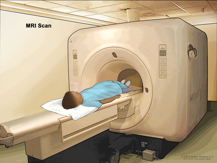
Childhood Soft Tissue Sarcoma Treatment (PDQ®)–Health Professional Version

Childhood Soft Tissue Sarcoma Treatment (PDQ®)–Health Professional Version

General Information About Childhood Soft Tissue Sarcoma

Dramatic improvements in survival have been achieved for children and adolescents with cancer. Between 1975 and 2020, childhood cancer mortality decreased by more than 50%.[13] Childhood and adolescent cancer survivors require close monitoring because cancer therapy side effects may persist or develop months or years after treatment. For information about the incidence, type, and monitoring of late effects in childhood and adolescent cancer survivors, see Late Effects of Treatment for Childhood Cancer.

Pediatric soft tissue sarcomas are a heterogenous group of malignant tumors that originate from primitive mesenchymal tissue and account for 6% of all childhood tumors (rhabdomyosarcomas, 3%; other soft tissue sarcomas, 3%).[2] For more information, see the Histopathological Classification of Childhood Soft Tissue Sarcoma section.

Rhabdomyosarcoma, a tumor of striated muscle, is the most common soft tissue sarcoma in children. It accounts for 50% of the soft tissue sarcomas in children aged 0 to 14 years.[2] For more information, see Childhood Rhabdomyosarcoma Treatment.

In pediatrics, the remaining soft tissue sarcomas are commonly referred to as nonrhabdomyosarcomatous soft tissue sarcomas (NRSTS) and account for approximately 3.5% of all childhood tumors.[2,4] This summary discusses the treatment of NRSTS.

NRSTS are often classified according to the normal tissue types from which they are derived. These types include various connective tissues, peripheral nervous system tissue, smooth muscle tissue, and vascular tissue. The classification also includes undifferentiated tumors that are not clearly related to specific tissue types. For more information about vascular tumors in children, see Childhood Vascular Tumors Treatment.

Incidence of Soft Tissue Sarcoma by Age and Histology

The distribution of soft tissue sarcomas by histology and age, based on the Surveillance, Epidemiology, and End Results (SEER) Program information from 2000 to 2015, is depicted in Table 1. The distribution of histological subtypes by age is also shown in Figure 2.

Table 1. Age Distribution of Soft Tissue Sarcomas in Children Aged 0 to 19 Years (SEER 2000–2015)a
  Age <5 y Age 5–9 y Age 10–14 y Age 15–19 y Age <20 y All Ages (Including Adults)
pPNET = peripheral primitive neuroectodermal tumors; SEER = Surveillance, Epidemiology, and End Results.
aSource: SEER database.[5]
All soft tissue and other extraosseous sarcomas 1,124 773 1,201 1,558 4,656 80,269
 
Rhabdomyosarcomas 668 417 382 327 1,794 3,284
Fibrosarcomas, peripheral nerve, and other fibrous neoplasms 137 64 112 181 494 6,645
  Fibroblastic and myofibroblastic tumors 114 33 41 77 265   4,228
  Nerve sheath tumors 23 31 70 102 226   2,303
  Other fibromatous neoplasms 0 0 1 2 3   114
Kaposi sarcoma 2 1 2 10 15 7,722
Other specified soft tissue sarcomas 237 238 559 865 1,899 49,004
  Ewing tumor and Askin tumor of soft tissue 37 36 72 113 258   596
  pPNET of soft tissue 24 23 42 56 145   402
  Extrarenal rhabdoid tumor 75 8 9 4 96   205
  Liposarcomas 4 6 37 79 126   10,749
  Fibrohistiocytic tumors 43 73 142 223 481   13,531
  Leiomyosarcomas 11 14 19 41 85   14,107
  Synovial sarcomas 12 39 141 210 402   2,608
  Blood vessel tumors 12 9 11 32 64   4,238
  Osseous and chondromatous neoplasms of soft tissue 1 6 16 14 37   1,018
  Alveolar soft parts sarcoma 4 5 22 33 64   211
  Miscellaneous soft tissue sarcomas 14 19 48 60 141   1,339
Unspecified soft tissue sarcomas 80 53 146 175 454 13,614

Soft tissue sarcomas include both rhabdomyosarcomas and NRSTS. NRSTS are more common in adolescents and adults.[6] Most of the information regarding treatment and natural history of the disease in younger patients has been based on studies in adult patients. The distributions of soft tissue sarcomas by age according to stage (Figure 1), histological subtype (Figure 2), and tumor site (Figure 3) are shown below.[7]

EnlargeChart showing the distribution of nonrhabdomyosarcomatous soft tissue sarcomas by age according to stage.
Figure 1. The distribution of soft tissue sarcomas by age according to stage.
EnlargeChart showing the distribution of nonrhabdomyosarcomatous soft tissue sarcomas by age according to histologic subtype.
Figure 2. The distribution of soft tissue sarcomas by age according to histological subtype.
EnlargeChart showing the distribution of nonrhabdomyosarcomatous soft tissue sarcomas by age according to tumor site.
Figure 3. The distribution of soft tissue sarcomas by age according to tumor site.

Risk Factors

Some genetic factors and external exposures have been associated with the development of NRSTS, including the following:

  • Genetic factors:
    • Li-Fraumeni syndrome: Patients with Li-Fraumeni syndrome (usually resulting from heritable cancer-associated changes of the TP53 tumor suppressor gene) have an increased risk of developing soft tissue tumors (mostly NRSTS), bone sarcomas, breast cancer, brain tumors, and acute leukemia.[8,9]
    • Familial adenomatous polyposis: Patients with familial adenomatous polyposis are at increased risk of developing desmoid-type fibromatosis.[10]
    • RB1 gene: Germline pathogenic variants of the RB1 gene have been associated with an increased risk of developing soft tissue sarcoma, particularly leiomyosarcoma, and the risk appears higher among those younger than 1 year who were treated with alkylating agents.[11,12]
    • SMARCB1 gene: Germline pathogenic variants or deletions of the SMARCB1 gene are associated with an increased risk of developing extrarenal rhabdoid tumors.[13] For more information about SMARCB1 and rhabdoid tumor predisposition syndrome type 1, see Rhabdoid Tumor Predisposition Syndrome Type 1.
    • Neurofibromatosis type 1: Approximately 4% of patients with neurofibromatosis type 1 develop malignant peripheral nerve sheath tumors, which usually develop after a long latency. Some patients develop multiple lesions.[1416]
    • Werner syndrome: Werner syndrome is characterized by spontaneous chromosomal instability, resulting in increased susceptibility to cancer and premature aging. An excess of soft tissue sarcomas has been reported in patients with Werner syndrome.[17]
    • Tuberous sclerosis complex: Tuberous sclerosis complex is associated with the development of various tumors showing perivascular epithelioid cell differentiation (PEComas), including lymphangioleiomyomatosis and hepatic and renal angiomyolipomas.[1820]
    • Adenosine deaminase–deficient severe combined immunodeficiency: Patients with adenosine deaminase–deficient severe combined immunodeficiency are at increased risk of developing multicentric dermatofibrosarcoma protuberans, which usually presents at an average age of 8.9 years.[21]
  • External exposures:
    • Radiation: Some NRSTS (particularly malignant fibrous histiocytoma) can develop within a previously irradiated site.[2226]
    • Epstein-Barr virus (EBV) infection in patients with AIDS: Some NRSTS (e.g., leiomyosarcoma) have been linked to EBV infection in patients with AIDS.[22,27]

Clinical Presentation

NRSTS can develop in any part of the body, but they arise most commonly in the trunk and extremities.[2830] Although rare, these tumors can arise in brain tissue and are treated according to the histological type.[31]

NRSTS can present initially as an asymptomatic solid mass, or they may be symptomatic because of local invasion or impact on adjacent anatomical structures. Systemic symptoms (e.g., fever, weight loss, and night sweats) are rare. Hypoglycemia and hypophosphatemic rickets have been reported in cases of hemangiopericytoma, which was identified as a solitary fibrous tumor and is now included within myofibroma in the revised World Health Organization (WHO) classification. Hyperglycemia has been noted in patients with fibrosarcoma of the lung.[32]

Diagnostic and Staging Evaluation

When a suspicious lesion is identified, it is crucial to perform a complete workup, followed by adequate biopsy. The lesion is imaged before initiating any intervention using the following procedures:

  • Plain films. Plain films can be used to rule out bone involvement and detect calcifications that may be seen in soft tissue tumors such as extraskeletal osteosarcoma or synovial sarcoma.
  • Computed tomography (CT). Chest CT is essential to assess the presence of metastases. An abdominal CT can be used to image intra-abdominal tumors, such as liposarcoma. Patients with NRSTS who were treated in 11 centers as part of the European paediatric Soft Tissue Sarcoma Study Group (EpSSG) were retrospectively assessed to evaluate the impact of indeterminate pulmonary nodules identified on chest CT.[33] Of the 206 patients examined, 109 (52.9%) did not have any nodules, 78 (38%) had at least one indeterminate nodule, and 19 (9.2%) had nodules meeting the definition of metastases. The 5-year event-free survival (EFS) rate was 78.5% (95% confidence interval [CI], 69.4%–85.1%) for patients without nodules and 69.6% (95% CI, 57.9%–78.7%) for patients with indeterminate nodules (P = .135). The 5-year overall survival (OS) rate was 87.4% (95% CI, 79.3%–92.5%) for patients without nodules and 79.0% (95% CI, 67.5%–86.8%) for patients with indeterminate nodules (P = .086).
  • Magnetic resonance imaging (MRI). MRI may be essential for a surgeon to achieve adequate surgical margins. MRI can be used to image intra-abdominal tumors, such as liposarcoma, and is essential for extremity lesions.
  • Positron emission tomography (PET) scan and bone scan. In a retrospective study, 46 PET scans were completed in 25 pediatric patients with soft tissue sarcoma.[34] The positive predictive value of finding metastatic disease was 89%, and the negative predictive value was 67%. A small retrospective study of nine patients with NRSTS suggested that PET-CT was more accurate and cost-effective than either modality alone in identifying distant metastatic disease.[35] The use of this modality in pediatric NRSTS has not been studied prospectively.

The imaging characteristics of some tumors can be highly suggestive of that particular diagnosis. For example, the imaging characteristics of pediatric low-grade fibromyxoid sarcoma and alveolar soft part sarcoma have been described and can aid in the diagnosis of these rare neoplasms.[36]

Biopsy strategies

Although NRSTS are pathologically distinct from rhabdomyosarcoma and Ewing sarcoma, the classification of childhood NRSTS type is often difficult. Core-needle biopsy, incisional biopsy, or excisional biopsy can be used to diagnose NRSTS. If possible, the surgeon who will perform the definitive resection needs to be involved in the biopsy decision. Poorly placed incisional or needle biopsies may adversely affect the ability to achieve negative margins.

Needle biopsy techniques must ensure adequate tissue sampling. Given the diagnostic importance of translocations and other molecular changes, a core-needle biopsy or small incisional biopsy that obtains adequate tumor tissue is crucial to allow for conventional histological and immunocytochemical analysis and other studies such as light and electron microscopy, cytogenetics, fluorescence in situ hybridization, and molecular pathology.[37,38]

The acquisition of multiple cores of tissue may be required. Of 530 suspected soft tissue masses in (largely adult) patients who underwent core-needle biopsies, 426 (80%) were proven to be soft tissue tumors, 225 (52.8%) of which were malignant. Core-needle biopsy was able to differentiate soft tissue sarcomas from benign lesions with a sensitivity of 96.3% and a specificity of 99.4%. Tumor subtype was accurately assigned in 89.5% of benign lesions and in 88% of soft tissue sarcomas. The biopsy complication rate was 0.4%.[39]

Considerations related to a biopsy procedure are as follows:

  • Core-needle biopsy for a deep-seated tumor can lead to formation of a hematoma, which affects subsequent resection and/or radiation (because the hematoma should be covered in the irradiated volume).
  • Fine-needle biopsy is usually not recommended because it is difficult to determine the accurate histological diagnosis and grade of the tumor in this heterogeneous group of tumors.
  • Image guidance using ultrasonography, CT scan, or MRI may be necessary to ensure a representative biopsy.[40] Image guidance is particularly helpful in deep lesions and to avoid cystic changes or necrotic tumors.[41]
  • Incisional biopsies must not compromise subsequent wide local excision.
  • Excisional biopsy of the lesion is only appropriate for small superficial lesions (<3 cm in size) and are discouraged.[42,43] If an excisional biopsy is contemplated, then MRI of the area is recommended to define the area of involvement as subsequent surgery or radiation therapy may be needed.
  • Various institutional series have demonstrated the feasibility and effectiveness of sentinel lymph node biopsy as a staging procedure in pediatric patients with soft tissue sarcomas.[4449] The utility of sentinel node biopsy is currently limited to epithelioid sarcoma, clear cell sarcoma, and rhabdomyosarcoma of the trunk and extremities.[50]

    In a prospective study of pediatric patients with sarcoma who underwent sentinel lymph node biopsy, 28 patients were examined. Sentinel lymph node biopsy was positive in 7 of the 28 patients, including 3 patients (43%) who had negative PET-CT scans. PET-CT overestimated and suggested nodal involvement in 14 patients, more than what was confirmed by sentinel lymph node biopsy. The findings from the sentinel lymph node biopsies resulted in altering therapy for all seven patients who were determined to have metastatic disease. As indicated by previous reports, epithelioid sarcoma and clear cell sarcoma were the two NRSTS included in this study.[50]

  • In the ARST0332 (NCT00346164) study, patients with epithelioid sarcoma, clear cell sarcoma, or radiographically enlarged nodes underwent regional node sampling. Nodal metastases were identified in 20 patients (3.8%), and all but one of these patients had radiographic evidence of nodal involvement. The most common histologies included epithelioid sarcoma (18%), angiosarcoma (17%), and clear cell sarcoma (14%). Patients with isolated nodal metastases had a similar outcome to those who did not have distant metastases (5-year OS rates, 85% vs. 87%). Sentinel lymph node biopsies were encouraged but not required for this study. A sentinel lymph node biopsy was not done in most patients because they had clinically enlarged nodes. Of note, three patients without clinical evidence of lymph node metastasis at study entry experienced lymph node basin failure. One of these patients had three lymph nodes in two different lymph node basins sampled by sentinel lymph node biopsy that were pathologically negative.[51]

    Transverse extremity incisions are avoided to reduce skin loss at re-excision and because they require a greater cross-sectional volume of tissue to be covered in the radiation field. Other extensive surgical procedures are also avoided before definitive diagnosis.

For these reasons, open biopsy or multiple core-needle biopsies are strongly encouraged so that adequate tumor tissue can be obtained to allow crucial studies to be performed and to avoid limiting future treatment options.

Unplanned resection

In children with unplanned resection of NRSTS, primary re-excision is frequently recommended because many patients will have tumor present in the re-excision specimen.[52,53] A single-institution analysis of adolescents and adults compared patients who had unplanned excisions of soft tissue sarcoma to stage-matched controls. In this retrospective analysis, unplanned initial excision of soft tissue sarcoma resulted in increased risk of local recurrence, metastasis, and death. This increased risk was greatest for high-grade tumors.[54][Level of evidence C1] In this case, a second resection is expected.

Chromosomal abnormalities

Many NRSTS are characterized by chromosomal abnormalities. Some of these chromosomal translocations lead to a fusion of two disparate genes. The resulting fusion transcript can be readily detected by using polymerase chain reaction–based techniques, thus facilitating the diagnosis of those neoplasms that have translocations.

Some of the most frequent aberrations seen in NRSTS are listed in Table 2.

Table 2. Frequent Chromosomal Aberrations Seen in Nonrhabdomyosarcomatous Soft Tissue Sarcomaa
Histology Chromosomal Aberrations Genes Involved
aAdapted from Sandberg,[55] Slater et al.,[56] Mertens et al.,[57] Romeo,[58] and Schaefer et al.[59]
Alveolar soft part sarcoma t(x;17)(p11.2;q25) ASPSCR1::TFE3 [6062]
Angiomatoid fibrous histiocytoma t(12;16)(q13;p11), t(2;22)(q33;q12), t(12;22)(q13;q12) FUS::ATF1, EWSR1::CREB1,[63] EWSR1::ATF1
BCOR-rearranged sarcomas inv(X)(p11.4;p11.2) BCOR::CCNB3
CIC-rearranged sarcomas t(4;19)(q35;q13), t(10;19)(q26;q13) CIC::DUX4
Clear cell sarcoma t(12;22)(q13;q12), t(2;22)(q33;q12) EWSR1::ATF1, EWSR1::CREB1 [64]
Congenital (infantile) fibrosarcoma/mesoblastic nephroma t(12;15)(p13;q25) ETV6::NTRK3
Dermatofibrosarcoma protuberans t(17;22)(q22;q13) COL1A1::PDGFB
Desmoid fibromatosis Trisomy 8 or 20, loss of 5q21 CTNNB1 or APC variants
Desmoplastic small round cell tumors t(11;22)(p13;q12) EWSR1::WT1 [65,66]
Epithelioid hemangioendothelioma t(1;3)(p36;q25) [67] WWTR1::CAMTA1
Epithelioid sarcoma Inactivation of SMARCB1 SMARCB1
Extraskeletal myxoid chondrosarcoma t(9;22)(q22;q12), t(9;17)(q22;q11), t(9;15)(q22;q21), t(3;9)(q11;q22) EWSR1::NR4A3, TAF2N::NR4A3, TCF12::NR4A3, TFG::NR4A3
Hemangiopericytoma (myofibroma) t(12;19)(q13;q13.3) and t(13;22)(q22;q13.3) LMNA::NTRK1 [68]
Infantile fibrosarcoma t(12;15)(p13;q25) ETV6::NTRK3
Inflammatory myofibroblastic tumor t(1;2)(q23;q23), t(2;19)(q23;q13), t(2;17)(q23;q23), t(2;2)(p23;q13), t(2;11)(p23;p15) [69] TPM3::ALK, TPM4::ALK, CLTC::ALK, RANBP2::ALK, CARS1::ALK, RAS, ROS1 [70,71]
Infantile myofibromatosis Gain-of-function variants PDGFRB [72]
Low-grade fibromyxoid sarcoma t(7;16)(q33;p11), t(11;16)(p11;p11) FUS::CREB3L2, FUS::CREB3L1
Malignant peripheral nerve sheath tumor 17q11.2, loss or rearrangement of 10p, 11q, 17q, 22q NF1
Mesenchymal chondrosarcoma Del(8)(q13.3q21.1) HEY1::NCOA2
Myoepithelioma t(19;22)(q13;q12), t(1;22)(q23;q12), t(6;22)(p21;q12) EWSR1::ZNF44, EWSR1::PBX1, EWSR1::POU5F1
Myxoid/round cell liposarcoma t(12;16)(q13;p11), t(12;22)(q13;q12) FUS::DDIT3, EWSR1::DDIT3
Primitive myxoid mesenchymal tumor of infancy Internal tandem duplication BCOR
Rhabdoid tumor Inactivation of SMARCB1 SMARCB1
Sclerosing epithelioid fibrosarcoma t(11;22)(p11;q12), t(19;22)(p13;q12) EWSR1::CREB3L1, EWSR1::CREB3L3
Solitary fibrous tumor inv(12)(q13q13) NAB2::STAT6
Synovial sarcoma t(x;18)(p11.2;q11.2) SS18::SSX
Tenosynovial giant cell tumor t(1;2)(p13;q35) COL6A3::CSF1

Prognosis and Prognostic Factors

The prognosis of NRSTS varies greatly depending on the following factors:[7375]

  • Site of the primary tumor.
  • Tumor size.
  • Tumor grade. For more information, see the Soft Tissue Sarcoma Tumor Pathological Grading System section.
  • Tumor histology.
  • Depth of tumor invasion.
  • Presence of metastases and site of the metastatic tumor.
  • Resectability of the tumor.
  • Use of radiation therapy.

In a review of a large adult series of NRSTS, patients with superficial extremity sarcomas had a better prognosis than did patients with deep tumors. This may be a reflection of differences in resectability. Thus, in addition to grade and size, the depth of invasion of the tumor should be considered.[76]

Data specific to NRSTS in children and adolescents are difficult to separate. Several adult and pediatric series have shown that patients with large or invasive tumors have a significantly worse prognosis than do those with small, noninvasive tumors. A retrospective review of soft tissue sarcomas (rhabdomyosarcoma and NRSTS) in children and adolescents suggests that the 5-cm cutoff used for adults with soft tissue sarcoma may not be ideal for smaller children, especially infants. The review identified an interaction between tumor diameter and body surface area.[77] This relationship has been questioned in a rhabdomyosarcoma study and requires further study to determine the therapeutic implications of the observation.[78]

Some pediatric NRSTS are associated with a better outcome. For instance, patients with infantile fibrosarcoma who present at age 4 years or younger have an excellent prognosis. This excellent outcome occurs because surgery alone can cure a significant number of these patients and infantile fibrosarcoma is highly chemosensitive. This tumor also responds well to larotrectinib, a specific tropomyosin receptor kinase inhibitor.[22,79]

Prognosis based on the Children’s Oncology Group (COG) ARST0332 trial

Soft tissue sarcomas in older children and adolescents often behave similarly to those in adult patients.[22,80] A large, prospective, multinational COG study (ARST0332 [NCT00346164]) enrolled newly diagnosed patients younger than 30 years. Patients were assigned to treatment based on their risk group. Risk groups were defined by the presence of metastasis, tumor resectability and margins, and tumor size and grade. For more information, see Figure 4.[81][Level of evidence B4]

EnlargeDiagram showing risk group and treatment assignment for the Children’s Oncology Group ARST0332 trial.
Figure 4. Risk group and treatment assignment for the Children’s Oncology Group ARST0332 trial. Reprinted from The Lancet Oncology, Volume 21 (Issue 1), Spunt SL, Million L, Chi YY, et al., A risk-based treatment strategy for non-rhabdomyosarcoma soft-tissue sarcomas in patients younger than 30 years (ARST0332): a Children’s Oncology Group prospective study, Pages 145–161, Copyright © 2020, with permission from Elsevier.

Each patient was assigned to one of three risk groups and one of four treatment groups. The risk groups were as follows:[81]

  1. Low risk: Nonmetastatic R0 (resection was complete with negative microscopic margins) or R1 (microscopically positive margins) low-grade tumor, or ≤5 cm R1 high-grade tumor.
  2. Intermediate risk: Nonmetastatic R0 or R1 >5 cm high-grade tumor, or unresected tumor of any size or grade.
  3. High risk: Metastatic tumor.

The treatment groups were as follows:

  1. Surgery alone (n = 205).
  2. Radiation therapy (55.8 Gy) (n = 17).
  3. Chemoradiation therapy (chemotherapy and 55.8 Gy radiation therapy) (n = 111).
  4. Neoadjuvant chemoradiation therapy (chemotherapy and 45 Gy radiation therapy, then surgery and radiation therapy boost based on margins with continued chemotherapy) (n = 196).

Chemotherapy included six cycles of ifosfamide (3 g/m2 per dose) given intravenously on days 1 through 3 and five cycles of doxorubicin (37.5 mg/m2 per dose) given intravenously on days 1 to 2 every 3 weeks, with the sequence adjusted based on the timing of surgery or radiation therapy.

For the 550 patients enrolled, 529 evaluable patients were included in the analysis. At a median follow-up of 6.5 years (interquartile range [IQR], 4.9–7.9), the survival results are shown in Table 3.

Table 3. Survival Results for the Children’s Oncology Group ARST0332 Trial
  5-Year Event-Free Survival 5-Year Overall Survival
Risk Group Events/Patients Estimate, % (95% CI) Events/Patients Estimate, % (95% CI)
CI = confidence interval; R0 = completely excised with negative microscopic margins; R1 = grossly excised but with positive microscopic margins; R2 = less than complete gross excision.
Low 26/222 88.9 (84.0–93.8) 10/222 96.2 (93.2–99.2)
Intermediate 84/227 65.0 (58.2–71.8) 55/227 79.2 (73.4–85.0)
High 63/80 21.2 (11.4–31.1) 52/80 35.5 (23.6–47.4)
Surgical Margin
R0 44/252 83.6 (78.3–89.0) 22/252 92.8 (89.1–96.5)
R1 29/81 66.2 (54.8–77.5) 17/81 79.7 (70.0–89.5)
R2 100/196 49.2 (41.4–57.0) 78/196 62.7 (55.2–70.3)

The COG ARST0332 trial was a risk-based stratification study. Overall, local control after radiation therapy was as follows: R0, 106 of 109 patients (97%); R1, 51 of 60 patients (85%); and R2/unresectable, 2 of 6 patients (33%). Local recurrence predictors included extent of delayed resection (P < .001), imaging response before delayed surgery (P < .001), histological subtype (P < .001), and no radiation therapy (P = .046). The 5-year EFS was significantly lower for patients unable to undergo R0 or R1 resection (P = .0003).[82]

Pediatric patients with unresected localized NRSTS have a poor outcome. Only about one-third of patients treated with multimodality therapy remain disease free.[73,83]; [84,85][Level of evidence C1] In an Italian review of 30 patients with NRSTS at visceral sites, only ten patients survived at 5 years. Unfavorable prognostic factors included inability to achieve complete resection, large tumor size, tumor invasion, histological subtype, and lung-pleura sites.[86][Level of evidence C1]

Prognosis based on the European paediatric Soft Tissue Sarcoma Study Group (EpSSG) NRSTS 2005 study

The EpSSG conducted a prospective trial for patients younger than 21 years with NRSTS. They reported an analysis of 206 patients with synovial sarcoma and 363 with adult-type NRSTS. Patients were treated according to assigned risk groups. For more information, see Figure 5.[87] With a median follow-up of 80 months (interquartile range, 54.3–111.3) for the 467 surviving patients, the 5-year EFS rate was 73.7% (95% CI, 69.7%–77.2%), and the OS rate was 83.8% (95% CI, 80.3%–86.7%). The survival by treatment groups are shown in Table 4.[87]

EnlargeFigure showing a treatment plan for patients with synovial sarcoma or adult-type non-rhabdomyosarcoma soft tissue sarcomas.
Figure 5. Treatment plan for patients with synovial sarcoma or adult-type non-rhabdomyosarcoma soft tissue sarcomas. Patients were divided into four treatment groups based on surgical stage, tumour size, nodal involvement, tumour grade (according to the Fédération Nationale des Centres de Lutte Contre le Cancer grading system for adult-type non-rhabdomyosarcoma soft tissue sarcomas), and tumour site (for synovial sarcoma). I+D = ifosfamide (3.0 g/m2 per day intravenously for 3 days) plus doxorubicin (37.5 mg/m2 per day intravenously for 2 days). I = ifosfamide (3.0 g/m2 per day intravenously for 2 days). IRS = Intergroup Rhabdomyosarcoma Study. N1 = nodal involvement. S = delayed surgery. Reprinted from The Lancet Child & Adolescent Health, Volume 5, Issue 8, Ferrari A, van Noesel MM, Brennan B, et al., Paediatric non-rhabdomyosarcoma soft tissue sarcomas: the prospective NRSTS 2005 study by the European paediatric Soft Tissue Sarcoma Study Group (EpSSG), Pages 546-558, Copyright 2021, with permission from Elsevier.
Table 4. Survival Outcomes by Treatment Groups in the EpSSG NRSTS 2005 Study
Treatment Group 5-Year Event-Free Survival Rate (95% CI) 5-Year Overall Survival Rate (95% CI) Local Recurrence Rate
CI = confidence interval; EpSSG = European paediatric Soft Tissue Sarcoma Study Group; NRSTS = nonrhabdomyosarcomatous soft tissue sarcomas.
Surgery alone 91.4% (87.0%–94.4%) 98.1% (95.0%–99.3%) 7.6% (19/250)
Adjuvant radiation therapy alone (n = 17) 75.5% (46.9%–90.1%) 88.2% (60.6%–96.9%) 6.7% (1/15)
Adjuvant chemotherapy ± radiation therapy (n = 93) 65.6% (54.8%–74.5%) 75.8% (65.3%–83.5%) 10.8% (7/65)
Neoadjuvant chemotherapy ± radiation therapy (n = 209) 56.4% (49.3%–63.0%) 70.4% (63.3%–76.4%) 14.2% (16/113)

Treatment failures specifically for the neoadjuvant therapy treatment groups are shown in Table 5.[87]

Table 5. Treatment Failures for Specific Neoadjuvant Therapy Groups in the EpSSG NRSTS 2005 Studya
Treatment Local Failure (No. of Patients) Local + Metastatic Failure (No. of Patients) Metastatic Failure (No. of Patients)
EpSSG = European paediatric Soft Tissue Sarcoma Study Group; No. = number; NRSTS = nonrhabdomyosarcomatous soft tissue sarcomas.
aAdapted from Ferrari et al.[87]
Radiation therapy alone (n = 21) 7 2 4
Delayed surgery followed by radiation therapy (n = 104) 16 6 8
Delayed surgery alone (n = 48) 8 3 8
No local treatment (n = 16) 12 4 0
Preoperative radiation therapy followed by delayed surgery (n = 20) 4 0 6

The authors concluded that adjuvant therapy (radiation therapy and chemotherapy) could safely be omitted in the group of patients assigned to surgery alone. Their criteria included the following:[87]

  • Synovial cell: Intergroup Rhabdomyosarcoma Study (IRS) group I tumor size <5 cm.
  • Adult-type NRSTS: IRS group I tumor size <5 cm, any grade.
  • Adult-type NRSTS: IRS group I tumor size >5 cm, tumor grade I.
  • Adult-type NRSTS: IRS group II any tumor size, tumor grade I.

They also concluded that improving the outcome for patients with high-risk, initially resected, adult-type NRSTS and those with initially unresected disease remains a major clinical challenge.[87]

In a pooled analysis from U.S. and European pediatric centers, outcomes were better for patients whose tumor removal procedure was deemed complete than for patients whose tumor removal was incomplete. Outcomes were better for patients who received radiation therapy than for patients who did not.[84][Level of evidence C1]

Because long-term morbidity must be minimized while disease-free survival is maximized, the ideal therapy for each patient must be carefully and individually determined using these prognostic factors before initiating therapy.[29,8892]

References
  1. Smith MA, Altekruse SF, Adamson PC, et al.: Declining childhood and adolescent cancer mortality. Cancer 120 (16): 2497-506, 2014. [PUBMED Abstract]
  2. National Cancer Institute: NCCR*Explorer: An interactive website for NCCR cancer statistics. Bethesda, MD: National Cancer Institute. Available online. Last accessed February 25, 2025.
  3. Surveillance Research Program, National Cancer Institute: SEER*Explorer: An interactive website for SEER cancer statistics. Bethesda, MD: National Cancer Institute. Available online. Last accessed December 30, 2024.
  4. Hawkins DS, Black JO, Orbach D, et al.: Nonrhabdomyosarcoma soft-tissue sarcomas. In: Blaney SM, Helman LJ, Adamson PC, eds.: Pizzo and Poplack’s Pediatric Oncology. 8th ed. Wolters Kluwer, 2020, pp 721-46.
  5. Surveillance, Epidemiology, and End Results (SEER) Program: SEER*Stat Database: Incidence – SEER 18 Regs Research Data + Hurricane Katrina Impacted Louisiana Cases, Nov 2017 Sub (1973-2015 varying) – Linked To County Attributes – Total U.S., 1969-2016 Counties [Database]. National Cancer Institute, DCCPS, Surveillance Research Program, released April 2018, based on the November 2017 submission. Available online. Last accessed October 12, 2022.
  6. Weiss SW, Goldblum JR: General considerations. In: Weiss SW, Goldblum JR: Enzinger and Weiss’s Soft Tissue Tumors. 5th ed. Mosby, 2008, pp 1-14.
  7. Ferrari A, Sultan I, Huang TT, et al.: Soft tissue sarcoma across the age spectrum: a population-based study from the Surveillance Epidemiology and End Results database. Pediatr Blood Cancer 57 (6): 943-9, 2011. [PUBMED Abstract]
  8. Chang F, Syrjänen S, Syrjänen K: Implications of the p53 tumor-suppressor gene in clinical oncology. J Clin Oncol 13 (4): 1009-22, 1995. [PUBMED Abstract]
  9. Plon SE, Malkin D: Childhood cancer and hereditary. In: Pizzo PA, Poplack DG, eds.: Principles and Practice of Pediatric Oncology. 7th ed. Lippincott Williams and Wilkins, 2015, pp 13-31.
  10. Groen EJ, Roos A, Muntinghe FL, et al.: Extra-intestinal manifestations of familial adenomatous polyposis. Ann Surg Oncol 15 (9): 2439-50, 2008. [PUBMED Abstract]
  11. Kleinerman RA, Tucker MA, Abramson DH, et al.: Risk of soft tissue sarcomas by individual subtype in survivors of hereditary retinoblastoma. J Natl Cancer Inst 99 (1): 24-31, 2007. [PUBMED Abstract]
  12. Wong JR, Morton LM, Tucker MA, et al.: Risk of subsequent malignant neoplasms in long-term hereditary retinoblastoma survivors after chemotherapy and radiotherapy. J Clin Oncol 32 (29): 3284-90, 2014. [PUBMED Abstract]
  13. Eaton KW, Tooke LS, Wainwright LM, et al.: Spectrum of SMARCB1/INI1 mutations in familial and sporadic rhabdoid tumors. Pediatr Blood Cancer 56 (1): 7-15, 2011. [PUBMED Abstract]
  14. Weiss SW, Goldblum JR: Benign tumors of peripheral nerves. In: Weiss SW, Goldblum JR: Enzinger and Weiss’s Soft Tissue Tumors. 5th ed. Mosby, 2008, pp 825-901.
  15. deCou JM, Rao BN, Parham DM, et al.: Malignant peripheral nerve sheath tumors: the St. Jude Children’s Research Hospital experience. Ann Surg Oncol 2 (6): 524-9, 1995. [PUBMED Abstract]
  16. Stark AM, Buhl R, Hugo HH, et al.: Malignant peripheral nerve sheath tumours–report of 8 cases and review of the literature. Acta Neurochir (Wien) 143 (4): 357-63; discussion 363-4, 2001. [PUBMED Abstract]
  17. Goto M, Miller RW, Ishikawa Y, et al.: Excess of rare cancers in Werner syndrome (adult progeria). Cancer Epidemiol Biomarkers Prev 5 (4): 239-46, 1996. [PUBMED Abstract]
  18. Fricke BL, Donnelly LF, Casper KA, et al.: Frequency and imaging appearance of hepatic angiomyolipomas in pediatric and adult patients with tuberous sclerosis. AJR Am J Roentgenol 182 (4): 1027-30, 2004. [PUBMED Abstract]
  19. Adriaensen ME, Schaefer-Prokop CM, Duyndam DA, et al.: Radiological evidence of lymphangioleiomyomatosis in female and male patients with tuberous sclerosis complex. Clin Radiol 66 (7): 625-8, 2011. [PUBMED Abstract]
  20. Hornick JL, Fletcher CD: PEComa: what do we know so far? Histopathology 48 (1): 75-82, 2006. [PUBMED Abstract]
  21. Kesserwan C, Sokolic R, Cowen EW, et al.: Multicentric dermatofibrosarcoma protuberans in patients with adenosine deaminase-deficient severe combined immune deficiency. J Allergy Clin Immunol 129 (3): 762-769.e1, 2012. [PUBMED Abstract]
  22. Spunt SL, Million L, Coffin C: The nonrhabdomyosarcoma soft tissue sarcoma. In: Pizzo PA, Poplack DG, eds.: Principles and Practice of Pediatric Oncology. 7th ed. Lippincott Williams and Wilkins, 2015, pp 827-54.
  23. Weiss SW, Goldblum JR: Malignant fibrous histiocytoma (pleomorphic undifferentiated sarcoma). In: Weiss SW, Goldblum JR: Enzinger and Weiss’s Soft Tissue Tumors. 5th ed. Mosby, 2008, pp 403-27.
  24. Tukenova M, Guibout C, Hawkins M, et al.: Radiation therapy and late mortality from second sarcoma, carcinoma, and hematological malignancies after a solid cancer in childhood. Int J Radiat Oncol Biol Phys 80 (2): 339-46, 2011. [PUBMED Abstract]
  25. Bartkowiak D, Humble N, Suhr P, et al.: Second cancer after radiotherapy, 1981-2007. Radiother Oncol 105 (1): 122-6, 2012. [PUBMED Abstract]
  26. Casey DL, Friedman DN, Moskowitz CS, et al.: Second cancer risk in childhood cancer survivors treated with intensity-modulated radiation therapy (IMRT). Pediatr Blood Cancer 62 (2): 311-316, 2015. [PUBMED Abstract]
  27. McClain KL, Leach CT, Jenson HB, et al.: Association of Epstein-Barr virus with leiomyosarcomas in children with AIDS. N Engl J Med 332 (1): 12-8, 1995. [PUBMED Abstract]
  28. Dillon P, Maurer H, Jenkins J, et al.: A prospective study of nonrhabdomyosarcoma soft tissue sarcomas in the pediatric age group. J Pediatr Surg 27 (2): 241-4; discussion 244-5, 1992. [PUBMED Abstract]
  29. Rao BN: Nonrhabdomyosarcoma in children: prognostic factors influencing survival. Semin Surg Oncol 9 (6): 524-31, 1993 Nov-Dec. [PUBMED Abstract]
  30. Zeytoonjian T, Mankin HJ, Gebhardt MC, et al.: Distal lower extremity sarcomas: frequency of occurrence and patient survival rate. Foot Ankle Int 25 (5): 325-30, 2004. [PUBMED Abstract]
  31. Benesch M, von Bueren AO, Dantonello T, et al.: Primary intracranial soft tissue sarcoma in children and adolescents: a cooperative analysis of the European CWS and HIT study groups. J Neurooncol 111 (3): 337-45, 2013. [PUBMED Abstract]
  32. Weiss SW, Goldblum JR: Miscellaneous tumors of intermediate malignancy. In: Weiss SW, Goldblum JR: Enzinger and Weiss’s Soft Tissue Tumors. 5th ed. Mosby, 2008, pp 1093-1160.
  33. Giraudo C, Schoot R, Cardoen L, et al.: Indeterminate pulmonary nodules in non-rhabdomyosarcoma soft tissue sarcoma: A study of the European paediatric Soft Tissue Sarcoma Study Group. Cancer 130 (4): 597-608, 2024. [PUBMED Abstract]
  34. Mody RJ, Bui C, Hutchinson RJ, et al.: FDG PET imaging of childhood sarcomas. Pediatr Blood Cancer 54 (2): 222-7, 2010. [PUBMED Abstract]
  35. Tateishi U, Hosono A, Makimoto A, et al.: Accuracy of 18F fluorodeoxyglucose positron emission tomography/computed tomography in staging of pediatric sarcomas. J Pediatr Hematol Oncol 29 (9): 608-12, 2007. [PUBMED Abstract]
  36. Sargar K, Kao SC, Spunt SL, et al.: MRI and CT of Low-Grade Fibromyxoid Sarcoma in Children: A Report From Children’s Oncology Group Study ARST0332. AJR Am J Roentgenol 205 (2): 414-20, 2015. [PUBMED Abstract]
  37. Weiss SW, Goldblum JR: Enzinger and Weiss’s Soft Tissue Tumors. 5th ed. Mosby, 2008.
  38. Recommendations for the reporting of soft tissue sarcomas. Association of Directors of Anatomic and Surgical Pathology. Mod Pathol 11 (12): 1257-61, 1998. [PUBMED Abstract]
  39. Strauss DC, Qureshi YA, Hayes AJ, et al.: The role of core needle biopsy in the diagnosis of suspected soft tissue tumours. J Surg Oncol 102 (5): 523-9, 2010. [PUBMED Abstract]
  40. Chowdhury T, Barnacle A, Haque S, et al.: Ultrasound-guided core needle biopsy for the diagnosis of rhabdomyosarcoma in childhood. Pediatr Blood Cancer 53 (3): 356-60, 2009. [PUBMED Abstract]
  41. Tuttle R, Kane JM: Biopsy techniques for soft tissue and bowel sarcomas. J Surg Oncol 111 (5): 504-12, 2015. [PUBMED Abstract]
  42. Coffin CM, Dehner LP, O’Shea PA: Pediatric Soft Tissue Tumors: A Clinical, Pathological, and Therapeutic Approach. Williams and Wilkins, 1997.
  43. Smith LM, Watterson J, Scott SM: Medical and surgical management of pediatric soft tissue tumors. In: Coffin CM, Dehner LP, O’Shea PA: Pediatric Soft Tissue Tumors: A Clinical, Pathological, and Therapeutic Approach. Williams and Wilkins, 1997, pp 360-71.
  44. Neville HL, Andrassy RJ, Lally KP, et al.: Lymphatic mapping with sentinel node biopsy in pediatric patients. J Pediatr Surg 35 (6): 961-4, 2000. [PUBMED Abstract]
  45. Neville HL, Raney RB, Andrassy RJ, et al.: Multidisciplinary management of pediatric soft-tissue sarcoma. Oncology (Huntingt) 14 (10): 1471-81; discussion 1482-6, 1489-90, 2000. [PUBMED Abstract]
  46. Kayton ML, Delgado R, Busam K, et al.: Experience with 31 sentinel lymph node biopsies for sarcomas and carcinomas in pediatric patients. Cancer 112 (9): 2052-9, 2008. [PUBMED Abstract]
  47. Dall’Igna P, De Corti F, Alaggio R, et al.: Sentinel node biopsy in pediatric patients: the experience in a single institution. Eur J Pediatr Surg 24 (6): 482-7, 2014. [PUBMED Abstract]
  48. Parida L, Morrisson GT, Shammas A, et al.: Role of lymphoscintigraphy and sentinel lymph node biopsy in the management of pediatric melanoma and sarcoma. Pediatr Surg Int 28 (6): 571-8, 2012. [PUBMED Abstract]
  49. Alcorn KM, Deans KJ, Congeni A, et al.: Sentinel lymph node biopsy in pediatric soft tissue sarcoma patients: utility and concordance with imaging. J Pediatr Surg 48 (9): 1903-6, 2013. [PUBMED Abstract]
  50. Wagner LM, Kremer N, Gelfand MJ, et al.: Detection of lymph node metastases in pediatric and adolescent/young adult sarcoma: Sentinel lymph node biopsy versus fludeoxyglucose positron emission tomography imaging-A prospective trial. Cancer 123 (1): 155-160, 2017. [PUBMED Abstract]
  51. Alvarez E, He J, Spunt SL, et al.: Lymph node metastases in paediatric and young adult patients with non-rhabdomyosarcoma soft tissue sarcoma (NRSTS): Findings from Children’s Oncology Group (COG) study ARST0332. Eur J Cancer 180: 89-98, 2023. [PUBMED Abstract]
  52. Chui CH, Spunt SL, Liu T, et al.: Is reexcision in pediatric nonrhabdomyosarcoma soft tissue sarcoma necessary after an initial unplanned resection? J Pediatr Surg 37 (10): 1424-9, 2002. [PUBMED Abstract]
  53. Cecchetto G, Guglielmi M, Inserra A, et al.: Primary re-excision: the Italian experience in patients with localized soft-tissue sarcomas. Pediatr Surg Int 17 (7): 532-4, 2001. [PUBMED Abstract]
  54. Qureshi YA, Huddy JR, Miller JD, et al.: Unplanned excision of soft tissue sarcoma results in increased rates of local recurrence despite full further oncological treatment. Ann Surg Oncol 19 (3): 871-7, 2012. [PUBMED Abstract]
  55. Sandberg AA: Translocations in malignant tumors. Am J Pathol 159 (6): 1979-80, 2001. [PUBMED Abstract]
  56. Slater O, Shipley J: Clinical relevance of molecular genetics to paediatric sarcomas. J Clin Pathol 60 (11): 1187-94, 2007. [PUBMED Abstract]
  57. Mertens F, Antonescu CR, Hohenberger P, et al.: Translocation-related sarcomas. Semin Oncol 36 (4): 312-23, 2009. [PUBMED Abstract]
  58. Romeo S, Dei Tos AP: Clinical application of molecular pathology in sarcomas. Curr Opin Oncol 23 (4): 379-84, 2011. [PUBMED Abstract]
  59. Schaefer IM, Cote GM, Hornick JL: Contemporary Sarcoma Diagnosis, Genetics, and Genomics. J Clin Oncol 36 (2): 101-110, 2018. [PUBMED Abstract]
  60. Ladanyi M, Lui MY, Antonescu CR, et al.: The der(17)t(X;17)(p11;q25) of human alveolar soft part sarcoma fuses the TFE3 transcription factor gene to ASPL, a novel gene at 17q25. Oncogene 20 (1): 48-57, 2001. [PUBMED Abstract]
  61. Ladanyi M: The emerging molecular genetics of sarcoma translocations. Diagn Mol Pathol 4 (3): 162-73, 1995. [PUBMED Abstract]
  62. Williams A, Bartle G, Sumathi VP, et al.: Detection of ASPL/TFE3 fusion transcripts and the TFE3 antigen in formalin-fixed, paraffin-embedded tissue in a series of 18 cases of alveolar soft part sarcoma: useful diagnostic tools in cases with unusual histological features. Virchows Arch 458 (3): 291-300, 2011. [PUBMED Abstract]
  63. Antonescu CR, Dal Cin P, Nafa K, et al.: EWSR1-CREB1 is the predominant gene fusion in angiomatoid fibrous histiocytoma. Genes Chromosomes Cancer 46 (12): 1051-60, 2007. [PUBMED Abstract]
  64. Hisaoka M, Ishida T, Kuo TT, et al.: Clear cell sarcoma of soft tissue: a clinicopathologic, immunohistochemical, and molecular analysis of 33 cases. Am J Surg Pathol 32 (3): 452-60, 2008. [PUBMED Abstract]
  65. Barnoud R, Sabourin JC, Pasquier D, et al.: Immunohistochemical expression of WT1 by desmoplastic small round cell tumor: a comparative study with other small round cell tumors. Am J Surg Pathol 24 (6): 830-6, 2000. [PUBMED Abstract]
  66. Wang LL, Perlman EJ, Vujanic GM, et al.: Desmoplastic small round cell tumor of the kidney in childhood. Am J Surg Pathol 31 (4): 576-84, 2007. [PUBMED Abstract]
  67. Errani C, Zhang L, Sung YS, et al.: A novel WWTR1-CAMTA1 gene fusion is a consistent abnormality in epithelioid hemangioendothelioma of different anatomic sites. Genes Chromosomes Cancer 50 (8): 644-53, 2011. [PUBMED Abstract]
  68. Haller F, Knopf J, Ackermann A, et al.: Paediatric and adult soft tissue sarcomas with NTRK1 gene fusions: a subset of spindle cell sarcomas unified by a prominent myopericytic/haemangiopericytic pattern. J Pathol 238 (5): 700-10, 2016. [PUBMED Abstract]
  69. Jain S, Xu R, Prieto VG, et al.: Molecular classification of soft tissue sarcomas and its clinical applications. Int J Clin Exp Pathol 3 (4): 416-28, 2010. [PUBMED Abstract]
  70. Mariño-Enríquez A, Wang WL, Roy A, et al.: Epithelioid inflammatory myofibroblastic sarcoma: An aggressive intra-abdominal variant of inflammatory myofibroblastic tumor with nuclear membrane or perinuclear ALK. Am J Surg Pathol 35 (1): 135-44, 2011. [PUBMED Abstract]
  71. Lovly CM, Gupta A, Lipson D, et al.: Inflammatory myofibroblastic tumors harbor multiple potentially actionable kinase fusions. Cancer Discov 4 (8): 889-95, 2014. [PUBMED Abstract]
  72. Agaimy A, Bieg M, Michal M, et al.: Recurrent Somatic PDGFRB Mutations in Sporadic Infantile/Solitary Adult Myofibromas But Not in Angioleiomyomas and Myopericytomas. Am J Surg Pathol 41 (2): 195-203, 2017. [PUBMED Abstract]
  73. Spunt SL, Hill DA, Motosue AM, et al.: Clinical features and outcome of initially unresected nonmetastatic pediatric nonrhabdomyosarcoma soft tissue sarcoma. J Clin Oncol 20 (15): 3225-35, 2002. [PUBMED Abstract]
  74. Spunt SL, Poquette CA, Hurt YS, et al.: Prognostic factors for children and adolescents with surgically resected nonrhabdomyosarcoma soft tissue sarcoma: an analysis of 121 patients treated at St Jude Children’s Research Hospital. J Clin Oncol 17 (12): 3697-705, 1999. [PUBMED Abstract]
  75. Ferrari A, Casanova M, Collini P, et al.: Adult-type soft tissue sarcomas in pediatric-age patients: experience at the Istituto Nazionale Tumori in Milan. J Clin Oncol 23 (18): 4021-30, 2005. [PUBMED Abstract]
  76. Brooks AD, Heslin MJ, Leung DH, et al.: Superficial extremity soft tissue sarcoma: an analysis of prognostic factors. Ann Surg Oncol 5 (1): 41-7, 1998 Jan-Feb. [PUBMED Abstract]
  77. Ferrari A, Miceli R, Meazza C, et al.: Soft tissue sarcomas of childhood and adolescence: the prognostic role of tumor size in relation to patient body size. J Clin Oncol 27 (3): 371-6, 2009. [PUBMED Abstract]
  78. Rodeberg DA, Stoner JA, Garcia-Henriquez N, et al.: Tumor volume and patient weight as predictors of outcome in children with intermediate risk rhabdomyosarcoma: a report from the Children’s Oncology Group. Cancer 117 (11): 2541-50, 2011. [PUBMED Abstract]
  79. Hong DS, DuBois SG, Kummar S, et al.: Larotrectinib in patients with TRK fusion-positive solid tumours: a pooled analysis of three phase 1/2 clinical trials. Lancet Oncol 21 (4): 531-540, 2020. [PUBMED Abstract]
  80. Weiss SW, Goldblum JR: Enzinger and Weiss’s Soft Tissue Tumors. 4th ed. Mosby, 2001.
  81. Spunt SL, Million L, Chi YY, et al.: A risk-based treatment strategy for non-rhabdomyosarcoma soft-tissue sarcomas in patients younger than 30 years (ARST0332): a Children’s Oncology Group prospective study. Lancet Oncol 21 (1): 145-161, 2020. [PUBMED Abstract]
  82. Million L, Hayes-Jordan A, Chi YY, et al.: Local Control For High-Grade Nonrhabdomyosarcoma Soft Tissue Sarcoma Assigned to Radiation Therapy on ARST0332: A Report From the Childrens Oncology Group. Int J Radiat Oncol Biol Phys 110 (3): 821-830, 2021. [PUBMED Abstract]
  83. O’Sullivan B, Davis AM, Turcotte R, et al.: Preoperative versus postoperative radiotherapy in soft-tissue sarcoma of the limbs: a randomised trial. Lancet 359 (9325): 2235-41, 2002. [PUBMED Abstract]
  84. Ferrari A, Miceli R, Rey A, et al.: Non-metastatic unresected paediatric non-rhabdomyosarcoma soft tissue sarcomas: results of a pooled analysis from United States and European groups. Eur J Cancer 47 (5): 724-31, 2011. [PUBMED Abstract]
  85. Smith KB, Indelicato DJ, Knapik JA, et al.: Definitive radiotherapy for unresectable pediatric and young adult nonrhabdomyosarcoma soft tissue sarcoma. Pediatr Blood Cancer 57 (2): 247-51, 2011. [PUBMED Abstract]
  86. Ferrari A, Magni C, Bergamaschi L, et al.: Pediatric nonrhabdomyosarcoma soft tissue sarcomas arising at visceral sites. Pediatr Blood Cancer 64 (9): , 2017. [PUBMED Abstract]
  87. Ferrari A, van Noesel MM, Brennan B, et al.: Paediatric non-rhabdomyosarcoma soft tissue sarcomas: the prospective NRSTS 2005 study by the European Pediatric Soft Tissue Sarcoma Study Group (EpSSG). Lancet Child Adolesc Health 5 (8): 546-558, 2021. [PUBMED Abstract]
  88. Dillon PW, Whalen TV, Azizkhan RG, et al.: Neonatal soft tissue sarcomas: the influence of pathology on treatment and survival. Children’s Cancer Group Surgical Committee. J Pediatr Surg 30 (7): 1038-41, 1995. [PUBMED Abstract]
  89. Pappo AS, Fontanesi J, Luo X, et al.: Synovial sarcoma in children and adolescents: the St Jude Children’s Research Hospital experience. J Clin Oncol 12 (11): 2360-6, 1994. [PUBMED Abstract]
  90. Marcus KC, Grier HE, Shamberger RC, et al.: Childhood soft tissue sarcoma: a 20-year experience. J Pediatr 131 (4): 603-7, 1997. [PUBMED Abstract]
  91. Pratt CB, Pappo AS, Gieser P, et al.: Role of adjuvant chemotherapy in the treatment of surgically resected pediatric nonrhabdomyosarcomatous soft tissue sarcomas: A Pediatric Oncology Group Study. J Clin Oncol 17 (4): 1219, 1999. [PUBMED Abstract]
  92. Pratt CB, Maurer HM, Gieser P, et al.: Treatment of unresectable or metastatic pediatric soft tissue sarcomas with surgery, irradiation, and chemotherapy: a Pediatric Oncology Group study. Med Pediatr Oncol 30 (4): 201-9, 1998. [PUBMED Abstract]

Histopathological Classification of Childhood Soft Tissue Sarcoma

World Health Organization (WHO) Classification of Soft Tissue Tumors

The WHO classification system for cancer represents the common nomenclature for cancer worldwide. In the United States, it has been adopted by the American Joint Committee on Cancer (AJCC) for sarcoma staging and the College of American Pathologists (CAP) cancer protocols for bone and soft tissue sarcomas. The WHO published a revision to their classification of soft tissue and bone tumors in 2020. The classification had several updates to existing classification, nomenclature, grading, and risk stratification schemes. The revised classification includes newly described entities, and it uses molecular alterations in the classifications.[1]

The grading of soft tissue tumors has always been a controversial issue. The 2020 WHO classification represents the consensus of several soft tissue pathologists and geneticists, as well as a medical oncologist, radiologist, and surgeon. This edition further integrates morphological and genetic information into the classification. For example, a new category of tumors called NTRK-rearranged spindle cell neoplasms was included, but infantile fibrosarcoma was excluded from this group. Ewing sarcoma was removed from the bone tumor section and, instead, is in the undifferentiated small cell sarcomas of bone and soft tissue section. This classification reflects the variable presentation sites and the variety of translocations seen in Ewing sarcoma. This classification also separated Ewing sarcoma from entities such as CIC-rearranged sarcomas, BCOR-rearranged sarcomas, and EWSR1 gene fusions involving non-ETS partner genes.[1]

  1. Adipocytic tumors.
    1. Benign.
      • Lipoma not otherwise specified (NOS).
      • Lipomatosis.
      • Lipomatosis of nerve.
      • Lipoblastomatosis.
      • Angiolipoma NOS.
      • Myolipoma.
      • Chondroid lipoma.
      • Spindle cell lipoma.
      • Atypical spindle cell/pleomorphic lipomatous tumor.
      • Hibernoma.
    2. Intermediate (locally aggressive).
    3. Malignant.
  2. Chondro-osseous tumors.
    1. Benign.
      • Chondroma NOS.
    2. Malignant.
  3. Fibroblastic and myofibroblastic tumors.
    1. Benign.
      • Nodular fasciitis.
      • Proliferative fasciitis.
      • Proliferative myositis.
      • Myositis ossificans and fibro-osseous pseudotumor of digits.
      • Ischemic fasciitis.
      • Elastofibroma.
      • Fibrous hamartoma of infancy.
      • Fibromatosis colli.
      • Juvenile hyaline fibromatosis.
      • Inclusion body fibromatosis.
      • Fibroma of tendon sheath.
      • Desmoplastic fibroblastoma.
      • Myofibroblastoma.
      • Calcifying aponeurotic fibroma.
      • EWSR1::SMAD3-positive fibroblastic tumor (emerging).
      • Angiomyofibroblastoma.
      • Cellular angiofibroma.
      • Angiofibroma NOS.
      • Nuchal fibroma.
      • Acral fibromyxoma.
      • Gardner fibroma.
    2. Intermediate (locally aggressive).
      • Solitary fibrous tumor, benign.
      • Palmar/plantar-type fibromatosis.
      • Desmoid-type fibromatosis (previously called desmoid tumor or aggressive fibromatoses).
      • Lipofibromatosis.
      • Giant cell fibroblastoma.
    3. Intermediate (rarely metastasizing).
      • Dermatofibrosarcoma protuberans NOS.
        • Pigmented dermatofibrosarcoma protuberans.
        • Dermatofibrosarcoma protuberans, fibrosarcomatous.
        • Myxoid dermatofibrosarcoma protuberans.
        • Plaque-like dermatofibrosarcoma protuberans.
      • Solitary fibrous tumor NOS.
      • Inflammatory myofibroblastic tumor.
        • Epithelioid inflammatory myofibroblastic sarcoma.
      • Myofibroblastic sarcoma.
      • Superficial CD34-positive fibroblastic tumor.
      • Myxoinflammatory fibroblastic sarcoma.
      • Infantile fibrosarcoma.[2]
    4. Malignant.
  4. Skeletal muscle tumors.
    1. Benign.
      • Rhabdomyoma NOS.
    2. Malignant.
  5. Smooth muscle tumors.
    1. Benign and intermediate.
      • Leiomyoma NOS.
      • Smooth muscle tumor of uncertain malignant potential.
    2. Malignant.

      Angioleiomyoma was reclassified under perivascular tumors.

  6. So-called fibrohistiocytic tumors.
    1. Benign.
      • Tenosynovial giant cell tumor NOS.
        • Diffuse type.
      • Deep benign fibrous histiocytoma.
    2. Intermediate (rarely metastasizing).
    3. Malignant.
      • Malignant tenosynovial giant cell tumor.
  7. Peripheral nerve sheath tumors.
    1. Benign.
      • Schwannoma NOS (including variants).
      • Neurofibroma NOS (including variants).
        • Plexiform neurofibroma.
      • Perineurioma NOS.
      • Granular cell tumor NOS.
      • Nerve sheath myxoma.
      • Solitary circumscribed neuroma.
      • Meningioma NOS.
      • Benign triton tumor/neuromuscular choristoma.
      • Hybrid nerve sheath tumor.
    2. Malignant.
  8. Pericytic (perivascular) tumors.
    1. Benign and intermediate.
      • Glomus tumor NOS (including variants).
        • Glomangiomatosis.
      • Myopericytoma.
        • Myofibromatosis.
        • Myofibroma (hemangiopericytomas are now included in recent WHO classification).
        • Infantile myofibromatosis.
      • Angioleiomyoma.
    2. Malignant.
      • Glomus tumor, malignant.
  9. Tumors of uncertain differentiation.
    1. Benign.
      • Myxoma NOS.
      • Aggressive angiomyxoma.
      • Pleomorphic hyalinizing angiectatic tumor.
      • Phosphaturic mesenchymal tumor NOS.
      • Perivascular epithelioid tumor, benign.
      • Angiomyolipoma.
    2. Intermediate (locally aggressive).
      • Hemosiderotic fibrolipomatous tumor.
      • Angiomyolipoma, epithelioid.
    3. Intermediate (rarely metastasizing).
      • Atypical fibroxanthoma.
      • Angiomatoid fibrous histiocytoma.
      • Ossifying fibromyxoid tumor NOS.
      • Mixed tumor NOS.
      • Mixed tumor, malignant, NOS.
      • Myoepithelioma NOS.
    4. Malignant.
  10. Vascular tumors.
    1. Benign.
      • Hemangioma NOS. For more information, see Childhood Vascular Tumors Treatment.
      • Intramuscular hemangioma.
      • Arteriovenous hemangioma.
      • Venous hemangioma.
      • Epithelioid hemangioma.
      • Lymphangioma NOS.
      • Cystic lymphangioma.
      • Acquired tufted hemangioma.
    2. Intermediate (locally aggressive).
    3. Intermediate (rarely metastasizing).
    4. Malignant.

With the increased use of next-generation sequencing techniques and heightened awareness of recently approved tyrosine kinase inhibitors that target NTRK and other genes, newer subgroups of pediatric soft tissue lesions that are characterized by kinase fusions have been identified and share a similar morphological spectrum. Identifying these rare entities is important because some of them might be amenable to therapeutic targeting with novel agents. Some examples of these lesions are described below.[4]

  • Lipofibromatosis-like neural tumors are superficial tumors that commonly affect children, and the cells are immunoreactive for S100. These tumors commonly have NTRK1 fusions but rarely harbor RET or ALK fusions.
  • Spindle cell tumors with S100 and CD34 positivity that resemble intermediate-grade malignant peripheral nerve sheath tumors predominate in children and young adults and can affect bone and soft tissues. They have fusions in various genes, including RAF1, BRAF, NTRK1, and NTRK2.
  • Infantile fibrosarcoma–like lesions morphologically resemble infantile fibrosarcoma and most commonly affect patients younger than 2 years. They have a predilection for intraabdominal sites. They often exhibit alternate fusions, involving genes such as BRAF, NTRK1, and MET.
  • Spindle cell sarcomas with hemangiopericytic and myopericytic patterns can affect children and have NTRK1 fusions.
  • RAF1 fusion–positive spindle cell sarcomas can be seen in children and adults and often arise in the trunk. They rarely behave aggressively.
  • BRAF fusion–positive soft tissue tumors have been associated with infantile fibrosarcoma–like variants or spindle cell sarcomas that resemble malignant peripheral nerve sheath tumors. They have been reported in children and often involve the abdominal cavity.
  • RET fusion–positive tumors predominantly affect children and have a similar phenotype to NTRK fusion–positive tumors. They can display fibroblastic and neural-like differentiation. These tumors are sensitive to the highly selective small-molecule RET inhibitor selpercatinib.[5]
References
  1. WHO Classification of Tumours Editorial Board: WHO Classification of Tumours. Volume 3: Soft Tissue and Bone Tumours. 5th ed., IARC Press, 2020.
  2. Steelman C, Katzenstein H, Parham D, et al.: Unusual presentation of congenital infantile fibrosarcoma in seven infants with molecular-genetic analysis. Fetal Pediatr Pathol 30 (5): 329-37, 2011. [PUBMED Abstract]
  3. Evans HL: Low-grade fibromyxoid sarcoma: a clinicopathologic study of 33 cases with long-term follow-up. Am J Surg Pathol 35 (10): 1450-62, 2011. [PUBMED Abstract]
  4. Antonescu CR: Emerging soft tissue tumors with kinase fusions: An overview of the recent literature with an emphasis on diagnostic criteria. Genes Chromosomes Cancer 59 (8): 437-444, 2020. [PUBMED Abstract]
  5. Ortiz MV, Gerdemann U, Raju SG, et al.: Activity of the Highly Specific RET Inhibitor Selpercatinib (LOXO-292) in Pediatric Patients With Tumors Harboring RET Gene Alterations. JCO Precis Oncol 4: , 2020. [PUBMED Abstract]

Staging and Grading Systems for Childhood Soft Tissue Sarcoma

Assessment of disease extent has an important role in predicting the clinical outcome and determining the most effective therapy for pediatric soft tissue sarcomas. As yet, there is no well-accepted assessment system that is applicable to all childhood sarcomas. The system from the American Joint Committee on Cancer (AJCC) that is used for adults has not been validated in pediatric studies.

No standardized staging system for pediatric nonrhabdomyosarcomatous soft tissue sarcomas (NRSTS) exists, but two systems are currently used to assess disease extent:[1]

  • Surgico-pathological group system: The surgico-pathological group system used by the Intergroup Rhabdomyosarcoma Study is based on the amount, or extent, of tumor that remains after initial surgery and whether the disease has metastasized. This group system was used in early pediatric trials.[2] For more information, see the Intergroup Rhabdomyosarcoma Study Clinical Group System section.
  • TNM staging system: The TNM staging system is a collaborative effort between the AJCC (United States) and the International Union Against Cancer (worldwide). Staging is based on the extent of the tumor (T), the extent of spread to the lymph nodes (N), the presence of metastasis (M), and the tumor grade. For the staging of soft tissue sarcoma from the eighth edition of the AJCC Cancer Staging Manual, see Tables 6, 7, 8, and 9.[37] The last Children’s Oncology Group (COG) trial used the sixth edition AJCC Cancer Staging Manual for soft tissue sarcoma (with central pathology review).[1] A review of children with NRSTS was performed with data from the Surveillance, Epidemiology, and End Results (SEER) Program and identified 941 patients between 1988 and 2007.[8] The COG risk stratification was validated in this cohort.

Intergroup Rhabdomyosarcoma Study Clinical Group System

Nonmetastatic disease

  • Group I: Localized tumor completely resected with histologically negative margins.
  • Group II: Grossly resected tumor with microscopic residual tumor at the margin(s) and/or extension into regional lymph nodes.
    • Group IIA: Localized, grossly resected tumor with microscopic residual disease.
    • Group IIB: Regional disease with involved nodes completely resected with no microscopic disease. The most proximal (to the patient, most distal to the tumor) regional lymph node must be negative.
    • Group IIC: Regional disease with involved nodes grossly resected but with evidence of residual microscopic disease at the primary site and/or histologic involvement of the most proximal regional lymph node in the dissection.
  • Group III: Localized tumor, incompletely resected, or biopsy only, with gross residual tumor.

Metastatic disease

  • Group IV: Any localized or regional tumor with distant metastases present at the time of diagnosis. This includes the presence of malignant cells in effusions (pleural, peritoneal) and/or cerebrospinal fluid (rare).

Recurrent/progressive disease

  • Any soft tissue sarcoma that recurs after initial treatment or progresses after radiation therapy, chemotherapy, or initial surgery.

TNM Staging System

The eighth edition of the AJCC Cancer Staging Manual has designated staging by the four criteria of tumor size, nodal status, histological grade, and metastasis and by anatomical primary tumor site (head and neck; trunk and extremities; abdomen and thoracic visceral organs; retroperitoneum; and unusual histologies and sites) (see Tables 6, 7, 8, and 9).[37] For information about unusual histologies and sites, see the AJCC Cancer Staging Manual.[7]

Table 6. Definition of Primary Tumor (T) for Soft Tissue Sarcoma of the Trunk, Extremities, and Retroperitoneum; Head and Neck; and Abdomen and Thoracic Visceral Organsa
T Category Soft Tissue Sarcoma of the Trunk, Extremities, and Retroperitoneum Soft Tissue Sarcoma of the Head and Neck Soft Tissue Sarcoma of the Abdomen and Thoracic Visceral Organs
aAdapted from O’Sullivan et al.,[3] Yoon et al.,[4] Raut et al.,[5] and Pollock et al.[6]
TX Primary tumor cannot be assessed. Primary tumor cannot be assessed. Primary tumor cannot be assessed.
T0 No evidence of primary tumor.    
T1 Tumor ≤5 cm in greatest dimension. Tumor ≤2 cm. Organ confined.
T2 Tumor >5 cm and ≤10 cm in greatest dimension. Tumor >2 to ≤4 cm. Tumor extension into tissue beyond organ.
T2a     Invades serosa or visceral peritoneum.
T2b     Extension beyond serosa (mesentery).
T3 Tumor >10 cm and ≤15 cm in greatest dimension. Tumor >4 cm. Invades another organ.
T4 Tumor >15 cm in greatest dimension. Tumor with invasion of adjoining structures. Multifocal involvement.
T4a   Tumor with orbital invasion, skull base/dural invasion, invasion of central compartment viscera, involvement of facial skeleton, or invasion of pterygoid muscles. Multifocal (2 sites).
T4b   Tumor with brain parenchymal invasion, carotid artery encasement, prevertebral muscle invasion, or central nervous system involvement via perineural spread. Multifocal (3–5 sites).
T4c     Multifocal (>5 sites).
Table 7. Definition of Regional Lymph Node (N) for Soft Tissue Sarcoma of the Head and Neck; Trunk and Extremities; Abdomen and Thoracic Visceral Organs; and Retroperitoneuma
aAdapted from O’Sullivan et al.,[3] Yoon et al.,[4] Raut et al.,[5] and Pollock et al.[6]
bFor soft tissue sarcoma of the abdomen and thoracic visceral organs, N0 = no lymph node involvement or unknown lymph node status and N1 = lymph node involvement present.
N0 No regional lymph node metastasis or unknown lymph node status.b
N1 Regional lymph node metastasis.b
Table 8. Definition of Distant Metastasis (M) for Soft Tissue Sarcoma of the Head and Neck; Trunk and Extremities; Abdomen and Thoracic Visceral Organs; and Retroperitoneuma
aAdapted from O’Sullivan et al.,[3] Yoon et al.,[4] Raut et al.,[5] and Pollock et al.[6]
bFor soft tissue sarcoma of the abdomen and thoracic visceral organs, M0 = no metastases and M1 = metastases present.
M0 No distant metastasis.b
M1 Distant metastasis.b
Table 9. AJCC Prognostic Stage Groups for Soft Tissue Sarcoma of the Trunk, Extremities, and Retroperitoneuma
Stage T N M Grade
T = primary tumor; N = regional lymph node; M = distant metastasis.
aAdapted from Yoon et al. [4] and Pollock et al.[6]
bStage IIIB for soft tissue sarcoma of the retroperitoneum; stage IV for soft tissue sarcoma of the trunk and extremities.
IA T1 N0 M0 G1, GX
IB T2, T3, T4 N0 M0 G1, GX
II T1 N0 M0 G2, G3
IIIA T2 N0 M0 G2, G3
IIIB T3, T4 N0 M0 G2, G3
IIIB/IVb Any T N1 M0 Any G
IV Any T Any N M1 Any G

Soft Tissue Sarcoma Tumor Pathological Grading System

In most cases of soft tissue sarcomas, accurate histopathological classification alone does not yield optimal information about their clinical behavior. Therefore, several histological parameters are evaluated in the grading process, including the following:

  • Degree of cellularity.
  • Cellular pleomorphism.
  • Mitotic activity.
  • Degree of necrosis.
  • Invasive growth.

This process is used to improve the correlation between histological findings and clinical outcome.[9] In children, grading of soft tissue sarcoma is complicated by certain factors, such as prognosis, patient age, extent of surgical resection, and ability to metastasize. For example, children younger than 4 years with infantile fibrosarcoma and hemangiopericytoma have a good prognosis, and angiomatoid fibrous histiocytoma and dermatofibrosarcoma protuberans can recur locally if incompletely excised but usually do not metastasize.

Testing the validity of a grading system within the pediatric population is difficult because of the rarity of these neoplasms. In 1986, the Pediatric Oncology Group (POG) conducted a prospective study on pediatric NRSTS and devised the POG grading system. Analysis of outcomes for patients with localized NRSTS demonstrated that patients with grade 3 tumors fared significantly worse than those with grade 1 or grade 2 lesions. This finding suggests that this system can accurately predict the clinical behavior of NRSTS.[911]

The POG and Fédération Nationale des Centres de Lutte Contre Le Cancer (FNCLCC) grading systems have proven to be of prognostic value in pediatric and adult NRSTS.[1216] The COG uses the FNCLCC clinically. In a study of 130 tumors from children and adolescents with NRSTS enrolled in three prospective clinical trials, a correlation was found between the POG-assigned grade and the FNCLCC-assigned grade. However, grading did not correlate in all cases; 44 patients whose tumors received discrepant grades (POG grade 3, FNCLCC grade 1 or 2) had outcomes between concurrent grade 3 and grades 1 and 2. A mitotic index of 10 or greater emerged as an important prognostic factor.[17]

The COG ARST0332 (NCT00346164) trial compared the POG and FNCLCC pathological grading systems as part of a prospective risk-based strategy. The study found that, in addition to tumor depth and invasiveness, the FNCLCC grade was strongly associated with event-free survival and overall survival.[18] The closed COG ARST1321 (NCT02180867) trial used the FNCLCC system to assign histological grade.

The FNCLCC Sarcoma Group is described below. The POG grading system is no longer used.

FNCLCC grading system

The FNCLCC histological grading system was developed for adults with soft tissue sarcoma. The purpose of the grading system is to predict which patients will develop metastasis and subsequently benefit from postoperative chemotherapy.[19,20] For information about the FNCLCC histological grading system for adults, see the FNCLCC histological grade section in Soft Tissue Sarcoma Treatment.

References
  1. American Joint Committee on Cancer: AJCC Cancer Staging Manual. 6th ed. Springer, 2002.
  2. Maurer HM, Beltangady M, Gehan EA, et al.: The Intergroup Rhabdomyosarcoma Study-I. A final report. Cancer 61 (2): 209-20, 1988. [PUBMED Abstract]
  3. O’Sullivan B, Maki RG, Agulnik M, et al.: Soft tissue sarcoma of the head and neck. In: Amin MB, Edge SB, Greene FL, et al., eds.: AJCC Cancer Staging Manual. 8th ed. Springer; 2017, pp 499-505.
  4. Yoon SS, Maki RG, Asare EA, et al.: Soft tissue sarcoma of the trunk and extremities. In: Amin MB, Edge SB, Greene FL, et al., eds.: AJCC Cancer Staging Manual. 8th ed. Springer; 2017, pp 507-15.
  5. Raut CP, Maki RG, Baldini EH, et al.: Soft tissue sarcoma of the abdomen and thoracic visceral organs. In: Amin MB, Edge SB, Greene FL, et al., eds.: AJCC Cancer Staging Manual. 8th ed. Springer; 2017, pp 517-21.
  6. Pollock RE, Maki RG, Baldini EH, et al.: Soft tissue sarcoma of the retroperitoneum. In: Amin MB, Edge SB, Greene FL, et al., eds.: AJCC Cancer Staging Manual. 8th ed. Springer; 2017, pp 531-7.
  7. Maki RG, Folpe AL, Guadagnolo BA, et al.: Soft tissue sarcoma – unusual histologies and sites. In: Amin MB, Edge SB, Greene FL, et al., eds.: AJCC Cancer Staging Manual. 8th ed. Springer; 2017, pp 539-45.
  8. Waxweiler TV, Rusthoven CG, Proper MS, et al.: Non-Rhabdomyosarcoma Soft Tissue Sarcomas in Children: A Surveillance, Epidemiology, and End Results Analysis Validating COG Risk Stratifications. Int J Radiat Oncol Biol Phys 92 (2): 339-48, 2015. [PUBMED Abstract]
  9. Parham DM, Webber BL, Jenkins JJ, et al.: Nonrhabdomyosarcomatous soft tissue sarcomas of childhood: formulation of a simplified system for grading. Mod Pathol 8 (7): 705-10, 1995. [PUBMED Abstract]
  10. Recommendations for the reporting of soft tissue sarcomas. Association of Directors of Anatomic and Surgical Pathology. Mod Pathol 11 (12): 1257-61, 1998. [PUBMED Abstract]
  11. Skytting B, Meis-Kindblom JM, Larsson O, et al.: Synovial sarcoma–identification of favorable and unfavorable histologic types: a Scandinavian sarcoma group study of 104 cases. Acta Orthop Scand 70 (6): 543-54, 1999. [PUBMED Abstract]
  12. Rao BN: Nonrhabdomyosarcoma in children: prognostic factors influencing survival. Semin Surg Oncol 9 (6): 524-31, 1993 Nov-Dec. [PUBMED Abstract]
  13. Pisters PW, Leung DH, Woodruff J, et al.: Analysis of prognostic factors in 1,041 patients with localized soft tissue sarcomas of the extremities. J Clin Oncol 14 (5): 1679-89, 1996. [PUBMED Abstract]
  14. Coindre JM, Terrier P, Bui NB, et al.: Prognostic factors in adult patients with locally controlled soft tissue sarcoma. A study of 546 patients from the French Federation of Cancer Centers Sarcoma Group. J Clin Oncol 14 (3): 869-77, 1996. [PUBMED Abstract]
  15. Pappo AS, Fontanesi J, Luo X, et al.: Synovial sarcoma in children and adolescents: the St Jude Children’s Research Hospital experience. J Clin Oncol 12 (11): 2360-6, 1994. [PUBMED Abstract]
  16. Pratt CB, Maurer HM, Gieser P, et al.: Treatment of unresectable or metastatic pediatric soft tissue sarcomas with surgery, irradiation, and chemotherapy: a Pediatric Oncology Group study. Med Pediatr Oncol 30 (4): 201-9, 1998. [PUBMED Abstract]
  17. Khoury JD, Coffin CM, Spunt SL, et al.: Grading of nonrhabdomyosarcoma soft tissue sarcoma in children and adolescents: a comparison of parameters used for the Fédération Nationale des Centers de Lutte Contre le Cancer and Pediatric Oncology Group Systems. Cancer 116 (9): 2266-74, 2010. [PUBMED Abstract]
  18. Spunt SL, Million L, Chi YY, et al.: A risk-based treatment strategy for non-rhabdomyosarcoma soft-tissue sarcomas in patients younger than 30 years (ARST0332): a Children’s Oncology Group prospective study. Lancet Oncol 21 (1): 145-161, 2020. [PUBMED Abstract]
  19. Coindre JM, Terrier P, Guillou L, et al.: Predictive value of grade for metastasis development in the main histologic types of adult soft tissue sarcomas: a study of 1240 patients from the French Federation of Cancer Centers Sarcoma Group. Cancer 91 (10): 1914-26, 2001. [PUBMED Abstract]
  20. Guillou L, Coindre JM, Bonichon F, et al.: Comparative study of the National Cancer Institute and French Federation of Cancer Centers Sarcoma Group grading systems in a population of 410 adult patients with soft tissue sarcoma. J Clin Oncol 15 (1): 350-62, 1997. [PUBMED Abstract]

Treatment Option Overview for Childhood Soft Tissue Sarcoma

Because of the rarity of pediatric nonrhabdomyosarcomatous soft tissue sarcomas (NRSTS), treatment should be coordinated by a multidisciplinary team that includes oncologists (pediatric or medical), pathologists, surgeons, and radiation oncologists for all children, adolescents, and young adults with these tumors. In addition, to better define the tumors’ natural history and response to therapy, entry into national or institutional treatment protocols should be considered for children with rare neoplasms. Information about ongoing clinical trials is available from the NCI website.

The Children’s Oncology Group (COG) performed a prospective nonrandomized trial (ARST0332 [NCT00346164]) for patients with soft tissue sarcomas.[1]

Surgical resection of the primary tumor was classified as follows:

  • R0 if the resection was complete with negative microscopic margins.
  • R1 if the margins were microscopically positive.
  • R2 if the resection left macroscopic residual tumor.

Patients were assigned to one of the following three risk groups:

  1. Low risk: Nonmetastatic R0 or R1 low-grade, or ≤5 cm R1 high-grade tumor.
  2. Intermediate risk: Nonmetastatic R0 or R1 >5 cm high-grade, or unresected tumor of any size or grade.
  3. High risk: Metastatic tumor.

The treatment groups were as follows:

  1. Surgery alone.
  2. Radiation therapy (55.8 Gy).
  3. Chemoradiation therapy (chemotherapy and 55.8 Gy radiation therapy).
  4. Neoadjuvant chemoradiation therapy (chemotherapy and 45 Gy radiation therapy, then surgery and radiation therapy boost based on margins with continued chemotherapy).

Chemotherapy included six cycles of intravenous (IV) ifosfamide (3 g/m2 per dose) on days 1 through 3 and five cycles of IV doxorubicin (37.5 mg/m2 per dose) on days 1 to 2 every 3 weeks, with the sequence adjusted based on timing of surgery or radiation therapy.

The analysis included 529 evaluable patients: low risk (n = 222), intermediate risk (n = 227), and high risk (n = 80). Patients underwent surgery alone (n = 205), radiation therapy (n = 17), chemoradiation therapy (n = 111), and neoadjuvant chemoradiation therapy (n = 196).

At a median follow-up of 6.5 years (interquartile range [IQR], 4.9–7.9), the 5-year event-free survival (EFS) and overall survival (OS) rates, by risk group, were as follows:

  • Low-risk group: EFS rate, 88.9% (95% confidence interval [CI], 84.0%–93.8%) and OS rate, 96.2% (95% CI, 93.2%–99.2%).
  • Intermediate-risk group: EFS rate, 65.0% (95% CI, 58.2%–71.8%) and OS rate, 79.2% (95% CI, 73.4%–85.0%).
  • High-risk group: EFS rate, 21.2% (95% CI, 11.4%–31.1%) and OS rate, 35.5% (95% CI, 23.6%–47.4%).

The authors concluded that pretreatment clinical features can be used to effectively define treatment failure risk and stratify young patients with NRSTS for risk-adapted therapy. Most low-risk patients can be cured without adjuvant therapy, avoiding known long-term treatment complications. Survival remains suboptimal for intermediate-risk and high-risk patients, and novel therapies are needed for these patients.

Surgery

Surgical resection of the primary tumor is the predominant therapy for most NRSTS. In the COG ARST0332 (NCT00346164) study, approximately 37% of patients younger than 30 years were treated with surgery alone.[1] Another 36% of patients had surgical resection after neoadjuvant chemoradiation therapy. Involvement of a surgeon with special expertise in the resection of soft tissue sarcomas is highly desirable.

After an appropriate biopsy and pathological diagnosis, every attempt is made to resect the primary tumor. Completeness of resection predicts outcome. In the COG ARST0332 study, complete resections with negative microscopic margins (R0) resulted in the best outcomes.[1]

  • The 5-year EFS rates for patients treated with surgery and other modalities were the following:
    • 84% for patients who had R0 resections.
    • 66% for patients who had R1 resections.
    • 49% for patients who had R2 resections.
  • The 5-year OS rates for patients treated with surgery and other modalities were the following:
    • 93% for patients who had R0 resections.
    • 80% for patients who had R1 resections.
    • 63% for patients who had R2 resections.
  • The 5-year EFS rates for patients treated with surgery only were the following:
    • 96% for patients with low-grade tumors who had R0 resections.
    • 81% for patients with low-grade tumors who had R1 resections.
    • 84% for patients with high-grade tumors that were smaller than 5 cm and had R0 resections.

The COG reported results for the subset of patients with low-grade NRSTS enrolled in the ARST0332 study.[2] Low-risk patients were treated with surgery alone. Intermediate- and high-risk patients received ifosfamide/doxorubicin and radiation therapy, with definitive resection either before or after 12 weeks of chemotherapy and radiation therapy.

Table 10. Survival Results From the ARST0332 Study
Risk Group 5-Year Event-Free Survival Rate 5-Year Overall Survival Rate
Low risk 90% 100%
Intermediate risk 55% 78%
High risk 25% 25%
  • In low-risk patients, local-only recurrences were seen in 10% of patients. No patients with margins of resection greater than 1 mm had local recurrences.
  • Sixteen of 17 intermediate- and high-risk patients who completed neoadjuvant chemotherapy and radiation therapy underwent gross-total tumor resection, and 80% had negative margins.
  • In the intermediate- and high-risk groups, events included one local recurrence and seven metastatic recurrences.

The timing of surgery depends on an assessment of the feasibility and morbidity of surgery. In the COG ARST0332 study, if the central review surgeon deemed the tumor unresectable without loss of limb, form, or function, the patient was treated in arm C with neoadjuvant radiation therapy. Surgery was performed 4 to 6 weeks after the completion of radiation therapy. This early surgery allowed for decreased morbidity, better wound healing, and more complete surgical resection. The outcomes in the COG ARST0332 study were nearly identical for intermediate-risk patients who achieved an R0 or R1 resection with up-front surgery or surgery after neoadjuvant chemoradiation therapy (70% vs. 71%, respectively). An R0 resection was more likely to occur after neoadjuvant therapy.[1] These observations are true even for high-grade tumors, where the ability to achieve R0 or R1 resections was the major predictor of EFS. Treatment with neoadjuvant chemoradiation therapy resulted in lower doses of radiation therapy and achieved greater rates of R0 resections.[3] Resectability should be determined at the time of diagnosis. While there should be an emphasis on achieving negative margins without loss of form or function, given the variability of chemosensitivity of such diverse tumors, it may be better to tailor the resection to histology for each patient.

If the initial operation fails to achieve pathologically negative tissue margins or if the initial surgery was done without the knowledge that cancer was present, a re-excision of the affected area is performed to obtain clear, but not necessarily wide, margins.[47] This surgical tenet is true even if no mass is detected by magnetic resonance imaging after initial surgery.[8]; [9][Level of evidence C1]

Regional lymph node metastases at diagnosis are unusual and are most often seen in patients with epithelioid and clear cell sarcomas.[10,11] Sentinel lymph node biopsy as a staging procedure in soft tissue sarcoma remains controversial. However, it may help manage selected cases in adults with clear cell sarcoma and epithelioid sarcoma. There are insufficient data to support the use of sentinel lymph node biopsy in the management of pediatric patients with other NRSTS.[1217]

Radiation Therapy

Considerations for radiation therapy are based on the potential for surgery, with or without chemotherapy, to obtain local control without severe injury to critical organs, compromise of function, or significant cosmetic or psychological impairment. This will vary according to the following:

  • Patient variables (e.g., age and sex).
  • Tumor variables (e.g., histopathology, site, size, and grade).
  • Surgery and subsequent margin status.
  • Expectations for radiation-induced morbidities (e.g., impaired bone or muscle development, organ damage, or subsequent neoplasms).

Radiation therapy can be given preoperatively or postoperatively. It can also be used as definitive therapy in rare situations in which surgical resection is not performed.[18] Radiation field size and dose will be based on patient and tumor variables and the surgical procedure.[19] Radiation therapy is associated with improved OS compared with surgery alone when delivered preoperatively or postoperatively.[20]

Brachytherapy and intraoperative radiation may be applicable in select situations.[2123]; [24][Level of evidence C2]

Preoperative radiation therapy

Preoperative radiation therapy has been associated with excellent local control rates.[2527] The advantages of this approach include treating smaller tissue volumes without the need to treat a postsurgical bed and somewhat lower radiation doses because relative hypoxia from surgical disruption of vasculature and scarring is not present. Preoperative radiation therapy has been associated with an increased rate of wound complications in adults, primarily in lower extremity tumors. However, the degree of these complications is questionable.[28] Conversely, preoperative radiation therapy may lead to less fibrosis than with postoperative approaches, perhaps because of the smaller treatment volume and dose.[29] Radiation techniques, like proton-beam radiation therapy can facilitate normal tissue sparing. Compared with 3-dimensional conformal radiation therapy, intensity-modulated radiation therapy may decrease radiation dose to the skin and epiphysis when irradiating extremity sarcomas, which can translate into decreased fibrosis or growth impairment.[30,31]

Postoperative radiation therapy

Radiation therapy can also be given postoperatively. In general, radiation is indicated for patients with inadequate surgical margins and for larger, high-grade tumors.[32,33] This is particularly important in high-grade tumors with tumor margins smaller than 1 cm.[34,35]; [36][Level of evidence C3] With combined R0 (negative margin) surgery and radiation therapy, local control of the primary tumor can be achieved in about 90% of patients with extremity sarcomas, 70% to 75% of patients with retroperitoneal sarcomas, and 80% of patients overall.[21,3740]

Retroperitoneal sarcomas are unique in that the radiosensitivity of the bowel increases the risk of injury and makes postoperative radiation therapy less desirable.[41,42] Postoperative adhesions and bowel immobility can increase the risk of damage from any given radiation dose. This contrasts with the preoperative approach in which the tumor often displaces bowel outside of the radiation field, and any exposed bowel is more mobile, which decreases exposure to specific bowel segments.

Dose and volume

Radiation volume and dose depend on the patient, tumor, and surgical variables noted above, as well as the following:

  • Patient age and growth potential.
  • Ability to avoid critical organs, epiphyseal plates, and lymphatics (but not the neurovascular bundles that are relatively radiation tolerant).
  • Functional/cosmetic outcome.

Radiation doses are typically 45 Gy to 50 Gy preoperatively, and as high as 60 Gy to very small volumes at highest risk when postoperative resection margins are predicted to be microscopically or grossly positive. Planned brachytherapy is an option if the resection is predicted to be subtotal. This can be accomplished with a simultaneously integrated boost dose (i.e., higher dose area within the larger lower dose volume) or administered with a small field of radiation after the initial volume is treated with a dose of 45 Gy to 50 Gy. However, data documenting the efficacy of a postoperative boost to areas with microscopically positive margins are lacking.[43] The postoperative radiation dose is 55 Gy to 60 Gy for R0 resections, up to 65 Gy for R1 resections (microscopic positive margins), and higher when unresectable gross residual disease exists, depending on overall treatment goals (e.g., definitive local control vs. palliation).

The COG analyzed local recurrence (LR) for NRSTS after radiation therapy in patients treated in the ARST0332 trial.[3] Patients younger than 30 years with high-grade NRSTS received radiation therapy (55.8 Gy) for an R1, 5 cm or smaller tumor (arm B); radiation therapy (55.8 Gy) with chemotherapy for an R0/R1, larger than 5 cm tumor (arm C); or neoadjuvant radiation therapy (45 Gy) with chemotherapy plus delayed surgery, chemotherapy, and postoperative boost to 10.8 Gy for an R0, smaller than 5 mm margins tumor or R1 tumor, or 19.8 Gy for R2 or unresected tumors (arm D).

  • Of 193 eligible patients, 24 had local recurrences (arm B: 1 of 15 [6.7%], arm C: 7 of 65 [10.8%], arm D: 16 of 113 [14.2%]) with a median time to local recurrence of 1.1 years (range, 0.11–5.27 years).
  • Of 95 patients eligible for delayed surgery after neoadjuvant therapy, 89 (93.7%) achieved R0/R1 margins.
  • Overall local control after radiation therapy were as follows: R0, 106 of 109 (97%); R1, 51 of 60 (85%); and R2/unresectable, 2 of 6 (33%).
  • The authors concluded that risk-based treatment for young patients with high-grade NRSTS treated on ARST0332 produced very high local control, particularly after R0 resection (97%), despite lower-than-standard radiation therapy doses.

Radiation margins are typically 2 cm to 4 cm longitudinally and encompass fascial planes axially.[44,45]

Radiation therapy was used in the COG ARST1321 trial.

Chemotherapy

The role of postoperative chemotherapy remains unclear.[46]

Evidence (lack of clarity regarding postoperative chemotherapy):

  1. A meta-analysis of data from all randomized trials of adults with soft tissue sarcoma observed the following:[47]
    • Recurrence-free survival was better with postoperative chemotherapy for patients with high-grade tumors larger than 5 cm.
  2. In a European trial, adults with completely resected soft tissue sarcoma were randomly assigned to observation or postoperative chemotherapy with ifosfamide and doxorubicin.[48][Level of evidence A1]
    • Postoperative chemotherapy was not associated with improved EFS or OS.
    • It is difficult to extrapolate this trial to pediatric patients because the trial included: (1) a wide variety of histologies; (2) a relatively low dose of ifosfamide; (3) patients assigned to chemotherapy had definitive radiation delayed until completion of chemotherapy; and (4) almost one-half of the patients in the trial had intermediate-grade tumors.
    • In the discussion section, the authors merged their patients with previously published series, including those from the European meta-analysis, and concluded that the results suggested a benefit for postoperative chemotherapy.
  3. The largest prospective pediatric trial failed to demonstrate any benefit with postoperative vincristine, dactinomycin, cyclophosphamide, and doxorubicin.[37]
  4. Doxorubicin and ifosfamide were used in the risk-based COG ARST0332 (NCT00346164) trial.[1][Level of evidence C1]
    • Although this was not a randomized study, results at 2.6 years showed that patients with high-risk (>5 cm and high grade), grossly resected, nonmetastatic tumors who were treated with radiation therapy and postoperative doxorubicin and ifosfamide had a 5-year EFS rate of 67.2% and an OS rate of 78%. However, this study did not have a comparison group of patients who did not receive chemotherapy.
    • In patients with metastatic disease treated with preoperative chemotherapy and radiation therapy, the estimated 5-year EFS rate was 21.2%, and the OS rate was 35.5%.

Targeted Therapy

The use of angiogenesis and mammalian target of rapamycin (mTOR) inhibitors has been explored in the treatment of adult patients with soft tissue sarcomas but not in pediatric patients. Other targeted therapy agents are reported for specific tumor types in the following sections.

Evidence (targeted therapy in adults with soft tissue sarcoma):

  1. In a trial of 711 adult patients who achieved a response or stable disease after chemotherapy, patients were randomly assigned to receive ridaforolimus (a rapamycin inhibitor) or placebo.[49]
    • The administration of ridaforolimus was associated with a 3-week improvement in progression-free survival (PFS) when compared with placebo.
  2. In another trial of 371 randomly assigned adult patients with metastatic soft tissue sarcoma that progressed after chemotherapy, pazopanib (a multitargeted receptor tyrosine kinase inhibitor) was compared with placebo.[50]
    • The median PFS for the pazopanib arm was 4.6 months, compared with 1.6 months for the placebo arm. OS was not different between the two arms.
  3. In a study of 182 previously treated adult patients with recurrent liposarcoma, leiomyosarcoma, synovial sarcoma, and other sarcomas, patients were randomly assigned to receive either regorafenib or placebo.[51]
    • Patients with nonadipocytic tumors who were treated with regorafenib had significant improvements in PFS when compared with patients who were treated with placebo.
  4. The COG and NRG Oncology cancer consortia conducted a randomized trial of pazopanib added to neoadjuvant chemotherapy (doxorubicin and ifosfamide) and preoperative radiation therapy in pediatric (older than 2 years) and adult patients with NRSTS. Patients with intermediate- or high-grade disease whose tumors were larger than 5 cm were eligible. The end point of the trial was pathological tumor response after adjuvant therapy. Study entry was closed early because the planned interim analysis showed that the pathological response boundary was crossed. Eighty-one patients were enrolled, but only 42 (52%) were available for response data (17 patients from each group discontinued therapy for either progression, unacceptable toxicity, or patient or physician choice).[52,53]
    • Four of 18 patients (22%) in the control group had greater than 90% necrosis at resection, compared with 14 of 24 patients (58%) in the group treated with pazopanib, meeting the criteria for early stopping of the study.
    • Toxicity was greater in the pazopanib group, mainly resulting from increased myelosuppression. Wound complications were also more frequent in the pazopanib group.
    • With longer follow-up, the investigators were able to analyze the secondary objectives of OS and EFS.[54] At a median follow-up of 3.3 years (range, 0.1–5.8 years), the 3-year EFS rate for all patients in the intent-to-treat analysis was 52.5% for patients who received pazopanib and 50.6% for those who did not (log-rank P = .8677). The 3-year OS rate was 75.7% for patients who received pazopanib and 65.4% for the control group (log-rank P = .1919). However, the study was not powered to evaluate these end points.
References
  1. Spunt SL, Million L, Chi YY, et al.: A risk-based treatment strategy for non-rhabdomyosarcoma soft-tissue sarcomas in patients younger than 30 years (ARST0332): a Children’s Oncology Group prospective study. Lancet Oncol 21 (1): 145-161, 2020. [PUBMED Abstract]
  2. Douglass DP, Coffin CM, Randall RL, et al.: Clinical features and outcomes of young patients with low-grade non-rhabdomyosarcoma soft tissue sarcomas treated with a risk-based strategy: A report from Children’s Oncology Group study ARST0332. Pediatr Blood Cancer 71 (8): e31062, 2024. [PUBMED Abstract]
  3. Million L, Hayes-Jordan A, Chi YY, et al.: Local Control For High-Grade Nonrhabdomyosarcoma Soft Tissue Sarcoma Assigned to Radiation Therapy on ARST0332: A Report From the Childrens Oncology Group. Int J Radiat Oncol Biol Phys 110 (3): 821-830, 2021. [PUBMED Abstract]
  4. Sugiura H, Takahashi M, Katagiri H, et al.: Additional wide resection of malignant soft tissue tumors. Clin Orthop (394): 201-10, 2002. [PUBMED Abstract]
  5. Cecchetto G, Guglielmi M, Inserra A, et al.: Primary re-excision: the Italian experience in patients with localized soft-tissue sarcomas. Pediatr Surg Int 17 (7): 532-4, 2001. [PUBMED Abstract]
  6. Chui CH, Spunt SL, Liu T, et al.: Is reexcision in pediatric nonrhabdomyosarcoma soft tissue sarcoma necessary after an initial unplanned resection? J Pediatr Surg 37 (10): 1424-9, 2002. [PUBMED Abstract]
  7. Paulino AC, Ritchie J, Wen BC: The value of postoperative radiotherapy in childhood nonrhabdomyosarcoma soft tissue sarcoma. Pediatr Blood Cancer 43 (5): 587-93, 2004. [PUBMED Abstract]
  8. Kaste SC, Hill A, Conley L, et al.: Magnetic resonance imaging after incomplete resection of soft tissue sarcoma. Clin Orthop (397): 204-11, 2002. [PUBMED Abstract]
  9. Chandrasekar CR, Wafa H, Grimer RJ, et al.: The effect of an unplanned excision of a soft-tissue sarcoma on prognosis. J Bone Joint Surg Br 90 (2): 203-8, 2008. [PUBMED Abstract]
  10. Daigeler A, Kuhnen C, Moritz R, et al.: Lymph node metastases in soft tissue sarcomas: a single center analysis of 1,597 patients. Langenbecks Arch Surg 394 (2): 321-9, 2009. [PUBMED Abstract]
  11. Mazeron JJ, Suit HD: Lymph nodes as sites of metastases from sarcomas of soft tissue. Cancer 60 (8): 1800-8, 1987. [PUBMED Abstract]
  12. Neville HL, Andrassy RJ, Lally KP, et al.: Lymphatic mapping with sentinel node biopsy in pediatric patients. J Pediatr Surg 35 (6): 961-4, 2000. [PUBMED Abstract]
  13. Neville HL, Raney RB, Andrassy RJ, et al.: Multidisciplinary management of pediatric soft-tissue sarcoma. Oncology (Huntingt) 14 (10): 1471-81; discussion 1482-6, 1489-90, 2000. [PUBMED Abstract]
  14. Kayton ML, Delgado R, Busam K, et al.: Experience with 31 sentinel lymph node biopsies for sarcomas and carcinomas in pediatric patients. Cancer 112 (9): 2052-9, 2008. [PUBMED Abstract]
  15. Dall’Igna P, De Corti F, Alaggio R, et al.: Sentinel node biopsy in pediatric patients: the experience in a single institution. Eur J Pediatr Surg 24 (6): 482-7, 2014. [PUBMED Abstract]
  16. Parida L, Morrisson GT, Shammas A, et al.: Role of lymphoscintigraphy and sentinel lymph node biopsy in the management of pediatric melanoma and sarcoma. Pediatr Surg Int 28 (6): 571-8, 2012. [PUBMED Abstract]
  17. Alcorn KM, Deans KJ, Congeni A, et al.: Sentinel lymph node biopsy in pediatric soft tissue sarcoma patients: utility and concordance with imaging. J Pediatr Surg 48 (9): 1903-6, 2013. [PUBMED Abstract]
  18. Haas RL, Gronchi A, van de Sande MAJ, et al.: Perioperative Management of Extremity Soft Tissue Sarcomas. J Clin Oncol 36 (2): 118-124, 2018. [PUBMED Abstract]
  19. Crompton JG, Ogura K, Bernthal NM, et al.: Local Control of Soft Tissue and Bone Sarcomas. J Clin Oncol 36 (2): 111-117, 2018. [PUBMED Abstract]
  20. Nussbaum DP, Rushing CN, Lane WO, et al.: Preoperative or postoperative radiotherapy versus surgery alone for retroperitoneal sarcoma: a case-control, propensity score-matched analysis of a nationwide clinical oncology database. Lancet Oncol 17 (7): 966-975, 2016. [PUBMED Abstract]
  21. Merchant TE, Parsh N, del Valle PL, et al.: Brachytherapy for pediatric soft-tissue sarcoma. Int J Radiat Oncol Biol Phys 46 (2): 427-32, 2000. [PUBMED Abstract]
  22. Schomberg PJ, Gunderson LL, Moir CR, et al.: Intraoperative electron irradiation in the management of pediatric malignancies. Cancer 79 (11): 2251-6, 1997. [PUBMED Abstract]
  23. Nag S, Shasha D, Janjan N, et al.: The American Brachytherapy Society recommendations for brachytherapy of soft tissue sarcomas. Int J Radiat Oncol Biol Phys 49 (4): 1033-43, 2001. [PUBMED Abstract]
  24. Viani GA, Novaes PE, Jacinto AA, et al.: High-dose-rate brachytherapy for soft tissue sarcoma in children: a single institution experience. Radiat Oncol 3: 9, 2008. [PUBMED Abstract]
  25. Virkus WW, Mollabashy A, Reith JD, et al.: Preoperative radiotherapy in the treatment of soft tissue sarcomas. Clin Orthop (397): 177-89, 2002. [PUBMED Abstract]
  26. Zagars GK, Ballo MT, Pisters PW, et al.: Preoperative vs. postoperative radiation therapy for soft tissue sarcoma: a retrospective comparative evaluation of disease outcome. Int J Radiat Oncol Biol Phys 56 (2): 482-8, 2003. [PUBMED Abstract]
  27. Dickie C, Parent A, Griffin AM, et al.: The value of adaptive preoperative radiotherapy in management of soft tissue sarcoma. Radiother Oncol 122 (3): 458-463, 2017. [PUBMED Abstract]
  28. O’Sullivan B, Davis AM, Turcotte R, et al.: Preoperative versus postoperative radiotherapy in soft-tissue sarcoma of the limbs: a randomised trial. Lancet 359 (9325): 2235-41, 2002. [PUBMED Abstract]
  29. Davis AM, O’Sullivan B, Turcotte R, et al.: Late radiation morbidity following randomization to preoperative versus postoperative radiotherapy in extremity soft tissue sarcoma. Radiother Oncol 75 (1): 48-53, 2005. [PUBMED Abstract]
  30. Rao AD, Chen Q, Million L, et al.: Preoperative Intensity Modulated Radiation Therapy Compared to Three-Dimensional Conformal Radiation Therapy for High-Grade Extremity Sarcomas in Children: Analysis of the Children’s Oncology Group Study ARST0332. Int J Radiat Oncol Biol Phys 103 (1): 38-44, 2019. [PUBMED Abstract]
  31. Seddon B, Grange FL, Simões R, et al.: The IMRiS Trial: A Phase 2 Study of Intensity Modulated Radiation Therapy in Extremity Soft Tissue Sarcoma. Int J Radiat Oncol Biol Phys 120 (4): 978-989, 2024. [PUBMED Abstract]
  32. Marcus KC, Grier HE, Shamberger RC, et al.: Childhood soft tissue sarcoma: a 20-year experience. J Pediatr 131 (4): 603-7, 1997. [PUBMED Abstract]
  33. Delaney TF, Kepka L, Goldberg SI, et al.: Radiation therapy for control of soft-tissue sarcomas resected with positive margins. Int J Radiat Oncol Biol Phys 67 (5): 1460-9, 2007. [PUBMED Abstract]
  34. Blakely ML, Spurbeck WW, Pappo AS, et al.: The impact of margin of resection on outcome in pediatric nonrhabdomyosarcoma soft tissue sarcoma. J Pediatr Surg 34 (5): 672-5, 1999. [PUBMED Abstract]
  35. Skytting B: Synovial sarcoma. A Scandinavian Sarcoma Group project. Acta Orthop Scand Suppl 291: 1-28, 2000. [PUBMED Abstract]
  36. Hua C, Gray JM, Merchant TE, et al.: Treatment planning and delivery of external beam radiotherapy for pediatric sarcoma: the St. Jude Children’s Research Hospital experience. Int J Radiat Oncol Biol Phys 70 (5): 1598-606, 2008. [PUBMED Abstract]
  37. Pratt CB, Pappo AS, Gieser P, et al.: Role of adjuvant chemotherapy in the treatment of surgically resected pediatric nonrhabdomyosarcomatous soft tissue sarcomas: A Pediatric Oncology Group Study. J Clin Oncol 17 (4): 1219, 1999. [PUBMED Abstract]
  38. Karakousis CP, Driscoll DL: Treatment and local control of primary extremity soft tissue sarcomas. J Surg Oncol 71 (3): 155-61, 1999. [PUBMED Abstract]
  39. Zagars GK, Ballo MT, Pisters PW, et al.: Prognostic factors for disease-specific survival after first relapse of soft-tissue sarcoma: analysis of 402 patients with disease relapse after initial conservative surgery and radiotherapy. Int J Radiat Oncol Biol Phys 57 (3): 739-47, 2003. [PUBMED Abstract]
  40. Raut CP, Miceli R, Strauss DC, et al.: External validation of a multi-institutional retroperitoneal sarcoma nomogram. Cancer 122 (9): 1417-24, 2016. [PUBMED Abstract]
  41. Baldini EH, Wang D, Haas RL, et al.: Treatment Guidelines for Preoperative Radiation Therapy for Retroperitoneal Sarcoma: Preliminary Consensus of an International Expert Panel. Int J Radiat Oncol Biol Phys 92 (3): 602-12, 2015. [PUBMED Abstract]
  42. Bishop AJ, Zagars GK, Torres KE, et al.: Combined Modality Management of Retroperitoneal Sarcomas: A Single-Institution Series of 121 Patients. Int J Radiat Oncol Biol Phys 93 (1): 158-65, 2015. [PUBMED Abstract]
  43. Al Yami A, Griffin AM, Ferguson PC, et al.: Positive surgical margins in soft tissue sarcoma treated with preoperative radiation: is a postoperative boost necessary? Int J Radiat Oncol Biol Phys 77 (4): 1191-7, 2010. [PUBMED Abstract]
  44. Wang D, Bosch W, Kirsch DG, et al.: Variation in the gross tumor volume and clinical target volume for preoperative radiotherapy of primary large high-grade soft tissue sarcoma of the extremity among RTOG sarcoma radiation oncologists. Int J Radiat Oncol Biol Phys 81 (5): e775-80, 2011. [PUBMED Abstract]
  45. Bahig H, Roberge D, Bosch W, et al.: Agreement among RTOG sarcoma radiation oncologists in contouring suspicious peritumoral edema for preoperative radiation therapy of soft tissue sarcoma of the extremity. Int J Radiat Oncol Biol Phys 86 (2): 298-303, 2013. [PUBMED Abstract]
  46. Ferrari A: Role of chemotherapy in pediatric nonrhabdomyosarcoma soft-tissue sarcomas. Expert Rev Anticancer Ther 8 (6): 929-38, 2008. [PUBMED Abstract]
  47. Adjuvant chemotherapy for localised resectable soft-tissue sarcoma of adults: meta-analysis of individual data. Sarcoma Meta-analysis Collaboration. Lancet 350 (9092): 1647-54, 1997. [PUBMED Abstract]
  48. Woll PJ, Reichardt P, Le Cesne A, et al.: Adjuvant chemotherapy with doxorubicin, ifosfamide, and lenograstim for resected soft-tissue sarcoma (EORTC 62931): a multicentre randomised controlled trial. Lancet Oncol 13 (10): 1045-54, 2012. [PUBMED Abstract]
  49. Demetri GD, Chawla SP, Ray-Coquard I, et al.: Results of an international randomized phase III trial of the mammalian target of rapamycin inhibitor ridaforolimus versus placebo to control metastatic sarcomas in patients after benefit from prior chemotherapy. J Clin Oncol 31 (19): 2485-92, 2013. [PUBMED Abstract]
  50. van der Graaf WT, Blay JY, Chawla SP, et al.: Pazopanib for metastatic soft-tissue sarcoma (PALETTE): a randomised, double-blind, placebo-controlled phase 3 trial. Lancet 379 (9829): 1879-86, 2012. [PUBMED Abstract]
  51. Mir O, Brodowicz T, Italiano A, et al.: Safety and efficacy of regorafenib in patients with advanced soft tissue sarcoma (REGOSARC): a randomised, double-blind, placebo-controlled, phase 2 trial. Lancet Oncol 17 (12): 1732-1742, 2016. [PUBMED Abstract]
  52. Weiss AR, Chen YL, Scharschmidt TJ, et al.: Pathological response in children and adults with large unresected intermediate-grade or high-grade soft tissue sarcoma receiving preoperative chemoradiotherapy with or without pazopanib (ARST1321): a multicentre, randomised, open-label, phase 2 trial. Lancet Oncol 21 (8): 1110-1122, 2020. [PUBMED Abstract]
  53. Kayton ML, Weiss AR, Xue W, et al.: Neoadjuvant pazopanib in nonrhabdomyosarcoma soft tissue sarcomas (ARST1321): A report of major wound complications from the Children’s Oncology Group and NRG Oncology. J Surg Oncol 127 (5): 871-881, 2023. [PUBMED Abstract]
  54. Weiss AR, Chen YL, Scharschmidt TJ, et al.: Outcomes After Preoperative Chemoradiation With or Without Pazopanib in Non-Rhabdomyosarcoma Soft Tissue Sarcoma: A Report From Children’s Oncology Group and NRG Oncology. J Clin Oncol 41 (31): 4842-4848, 2023. [PUBMED Abstract]

Special Considerations for the Treatment of Children With Soft Tissue Sarcoma

Cancer in children and adolescents is rare, although the overall incidence has slowly increased since 1975.[1] Children and adolescents with cancer should be referred to medical centers that have a multidisciplinary team of cancer specialists with experience treating the cancers that occur during childhood and adolescence. This multidisciplinary team approach incorporates the skills of the following pediatric specialists and others to ensure that children receive treatment, supportive care, and rehabilitation to achieve optimal survival and quality of life:

  • Primary care physicians.
  • Pediatric surgeons.
  • Pathologists.
  • Pediatric radiation oncologists.
  • Pediatric medical oncologists and hematologists.
  • Rehabilitation specialists.
  • Pediatric oncology nurses.
  • Social workers.
  • Child-life professionals.
  • Psychologists.
  • Nutritionists.

For specific information about supportive care for children and adolescents with cancer, see the summaries on Supportive and Palliative Care.

The American Academy of Pediatrics has outlined guidelines for pediatric cancer centers and their role in the treatment of children and adolescents with cancer.[2] At these centers, clinical trials are available for most types of cancer that occur in children and adolescents, and the opportunity to participate is offered to most patients and their families. Multidisciplinary evaluation in pediatric cancer centers that have surgical and radiotherapeutic expertise is of critical importance to ensure the best clinical outcome for these patients. Although surgery with or without radiation therapy can be curative for a significant proportion of patients, the addition of chemotherapy might benefit subsets of children with the disease. Therefore, enrollment in clinical trials is encouraged. Clinical trials for children and adolescents with cancer are generally designed to compare potentially better therapy with current standard therapy. Other types of clinical trials test novel therapies when there is no standard therapy for a cancer diagnosis. Most of the progress in identifying curative therapies for childhood cancers has been achieved through clinical trials. Information about ongoing clinical trials is available from the NCI website.

Many therapeutic strategies for children and adolescents with soft tissue tumors are similar to those for adult patients, although there are important differences. For example, the biology of the neoplasm in pediatric patients may differ dramatically from that of the adult lesion. Additionally, limb-sparing procedures are more difficult to perform in pediatric patients. The morbidity associated with radiation therapy, particularly in infants and young children, may be much greater than that observed in adults.[3]

Improved outcomes with multimodality therapy in adults and children with soft tissue sarcomas over the past 20 years have caused increasing concern about the potential long-term side effects of this therapy in children. To maximize tumor control and minimize long-term morbidity, treatment must be individualized for children and adolescents with nonrhabdomyosarcomatous soft tissue sarcoma. These patients should be enrolled in prospective studies that accurately assess any potential complications.[4]

References
  1. Smith MA, Altekruse SF, Adamson PC, et al.: Declining childhood and adolescent cancer mortality. Cancer 120 (16): 2497-506, 2014. [PUBMED Abstract]
  2. American Academy of Pediatrics: Standards for pediatric cancer centers. Pediatrics 134 (2): 410-4, 2014. Also available online. Last accessed February 25, 2025.
  3. Suit H, Spiro I: Radiation as a therapeutic modality in sarcomas of the soft tissue. Hematol Oncol Clin North Am 9 (4): 733-46, 1995. [PUBMED Abstract]
  4. Hawkins DS, Black JO, Orbach D, et al.: Nonrhabdomyosarcoma soft-tissue sarcomas. In: Blaney SM, Helman LJ, Adamson PC, eds.: Pizzo and Poplack’s Pediatric Oncology. 8th ed. Wolters Kluwer, 2020, pp 721-46.

Treatment of Adipocytic Tumors

Adipocytic tumors account for less than 10% of soft tissue lesions in patients younger than 20 years. The most common adipocytic tumors in children are lipomas and lipoblastomas.

Table 11 summarizes the adipocytic neoplasms seen in pediatric patients and includes information about their corresponding clinico-pathological and molecular features.[1]

Table 11. Adipocytic Neoplasms in Pediatric Populationa
Adipocytic Tumors Frequency [2,3] Epidemiology Predilection Site(s) Histology Cytogenetic/Molecular Alterations
M = male; F = female; HGMA2 = high-mobility group AT-hook 2; PLAG1 = pleomorphic adenoma gene 1; MDM2 = mouse double minute 2 homolog; FUS = fused in sarcoma; DDIT3 = DNA damage inducible transcript 3.
aReprinted from Seminars in Diagnostic Pathology, Volume 36, Issue 2, Putra J, Al-Ibraheemi A, Adipocytic tumors in Children: A contemporary review, Pages 95–104, Copyright 2019, with permission from Elsevier.[1]
Benign
Lipoma 64%–70% (including variants) • Solitary: M = F Trunk. Monotonous sheets of mature adipocytes. Chromosomes 12q (HMGA2), 13q and 6p.
• Multiple: M > F
• Uncommon in the first 2 decades of life.
• Most common between age 40–60 years.
Angiolipoma 4%–28% • M > F Trunk and extremities. • Mature adipocytic proliferation.
• Most common in late teens or early twenties. • Vascular proliferation (capillary proliferation with fibrin thrombi).
Lipoblastoma 18%–30% • M > F Trunk and extremities. • Lobular architecture. Chromosome 8q (PLAG1) rearrangement.
• Zones of maturation.
• <3 years old (90%) • Primitive stellate cells.
• Multivacuolated lipoblasts.
• Myxoid area with prominent plexiform vessels.
Hibernoma 2% • M = F Back (scapular area), chest wall, axilla and inguinal regions. • Lobular architecture. Chromosome 11q13-21 rearrangement.
• Rare in the first 2 decades of life (5%). • Different types of cells: brown fat cells, multivacuolated lipoblasts, mature fat cells.
• 60% occur in the 3rd and 4th decades of life. • Prominent capillary network (less pronounced than lipoblastoma and myxoid liposarcoma).
Intermediate
Atypical lipomatous tumor/well-differentiated liposarcoma Rare • M = F Extremities, head and neck, trunk. • Mature adipocytic proliferation. Supernumerary ring and giant marker chromosome 12q14-15 (MDM2).
• Extremely rare in children (associated with Li-Fraumeni syndrome). • Significant variation in size.
• Peak incidence is 6th decade of life. • Hyperchromatic nuclei with atypia.
Malignant
Myxoid liposarcoma 4% • F > M Extremities, trunk, head and neck and abdominal regions. • Nodular architecture. Recurrent t(12;16)(q13;p11) resulting in FUS::DDIT3 gene fusion.
• Mixture of round to spindle nonlipogenic cells and lipoblasts.
• The most common liposarcoma in children (2nd decade of life), but less frequent than adults. • Prominent myxoid stroma with chicken-wire vasculature.
• Variants seen in children: pleomorphic and spindle cell subtypes.
• Peak incidence is 4th and 5th decades of life. • Progression to round cell morphology is uncommon in children.
Dedifferentiated liposarcoma Rare • Reported in an 8-year old with a well-differentiated liposarcoma.[4] • Lower extremity in a single case report of pediatric patient.[4] • Transition from a well-differentiated liposarcoma to nonlipogenic, high-grade sarcoma. Supernumerary ring and giant marker chromosome 12q14-15 (MDM2).
• Dedifferentiation occurs in up to 10% of well-differentiated liposarcomas in adults. • Retroperitoneum (adults). • Heterologous differentiation (5%–10%).
• Peak incidence is 6th decade of life.
Pleomorphic liposarcoma Rare/not reported • Peak incidence of pleomorphic liposarcoma is 7th decade of life. • Extremities (adults). • Pleomorphic lipoblasts.
• The subtype has been reported in the settings of Li-Fraumeni [5] and Muir-Torre syndromes.[6] • Background of a high-grade, pleomorphic sarcoma (non-lipogenic).

Liposarcoma, Well-Differentiated, Not Otherwise Specified (NOS)

Liposarcoma is rare in the pediatric population and accounts for 3% of soft tissue sarcoma in patients younger than 20 years (see Table 1).

In a review of 182 pediatric patients with adult-type sarcomas, only 14 had a diagnosis of liposarcoma.[7] One retrospective study identified 34 patients younger than 22 years from 1960 to 2011.[8] There were roughly equal numbers of male and female patients, and the median age was 18 years. In an international clinico-pathological review, the characteristics of 82 cases of pediatric liposarcoma were reported.[9] The median age was 15.5 years, and females were more commonly affected. In both reports, most patients had myxoid liposarcoma.[8,9]

A literature review of 275 cases of pediatric liposarcoma showed that:[10]

  • Myxoid liposarcoma was the most common histology (68%), followed by well-differentiated liposarcoma (10.5%).
  • Twelve percent of patients died of disease, and most of the deaths occurred in patients with the pleomorphic and myxoid pleomorphic subtypes.
  • About 70% of patients with myxoid and well-differentiated liposarcoma were treated with surgery only. The overall clinical outcomes for these groups of patients were excellent, with no evidence of disease in 114 of 127 patients.
  • In contrast, more than 50% of patients with pleomorphic liposarcoma received radiation therapy and chemotherapy in addition to surgery, and their clinical outcome was suboptimal, with no evidence of disease in only 5 of 10 patients.
  • Germline TP53 pathogenic variants were seen in two patients with myxoid pleomorphic liposarcoma and two patients with well-differentiated liposarcoma who had a family history compatible with Li-Fraumeni syndrome.

Clinical presentation

Most liposarcomas in the pediatric and adolescent age range are low grade and located subcutaneously. Metastasis to lymph nodes is uncommon, and most metastases are pulmonary. Tumors arising in the periphery are more likely to be low grade and myxoid. Tumors arising centrally are more likely to be high grade, pleomorphic, and present with metastasis or recur with metastasis.

Histopathological classification

The World Health Organization (WHO) classification for liposarcoma is as follows:[11]

  1. Intermediate (locally aggressive).
    • Atypical lipomatous tumor. These tumors do not metastasize unless they undergo dedifferentiation.
  2. Malignant.
    • Dedifferentiated liposarcoma.
    • Myxoid liposarcoma. Pure myxoid liposarcomas are characterized by a t(12;16)(q13;p11) translocation and can metastasize, but patients usually have an excellent outcome when they do not have a round cell component.[12] Myxoid liposarcoma is the most common subtype of liposarcoma in the pediatric population.[8,9]
    • Pleomorphic liposarcoma. This is an uncommon type of liposarcoma and primarily arises in older adults.
    • Myxoid pleomorphic liposarcoma. This rare entity occurs primarily in children, adolescents, and young adults. It commonly presents in the mediastinum and is clinically aggressive.
    • Liposarcoma, well-differentiated, NOS.

Genomic alterations

  • Atypical lipomatous tumor. This entity is characterized by supernumerary ring and giant marker chromosomes that contain chromosomal region 12q14-q15, which includes MDM2. MDM2 amplification can be detected in virtually all cases of atypical lipomatous tumor/well-differentiated liposarcoma, with nearby genes such as CDK4 and FRS2 commonly being coamplified with MDM2.[13]
  • Dedifferentiated liposarcoma. This entity, like atypical lipomatous tumor, is characterized by MDM2 amplification and the supernumerary ring and giant marker chromosomes containing the chromosomal region 12q14-q15. Dedifferentiated liposarcoma contains a high number of segmental copy number alterations, but has few gene variants.[14]
  • Myxoid liposarcoma. This entity is characterized by the t(12;16)(q13;p11) translocation that produces the FUS::DDIT3 gene fusion.[14] In a small percentage of cases, EWSR1 substitutes for FUS, producing the EWSR1::DDIT3 gene fusion (t(12;22)(q13;q12)). DDIT3 (previously called CHOP and GADD153) is a stress-induced gene that has an inhibitory effect on adipogenesis.[15] Myxoid liposarcoma is the most common subtype of liposarcoma in the pediatric population. Most pediatric cases show the FUS::DDIT3 gene fusion.[8,9,16]
  • Pleomorphic liposarcoma. This entity is primarily a disease of older adults and lacks either DDIT3 gene rearrangements or MDM2 amplification. Cases of pleomorphic liposarcoma typically have multiple chromosomal imbalances, including variants in TP53 and NF1 observed in some cases.[17]
  • Myxoid pleomorphic liposarcoma. This entity most commonly presents in the adolescent and young adult population and lacks the DDIT3 gene rearrangement of myxoid liposarcoma and the MDM2 amplification of atypical lipomatous tumor and dedifferentiated liposarcoma.[9,16,18] Instead, myxoid pleomorphic liposarcoma presents with multiple chromosomal gains and losses. Loss of Rb expression is commonly observed, sometimes in association with loss of chromosome 13q14 where RB1 is located.[18,19] Although most cases of myxoid pleomorphic liposarcoma lack TP53 variants, a minority have TP53 variants that are associated with Li-Fraumeni syndrome in some cases.[2022]

Prognosis

Higher grade or central tumors are associated with a significantly higher risk of death. In an international retrospective review, the 5-year survival rate was 42% for patients with central tumors. Seven of ten patients with pleomorphic myxoid liposarcoma died of their disease.[9] In a retrospective study of 14 patients, the 5-year survival rate was 78%. Tumor grade, histological subtype, and primary location correlated with survival.[8]

Treatment of liposarcoma

Treatment options for liposarcoma include the following:

  1. Surgery. If the tumor is not completely removed or locally recurs, a second surgery may be performed.[2325]
  2. Chemotherapy followed by surgery.
  3. Surgery and radiation therapy (evidence based on adult studies).[26,27]
  4. Targeted therapy (evidence based on adult studies).[28]
Surgery

Surgery is the most important treatment for liposarcoma. After complete surgical resection of well-differentiated or myxoid liposarcoma, the event-free survival (EFS) and overall survival (OS) rates are roughly 90%.[29] If initial surgery is incomplete, re-excision should be performed to achieve a wide margin of resection.[2325] Local recurrences have been seen and are controlled with a second resection of the tumor, particularly for low-grade liposarcomas.

Chemotherapy

Chemotherapy has been used to decrease the size of liposarcoma before surgery to facilitate complete resection, particularly in central tumors.[30,31] The role of postoperative chemotherapy for liposarcoma is poorly defined. Postoperative therapy for completely resected myxoid liposarcomas does not appear to be needed. Even with the use of postoperative chemotherapy, the survival of patients with pleomorphic liposarcomas remains poor.[32]

There are limited data to support the use of trabectedin in pediatric patients.[33] Trabectedin has produced encouraging responses in adults with advanced myxoid liposarcoma.[34] In one study, adult patients with recurrent liposarcoma and leiomyosarcoma were randomly assigned to treatment with either trabectedin or dacarbazine. Patients treated with trabectedin had a 45% reduction in disease progression.[35][Level of evidence B1]

Treatment with eribulin, a nontaxane microtubule dynamics inhibitor, significantly improved survival in adult patients with recurrent liposarcoma compared with dacarbazine. The median OS was 15.6 months for patients who received eribulin, versus 8.4 months for patients who received dacarbazine. Survival differences were more pronounced in patients with dedifferentiated and pleomorphic liposarcoma. Eribulin was effective in prolonging survival of patients with either high-grade or intermediate-grade tumors.[36][Level of evidence A1] A pediatric phase I trial of eribulin did not accrue any patients with liposarcoma.[37]

Surgery and radiation therapy

Radiation therapy is also considered either preoperatively or postoperatively, depending on the cosmetic/functional consequences of additional surgery and radiation therapy.[38,39]

Targeted therapy

In a phase II, single-arm, multicenter study, 41 adult patients with unresectable or metastatic high-grade or intermediate-grade liposarcoma were treated with pazopanib at a dose of 800 mg daily.[28][Level of evidence B4]

  • The progression-free survival (PFS) rate at 12 weeks was 68.3%, which was significantly greater than the null hypothesis value of 40%.
  • Forty-four percent of patients experienced tumor control. One patient had a partial response, and 17 patients had stable disease.
  • At 24 weeks, 39% of the patients remained progression free. The median PFS was 4.4 months, and median OS was 12.6 months.
References
  1. Putra J, Al-Ibraheemi A: Adipocytic tumors in Children: A contemporary review. Semin Diagn Pathol 36 (2): 95-104, 2019. [PUBMED Abstract]
  2. Coffin CM, Alaggio R: Adipose and myxoid tumors of childhood and adolescence. Pediatr Dev Pathol 15 (1 Suppl): 239-54, 2012. [PUBMED Abstract]
  3. Dehner LP, Gru AA: Nonepithelial Tumors and Tumor-like Lesions of the Skin and Subcutis in Children. Pediatr Dev Pathol 21 (2): 150-207, 2018 Mar-Apr. [PUBMED Abstract]
  4. Yozu M, Symmans P, Dray M, et al.: Muir-Torre syndrome-associated pleomorphic liposarcoma arising in a previous radiation field. Virchows Arch 462 (3): 355-60, 2013. [PUBMED Abstract]
  5. Palit A, Inamadar AC: Circumferential skin folds in a child: a case of Michelin tire baby syndrome. Indian J Dermatol Venereol Leprol 73 (1): 49-51, 2007 Jan-Feb. [PUBMED Abstract]
  6. Goucha S, Khaled A, Zéglaoui F, et al.: Nevus lipomatosus cutaneous superficialis: Report of eight cases. Dermatol Ther (Heidelb) 1 (2): 25-30, 2011. [PUBMED Abstract]
  7. Ferrari A, Casanova M, Collini P, et al.: Adult-type soft tissue sarcomas in pediatric-age patients: experience at the Istituto Nazionale Tumori in Milan. J Clin Oncol 23 (18): 4021-30, 2005. [PUBMED Abstract]
  8. Stanelle EJ, Christison-Lagay ER, Sidebotham EL, et al.: Prognostic factors and survival in pediatric and adolescent liposarcoma. Sarcoma 2012: 870910, 2012. [PUBMED Abstract]
  9. Alaggio R, Coffin CM, Weiss SW, et al.: Liposarcomas in young patients: a study of 82 cases occurring in patients younger than 22 years of age. Am J Surg Pathol 33 (5): 645-58, 2009. [PUBMED Abstract]
  10. Baday YI, Navai SA, Hicks MJ, et al.: Pediatric liposarcoma: A case series and literature review. Pediatr Blood Cancer 68 (12): e29327, 2021. [PUBMED Abstract]
  11. Fletcher CDM, Bridge JA, Hogendoorn P, et al., eds.: WHO Classification of Tumours of Soft Tissue and Bone. 4th ed. IARC Press, 2013.
  12. Sreekantaiah C, Karakousis CP, Leong SP, et al.: Cytogenetic findings in liposarcoma correlate with histopathologic subtypes. Cancer 69 (10): 2484-95, 1992. [PUBMED Abstract]
  13. Kanojia D, Nagata Y, Garg M, et al.: Genomic landscape of liposarcoma. Oncotarget 6 (40): 42429-44, 2015. [PUBMED Abstract]
  14. Powers MP, Wang WL, Hernandez VS, et al.: Detection of myxoid liposarcoma-associated FUS-DDIT3 rearrangement variants including a newly identified breakpoint using an optimized RT-PCR assay. Mod Pathol 23 (10): 1307-15, 2010. [PUBMED Abstract]
  15. Han J, Murthy R, Wood B, et al.: ER stress signalling through eIF2α and CHOP, but not IRE1α, attenuates adipogenesis in mice. Diabetologia 56 (4): 911-24, 2013. [PUBMED Abstract]
  16. Peng R, Li N, Lan T, et al.: Liposarcoma in children and young adults: a clinicopathologic and molecular study of 23 cases in one of the largest institutions of China. Virchows Arch 479 (3): 537-549, 2021. [PUBMED Abstract]
  17. Barretina J, Taylor BS, Banerji S, et al.: Subtype-specific genomic alterations define new targets for soft-tissue sarcoma therapy. Nat Genet 42 (8): 715-21, 2010. [PUBMED Abstract]
  18. Creytens D, Folpe AL, Koelsche C, et al.: Myxoid pleomorphic liposarcoma-a clinicopathologic, immunohistochemical, molecular genetic and epigenetic study of 12 cases, suggesting a possible relationship with conventional pleomorphic liposarcoma. Mod Pathol 34 (11): 2043-2049, 2021. [PUBMED Abstract]
  19. Hofvander J, Jo VY, Ghanei I, et al.: Comprehensive genetic analysis of a paediatric pleomorphic myxoid liposarcoma reveals near-haploidization and loss of the RB1 gene. Histopathology 69 (1): 141-147, 2016. [PUBMED Abstract]
  20. Sinclair TJ, Thorson CM, Alvarez E, et al.: Pleomorphic myxoid liposarcoma in an adolescent with Li-Fraumeni syndrome. Pediatr Surg Int 33 (5): 631-635, 2017. [PUBMED Abstract]
  21. Francom CR, Leoniak SM, Lovell MA, et al.: Head and neck pleomorphic myxoid liposarcoma in a child with Li-Fraumeni syndrome. Int J Pediatr Otorhinolaryngol 123: 191-194, 2019. [PUBMED Abstract]
  22. Zare SY, Leivo M, Fadare O: Recurrent Pleomorphic Myxoid Liposarcoma in a Patient With Li-Fraumeni Syndrome. Int J Surg Pathol 28 (2): 225-228, 2020. [PUBMED Abstract]
  23. Sugiura H, Takahashi M, Katagiri H, et al.: Additional wide resection of malignant soft tissue tumors. Clin Orthop (394): 201-10, 2002. [PUBMED Abstract]
  24. Cecchetto G, Guglielmi M, Inserra A, et al.: Primary re-excision: the Italian experience in patients with localized soft-tissue sarcomas. Pediatr Surg Int 17 (7): 532-4, 2001. [PUBMED Abstract]
  25. Chui CH, Spunt SL, Liu T, et al.: Is reexcision in pediatric nonrhabdomyosarcoma soft tissue sarcoma necessary after an initial unplanned resection? J Pediatr Surg 37 (10): 1424-9, 2002. [PUBMED Abstract]
  26. Bahig H, Roberge D, Bosch W, et al.: Agreement among RTOG sarcoma radiation oncologists in contouring suspicious peritumoral edema for preoperative radiation therapy of soft tissue sarcoma of the extremity. Int J Radiat Oncol Biol Phys 86 (2): 298-303, 2013. [PUBMED Abstract]
  27. Baldini EH, Wang D, Haas RL, et al.: Treatment Guidelines for Preoperative Radiation Therapy for Retroperitoneal Sarcoma: Preliminary Consensus of an International Expert Panel. Int J Radiat Oncol Biol Phys 92 (3): 602-12, 2015. [PUBMED Abstract]
  28. Samuels BL, Chawla SP, Somaiah N, et al.: Results of a prospective phase 2 study of pazopanib in patients with advanced intermediate-grade or high-grade liposarcoma. Cancer 123 (23): 4640-4647, 2017. [PUBMED Abstract]
  29. La Quaglia MP, Spiro SA, Ghavimi F, et al.: Liposarcoma in patients younger than or equal to 22 years of age. Cancer 72 (10): 3114-9, 1993. [PUBMED Abstract]
  30. Ferrari A, Casanova M, Spreafico F, et al.: Childhood liposarcoma: a single-institutional twenty-year experience. Pediatr Hematol Oncol 16 (5): 415-21, 1999 Sep-Oct. [PUBMED Abstract]
  31. Cecchetto G, Alaggio R, Dall’Igna P, et al.: Localized unresectable non-rhabdo soft tissue sarcomas of the extremities in pediatric age: results from the Italian studies. Cancer 104 (9): 2006-12, 2005. [PUBMED Abstract]
  32. Huh WW, Yuen C, Munsell M, et al.: Liposarcoma in children and young adults: a multi-institutional experience. Pediatr Blood Cancer 57 (7): 1142-6, 2011. [PUBMED Abstract]
  33. Baruchel S, Pappo A, Krailo M, et al.: A phase 2 trial of trabectedin in children with recurrent rhabdomyosarcoma, Ewing sarcoma and non-rhabdomyosarcoma soft tissue sarcomas: a report from the Children’s Oncology Group. Eur J Cancer 48 (4): 579-85, 2012. [PUBMED Abstract]
  34. Gronchi A, Bui BN, Bonvalot S, et al.: Phase II clinical trial of neoadjuvant trabectedin in patients with advanced localized myxoid liposarcoma. Ann Oncol 23 (3): 771-6, 2012. [PUBMED Abstract]
  35. Demetri GD, von Mehren M, Jones RL, et al.: Efficacy and Safety of Trabectedin or Dacarbazine for Metastatic Liposarcoma or Leiomyosarcoma After Failure of Conventional Chemotherapy: Results of a Phase III Randomized Multicenter Clinical Trial. J Clin Oncol 34 (8): 786-93, 2016. [PUBMED Abstract]
  36. Demetri GD, Schöffski P, Grignani G, et al.: Activity of Eribulin in Patients With Advanced Liposarcoma Demonstrated in a Subgroup Analysis From a Randomized Phase III Study of Eribulin Versus Dacarbazine. J Clin Oncol 35 (30): 3433-3439, 2017. [PUBMED Abstract]
  37. Schafer ES, Rau RE, Berg S, et al.: A phase 1 study of eribulin mesylate (E7389), a novel microtubule-targeting chemotherapeutic agent, in children with refractory or recurrent solid tumors: A Children’s Oncology Group Phase 1 Consortium study (ADVL1314). Pediatr Blood Cancer 65 (8): e27066, 2018. [PUBMED Abstract]
  38. Lee ATJ, Thway K, Huang PH, et al.: Clinical and Molecular Spectrum of Liposarcoma. J Clin Oncol 36 (2): 151-159, 2018. [PUBMED Abstract]
  39. Beane JD, Yang JC, White D, et al.: Efficacy of adjuvant radiation therapy in the treatment of soft tissue sarcoma of the extremity: 20-year follow-up of a randomized prospective trial. Ann Surg Oncol 21 (8): 2484-9, 2014. [PUBMED Abstract]

Treatment of Chondro-osseous Tumors

Chondro-osseous tumors have several subtypes, including the following:

Extraskeletal Mesenchymal Chondrosarcoma

Osseous and chondromatous neoplasms account for 0.8% of soft tissue sarcomas in patients younger than 20 years (see Table 1). Mesenchymal chondrosarcoma is more common in the head and neck region.

Histopathological features and genomic alterations

Mesenchymal chondrosarcoma is a rare tumor characterized by small round cells and hyaline cartilage, and it more commonly affects young adults.

Mesenchymal chondrosarcoma has been associated with a consistent chromosomal rearrangement. A retrospective analysis of cases of mesenchymal chondrosarcoma identified a HEY1::NCOA2 gene fusion in 10 of 15 tested specimens.[1] This gene fusion was not associated with chromosomal changes that could be detected by karyotyping. In one instance, translocation t(1;5)(q42;q32) was identified in a case of mesenchymal chondrosarcoma and shown to be associated with a novel IRF2BP::CDX1 gene fusion.[2]

A retrospective study analyzed 13 patients with mesenchymal chondrosarcoma, all with confirmed HEY1::NCOA2 gene fusions.[3]

  • The median age of presentation was 19 years.
  • Five patients with mesenchymal chondrosarcomas (39%) had an intraosseous presentation (skull, maxilla, palate, and mandible), while the remaining eight cases occurred in the brain/meninges, orbit, and nasal cavity.
  • Microscopically, head and neck mesenchymal chondrosarcomas were characterized by primitive round cells arranged in a distinctive nested architecture and a rich staghorn vasculature.
  • A cartilaginous component of hyaline cartilage islands and/or single chondrocytes were present in 69% of cases.
  • A combined immunoprofile of CD99(+)/SATB2(+)/CD34(-)/STAT6(-) was typically noted.

Prognostic factors and prognosis

A retrospective survey of European institutions identified 113 children and adults with mesenchymal chondrosarcoma. Factors associated with better outcomes included the following:[4][Level of evidence C1]

  • Lack of metastatic disease at initial presentation.
  • Clear resection margins.
  • Administration of postoperative chemotherapy after resection for patients with initially localized disease.

A retrospective analysis of Surveillance, Epidemiology, and End Results (SEER) Program data from 1973 to 2011 identified 205 patients with mesenchymal chondrosarcoma; 82 patients had skeletal primary tumors, and 123 patients had extraskeletal tumors.[5] The outcomes of patients with skeletal and extraskeletal primary tumors were the same. Factors associated with outcomes included the following:

  • Primary site: The 5-year overall survival (OS) rate was 50% for patients with appendicular tumors, 37% for patients with axial tumors, and 74% for patients with cranial tumors.
  • Metastases and tumor size: Presence of metastatic disease and larger tumor size were independently associated with an increased risk of death.

A single-institution retrospective review identified 43 cases of mesenchymal chondrosarcoma from 1979 to 2010.[6] Thirty patients with localized disease were evaluated. The mean age at diagnosis was 33 years (range, 11–65 years).

  • The 5-year OS rate was 51%, and the 10-year OS rate was 37%.
  • Younger age (<30 years) and male sex were associated with poorer OS and disease-free survival (DFS).
  • Patients who did not receive adjuvant radiation therapy were more likely to have a local recurrence.

Treatment of extraskeletal mesenchymal chondrosarcoma

Treatment options for extraskeletal mesenchymal chondrosarcoma include the following:

  1. Surgery.
  2. Surgery preceded or followed by radiation therapy.[7,8]
  3. Chemotherapy followed by surgery and additional chemotherapy. Radiation therapy may also be given.

A review of 15 patients younger than 26 years included 11 patients with soft tissue lesions from the German Cooperative Soft Tissue Sarcoma Study Group and 4 patients with primary bone lesions from the German-Austrian-Swiss Cooperative Osteosarcoma Study Group protocols. The review suggested that complete surgical removal, or incomplete resection followed by radiation therapy, was necessary for local control.[9][Level of evidence C1]

A single-institution, retrospective review identified 12 pediatric patients with mesenchymal chondrosarcoma.[10] Eleven patients presented with localized disease, and one patient presented with pulmonary nodules. Six patients received preoperative chemotherapy. All patients received postoperative chemotherapy (most commonly ifosfamide/doxorubicin) with or without radiation therapy (median dose, 59.4 Gy).

  • The NCOA2 rearrangement was documented in these patients’ tumors.
  • The study confirmed that surgical resection is necessary for cure.
  • At a median follow-up of 4.8 years, the 5-year DFS rate was 68.2% (95% confidence interval [CI], 39.8%–96.6%), and the OS rate was 88.9% (95% CI, 66.9%–100%).

A Japanese study of patients with extraskeletal myxoid chondrosarcoma and mesenchymal chondrosarcoma randomly assigned patients to treatment with either trabectedin or best supportive care.[11] The median age of patients was 38 years (range, 21–77 years).

  • The OS was higher for the patients assigned to receive trabectedin than for patients assigned to receive best supportive care.

Osteosarcoma, Extraskeletal

Extraskeletal osteosarcoma is extremely rare in the pediatric and adolescent population. Osseous and chondromatous neoplasms account for 0.8% of soft tissue sarcomas in patients younger than 20 years (see Table 1).

Genomic alterations

A review of 32 adult patients with extraskeletal osteosarcomas consistently revealed several alterations.[12] Frequent genomic alterations included copy number losses in CDKN2A (70%), TP53 (56%), and RB1 (49%). Variants were identified that affected methylation/demethylation (40%), chromatin remodeling (27%), and the WNT/SHH pathways (27%). Cases with simultaneous TP53 and RB1 biallelic copy number losses were associated with worse DFS and OS.

Prognostic factors and prognosis

Extraskeletal osteosarcoma is associated with a high risk of local recurrence and pulmonary metastasis.[13]

A single-institution retrospective review identified 43 patients with extraskeletal osteosarcoma; 37 patients had localized disease, and 6 patients presented with metastatic disease. The median age was 55 years (range, 7–81 years). Seventy-five percent of patients received chemotherapy.[14]

  • The median progression-free survival (PFS) was 21 months.
  • The median OS was 50 months.
  • There was a trend toward better survival for patients who received chemotherapy, and a statistically significant improvement in survival for patients who received chemotherapy that included cisplatin.

In a review of 274 patients with extraskeletal osteosarcoma, the median age at diagnosis was 57 years (range, 12–91 years).[15][Level of evidence C1]

  • The 5-year DFS and OS rates were significantly better for those who received chemotherapy.
  • The use of an osteosarcoma-type regimen was associated with improved response rates.

The European Musculoskeletal Oncology Society performed a retrospective analysis of 266 eligible patients with extraskeletal osteosarcoma treated between 1981 and 2014. Fifty patients (19%) presented with metastatic disease.[15]

  • An analysis of the 211 patients who achieved complete remission after surgical resection of the primary tumor showed a 5-year OS rate of 51% and a DFS rate of 43%.
  • There was a favorable trend for survival among patients who were treated with chemotherapy that is usually employed for patients with osseous osteosarcoma.
  • In a multivariable analysis, factors associated with better prognosis included younger age (<40 years), smaller tumors, and use of chemotherapy.

An analysis of SEER Program data from 1973 to 2009 identified 256 patients (6%) with extraskeletal osteosarcoma among 4,173 patients with high-grade osteosarcoma.[16]

  • Compared with skeletal osteosarcoma, patients with extraskeletal osteosarcoma were more likely to be older, female, have an axial primary tumor, and have regional lymph node involvement.
  • Adverse prognostic features included presence of metastatic disease, larger tumor size, older age, and axial primary tumor site.

Treatment of extraskeletal osteosarcoma

Treatment options for extraskeletal osteosarcoma include the following:

  1. Surgery followed by chemotherapy.[1315]

Typical chemotherapy regimens used for osteosarcoma include some combination of cisplatin, doxorubicin, high-dose methotrexate, and ifosfamide.[1315]

For more information about the treatment of extraosseous osteosarcoma, including chemotherapy options, see Osteosarcoma and Undifferentiated Pleomorphic Sarcoma of Bone Treatment.

References
  1. Wang L, Motoi T, Khanin R, et al.: Identification of a novel, recurrent HEY1-NCOA2 fusion in mesenchymal chondrosarcoma based on a genome-wide screen of exon-level expression data. Genes Chromosomes Cancer 51 (2): 127-39, 2012. [PUBMED Abstract]
  2. Nyquist KB, Panagopoulos I, Thorsen J, et al.: Whole-transcriptome sequencing identifies novel IRF2BP2-CDX1 fusion gene brought about by translocation t(1;5)(q42;q32) in mesenchymal chondrosarcoma. PLoS One 7 (11): e49705, 2012. [PUBMED Abstract]
  3. Xu B, Rooper LM, Dermawan JK, et al.: Mesenchymal chondrosarcoma of the head and neck with HEY1::NCOA2 fusion: A clinicopathologic and molecular study of 13 cases with emphasis on diagnostic pitfalls. Genes Chromosomes Cancer 61 (11): 670-677, 2022. [PUBMED Abstract]
  4. Frezza AM, Cesari M, Baumhoer D, et al.: Mesenchymal chondrosarcoma: prognostic factors and outcome in 113 patients. A European Musculoskeletal Oncology Society study. Eur J Cancer 51 (3): 374-81, 2015. [PUBMED Abstract]
  5. Schneiderman BA, Kliethermes SA, Nystrom LM: Survival in Mesenchymal Chondrosarcoma Varies Based on Age and Tumor Location: A Survival Analysis of the SEER Database. Clin Orthop Relat Res 475 (3): 799-805, 2017. [PUBMED Abstract]
  6. Kawaguchi S, Weiss I, Lin PP, et al.: Radiation therapy is associated with fewer recurrences in mesenchymal chondrosarcoma. Clin Orthop Relat Res 472 (3): 856-64, 2014. [PUBMED Abstract]
  7. Bahig H, Roberge D, Bosch W, et al.: Agreement among RTOG sarcoma radiation oncologists in contouring suspicious peritumoral edema for preoperative radiation therapy of soft tissue sarcoma of the extremity. Int J Radiat Oncol Biol Phys 86 (2): 298-303, 2013. [PUBMED Abstract]
  8. Baldini EH, Wang D, Haas RL, et al.: Treatment Guidelines for Preoperative Radiation Therapy for Retroperitoneal Sarcoma: Preliminary Consensus of an International Expert Panel. Int J Radiat Oncol Biol Phys 92 (3): 602-12, 2015. [PUBMED Abstract]
  9. Dantonello TM, Int-Veen C, Leuschner I, et al.: Mesenchymal chondrosarcoma of soft tissues and bone in children, adolescents, and young adults: experiences of the CWS and COSS study groups. Cancer 112 (11): 2424-31, 2008. [PUBMED Abstract]
  10. Bishop MW, Somerville JM, Bahrami A, et al.: Mesenchymal Chondrosarcoma in Children and Young Adults: A Single Institution Retrospective Review. Sarcoma 2015: 608279, 2015. [PUBMED Abstract]
  11. Morioka H, Takahashi S, Araki N, et al.: Results of sub-analysis of a phase 2 study on trabectedin treatment for extraskeletal myxoid chondrosarcoma and mesenchymal chondrosarcoma. BMC Cancer 16: 479, 2016. [PUBMED Abstract]
  12. Jour G, Wang L, Middha S, et al.: The molecular landscape of extraskeletal osteosarcoma: A clinicopathological and molecular biomarker study. J Pathol Clin Res 2 (1): 9-20, 2016. [PUBMED Abstract]
  13. Sordillo PP, Hajdu SI, Magill GB, et al.: Extraosseous osteogenic sarcoma. A review of 48 patients. Cancer 51 (4): 727-34, 1983. [PUBMED Abstract]
  14. Paludo J, Fritchie K, Haddox CL, et al.: Extraskeletal Osteosarcoma: Outcomes and the Role of Chemotherapy. Am J Clin Oncol 41 (9): 832-837, 2018. [PUBMED Abstract]
  15. Longhi A, Bielack SS, Grimer R, et al.: Extraskeletal osteosarcoma: A European Musculoskeletal Oncology Society study on 266 patients. Eur J Cancer 74: 9-16, 2017. [PUBMED Abstract]
  16. Thampi S, Matthay KK, Boscardin WJ, et al.: Clinical Features and Outcomes Differ between Skeletal and Extraskeletal Osteosarcoma. Sarcoma 2014: 902620, 2014. [PUBMED Abstract]

Treatment of Fibroblastic and Myofibroblastic Tumors

Fibroblastic and myofibroblastic tumors have several subtypes, including the following:

  1. Intermediate (locally aggressive).
  2. Intermediate (rarely metastasizing).
  3. Malignant.

Desmoid-Type Fibromatosis

Desmoid-type fibromatosis has previously been called desmoid tumor or aggressive fibromatosis.

Desmoid-type fibromatosis has an extremely low potential to metastasize. The tumors are locally infiltrating, and surgical control can be challenging because of difficulty obtaining margins of resection that contain the entire infiltrating tumor.

Desmoid-type fibromatosis has a high potential for local recurrence. These tumors also have a highly variable natural history, including well documented examples of spontaneous regression.[1,2]

Genomic alterations

Most desmoid tumors are sporadic, but a small proportion may occur in association with a variant in the APC gene (associated with intestinal polyps and a high incidence of colon cancer). In a study of 519 patients older than 10 years with a diagnosis of desmoid-type fibromatosis, 39 patients (7.5%, a possible underestimation) were found to have familial adenomatous polyposis (FAP).[3] The patients with FAP and desmoid-type fibromatosis were younger, more often male, and had more abdominal wall or mesenteric tumors than did patients with desmoid-type fibromatosis without FAP.

Variants in exon 3 of the CTNNB1 gene are seen in more than 80% of desmoid-type fibromatosis cases. The 45F variant in exon 3 of the CTNNB1 gene has been associated with an increased risk of disease recurrence.[4]

Currently, there are no general recommendations for genetic testing in children with desmoid-type fibromatosis. Pathological and molecular characteristics of the tumor only provide guidance for screening.

A patient should be referred to a genetic counselor if there is a family history of colon cancer, congenital hyperplasia of the retinal pigment epithelium is present,[5,6] or the desmoid-type fibromatosis occurs in the abdomen or abdominal wall.[3] If the tumor has a somatic CTNNB1 variant, screening is not necessary, because the APC gene variant has not been described in this setting. If a CTNNB1 variant is not identified, screening for the APC variant may be warranted.[7,8]

Pediatric desmoid tumors can harbor additional variants in the AKT, BRAF V600E, TP53, and RET genes.[9] For more information, see the Familial Adenomatous Polyposis (FAP) section in Genetics of Colorectal Cancer.

Treatment of desmoid-type fibromatosis

Evaluating the benefit of treatment interventions for desmoid-type fibromatosis has been extremely difficult, because desmoid-type fibromatosis has a highly variable natural history, with partial regressions seen in up to 20% of patients.[2] Large adult series and smaller pediatric series have reported long periods of disease stabilization and even regression without systemic therapy.[10,11]; [12][Level of evidence C2] For instance, in a large placebo-controlled trial of sorafenib in adult patients with desmoid tumors, the patients who received no therapy (observation/placebo) demonstrated a 20% partial regression rate, and 46% of the patients in the placebo group had no progression at 1 year.[2]

Treatment options for desmoid-type fibromatosis include the following:

Observation

Because of the variable natural history of desmoid tumors, as outlined above, observation is sometimes a viable option. This is particularly the case for lesions that are asymptomatic, do not pose a danger to vital organs, or are incompletely resected.[11,1319]

A global consensus meeting that involved sarcoma experts with experience in both adult and pediatric desmoid tumor was organized to define the appropriate management of these tumors. The Desmoid Tumor Working Group suggested that an initial active surveillance approach does not influence the efficacy of subsequent treatments. They suggested that active therapy should only be considered in cases of persistent progression or symptoms. Active surveillance includes continuous monitoring with a first magnetic resonance imaging within 1 to 2 months of diagnosis, followed by scans in 3- to 6-month intervals. When the disease is located in critical structures that may pose significant morbidity, such as the mesentery and head and neck region, earlier treatment decisions should be made.[20]

Evidence (observation vs. initial surgery):

  1. A nonrandomized prospective cohort study included 771 patients with desmoid-type fibromatosis who were referred for second opinion and molecular analysis from 22 referral centers in France. The study compared initial surgery with initial observation.[21][Level of evidence C2]
    • There was no difference in event-free survival (EFS) rates between the two groups (53% vs. 58%; P = .415).
    • Among patients with favorable tumor locations (defined as abdominal wall, intra-abdominal, breast, digestive viscera, and lower limb), the 2-year EFS rate was similar in patients who underwent surgery (70%) or were observed (63%; P = .41).
    • Among patients with tumors in unfavorable locations (defined as chest wall, head and neck, and upper limb), the 2-year EFS rate was significantly better for those treated nonsurgically (52%) compared with those who underwent initial surgery (25%; P = .001).
  2. There were 173 patients with desmoid-type fibromatosis who were treated on European paediatric Soft Tissue Sarcoma Study Group (EpSSG) studies since 2005. Thirteen patients (8%) had biopsies only (no further treatment), 65 patients (42%) received chemotherapy only, 31 patients (20%) underwent surgery only, 36 patients (23%) had both chemotherapy and surgery, and 9 patients (6%) received radiation therapy in addition to other therapies.[22][Level of evidence C2]
    • All patients were alive at the time of analysis.
    • The authors concluded that the conservative nonsurgical approach did not compromise outcome in pediatric patients.
Chemotherapy, for unresectable or recurrent tumors

The following chemotherapy regimens have been used to treat desmoid-type fibromatosis:

  • Methotrexate and vinblastine: This combination produced objective responses in about one-third of patients with unresectable or recurrent desmoid-type fibromatosis.[23]
  • Doxorubicin and dacarbazine: A series of mainly adult patients with FAP and unresectable desmoid-type fibromatosis that were unresponsive to hormone therapy showed that doxorubicin plus dacarbazine followed by meloxicam (an NSAID) can be safely administered and can induce responses.[24]
  • Pegylated liposomal doxorubicin: In a study of 11 patients, 4 patients achieved an objective response and 7 patients had stable disease.[25] In a series of five patients, a median progression-free interval of 29 months was reported.[26]
  • Hydroxyurea: A retrospective analysis reported the results of 16 children with previously treated desmoid tumors who were treated with hydroxyurea. Before hydroxyurea, seven patients had tumor progression, two patients had increased pain, and seven patients had both. Tumor shrinkage occurred in 37.5% of patients (with 18.7% partial remissions), and symptom improvement occurred in 68.7% of patients.[27]
  • Vinorelbine: A retrospective review of 24 patients with desmoid-type fibromatosis (median age, 13.9 years; range, 1–23 years) received oral vinorelbine at eight centers of the Société Française des Cancers de l’Enfant between 2005 and 2020. For the 23 evaluable patients, 13% had partial responses, 78% had disease stabilization, and 9% had disease progression. The progression-free survival (PFS) rate was 89.3% at 24 months.[28]
Tyrosine kinase inhibitors

Targeted therapy has been used to treat children and adults with desmoid-type fibromatosis.

Evidence (sorafenib):

  1. An international, prospective, phase III, double-blind study was conducted through the National Clinical Trials Network to evaluate the efficacy of sorafenib in patients with unresectable progressive or symptomatic desmoid tumors. Eighty-seven patients were enrolled (aged 18–72 years). Patients were randomly assigned in a 2:1 fashion to receive either sorafenib or placebo. Crossover to sorafenib was permitted after disease progression.[2][Level of evidence B1]
    • The objective response rate was 33% (95% confidence interval [CI], 20%–48%) in the sorafenib arm and 20% (95% CI, 8%–38%) in the placebo arm.
    • The median time to objective response was 9.5 months for patients treated with sorafenib and 13.3 months for patients who received the placebo.
    • The 2-year PFS rate was 81% for patients treated with sorafenib, compared with 36% for patients who received the placebo.

Evidence (pazopanib):

  1. One small series included six patients (aged 3–21 years) with desmoid-type fibromatosis who were treated with pazopanib.[29]
    • Symptomatic improvement and stable disease were reported in all patients.
  2. A randomized noncomparative study included adult patients with desmoid tumors who were treated with either pazopanib or methotrexate/vinblastine.[30]
    • About 84% of the patients who received pazopanib had no progression at 6 months.
NOTCH pathway/gamma-secretase inhibitors

The NOTCH pathway has been implicated in the development of desmoid tumors.[31] The NOTCH pathway/gamma-secretase inhibitor nirogacestat has been evaluated in adult and pediatric patients.

Evidence (nirogacestat):

  1. One study included 17 adult patients with desmoid tumors, 15 of whom had variants in the APC or CTNNB1 genes.[32][Level of evidence C3]
    • Five patients (29%) achieved a confirmed partial response to nirogacestat.
    • Four adult patients experienced grade 1 irregular menstruation.
    • In a clinical trial (NCT03785964), 75% of women of childbearing potential reported events related to ovarian dysfunction.[33]
  2. A small series included four patients younger than 20 years who received nirogacestat on a compassionate basis.[34][Level of evidence C3]
    • Three patients had a durable benefit, defined as a complete response (n = 1), partial response (n = 1), or stable disease (n = 1).
    • No patients experienced grades 3 or 4 adverse events.
  3. The U.S. Food and Drug Administration (FDA) approved nirogacestat for the treatment of patients aged 18 years and older with progressive desmoid tumors who require systemic therapy. The approval was based on a prospective, randomized, placebo-controlled trial conducted by a consortium.[33]
    • In 142 patients, nirogacestat had a significant PFS benefit over placebo (hazard ratio for disease progression or death, 0.29; 95% CI, 0.15–0.55; P < .001).
    • The likelihood of being event free at 2 years was 76% with nirogacestat and 44% with placebo.
    • Nirogacestat was associated with significant benefits related to PFS, objective response, pain, symptom burden, physical functioning, role functioning, and health-related quality of life in adults with progressing desmoid tumors.
    • Nirogacestat was associated with a significant risk of ovarian failure.
NSAIDs

NSAIDs such as sulindac have been used in single cases for desmoid-type fibromatosis. The responses seen were usually disease stabilization.[35]

Antiestrogen treatment

Antiestrogen treatment, usually tamoxifen, plus sulindac has also resulted in disease stabilization.[36] A prospective trial of the combination of tamoxifen and sulindac reported few side effects, although asymptomatic ovarian cysts were common in girls. This combination showed relatively little activity, as measured by rates of response and PFS.[37][Level of evidence B4]

Surgery

Surgical resection should be used judiciously in patients with desmoid tumors because spontaneous regression can occur in up to 20% of cases. Surgical resection is recommended when tumor enlargement threatens the airway or when symptoms such as pain are persistent. A watch-and-wait strategy is otherwise preferred.

If surgery is chosen, the intent is to achieve clear margins. However, a retrospective review of children who underwent surgery for desmoid-type fibromatosis at St. Jude Children’s Research Hospital (SJCRH) reported no correlation between surgical margins and risk of recurrence.[19] In this study, 10 of 39 patients experienced a recurrence after surgery, with a median interval time of 2.5 years.

Radiation therapy

Radiation has been used for unresectable and symptomatic desmoid-type fibromatosis or postoperatively for tumors with inadequate resections if progression would have morbid consequences. The potential long-term complications of radiation therapy, especially subsequent neoplasms, make this modality less appealing in younger patients.[38]

Postoperative radiation therapy can be considered when recurrence or progression would entail additional surgery that might cause functional or cosmetic compromise and if radiation is considered acceptable in terms of morbidities.

Treatment options under clinical evaluation

Information about National Cancer Institute (NCI)-supported clinical trials can be found on the NCI website. For information about clinical trials sponsored by other organizations, see the ClinicalTrials.gov website.

Dermatofibrosarcoma Protuberans NOS

Dermatofibrosarcoma is a rare tumor that can be present in all age groups, but many of the reported cases arise in children.[3941] A review of 451 cases in children younger than 20 years in the SEER Program database found that the incidence was 1 case per 1 million. The incidence was highest among Black patients aged 15 to 19 years. The most common sites were the trunk and extremities, which is similar to what is found in adults.

Ninety-five percent of patients underwent surgery. The overall survival (OS) rate was 100% at 5 years, 98% at 15 years, and 97% at 30 years. Male patients had decreased survival compared with female patients (P < .05).[42][Level of evidence C1]

Genomic alterations

The tumor has a consistent chromosomal translocation t(17;22)(q22;q13) that juxtaposes the COL1A1 gene with the PDGFRB gene.

Treatment of dermatofibrosarcoma protuberans

Guidelines for workup and management of dermatofibrosarcoma protuberans have been published.[43]

Treatment options for dermatofibrosarcoma protuberans include the following:

Surgery with or without radiation therapy

Most patients with dermatofibrosarcoma tumors can be cured by complete surgical resection. Wide excision with negative margins or Mohs/modified-Mohs surgery will prevent most tumors from recurring.[44] Despite the locally aggressive behavior of the tumor, lymph node or visceral metastasis rarely occurs.

Evidence (surgery):

  1. The EpSSG prospective NRSTS 2005 (NCT00334854) trial identified 46 patients with dermatofibrosarcoma protuberans.[45] The median age at diagnosis was 6.9 years (range, 0.4–17.5 years). All patients had localized disease, 93% of patients had small tumors (<5 cm), and 76% of patients had Intergroup Rhabdomyosarcoma Study (IRS) group I tumors.
    • All patients underwent up-front surgery, and 32 patients required two procedures.
    • There were 11 patients with IRS group II tumors, 2 of whom went on to have a local recurrence.
    • After a median follow-up of 49 months (range, 4.2–130.9 months), all patients were alive at the time of this report.
    • The 5-year EFS rate was 92.6% (95% CI, 78.8%–97.6%), and the OS rate was 100%.

In retrospective reviews, postoperative radiation therapy after incomplete excision may have decreased the likelihood of recurrence.[46,47] Metastatic disease is more likely after multiple recurrences, and radiation or other adjuvant therapy should be considered in patients with recurrences that cannot be managed surgically.[40,42]

Targeted therapy (imatinib)

When surgical resection cannot be accomplished or the tumor is recurrent, treatment with imatinib (a tyrosine kinase inhibitor) has been effective in adult patients.[4850]

Evidence (imatinib):

  1. A systematic review of nine studies examined 152 adult patients with histologically proven dermatofibrosarcoma protuberans who were treated with imatinib.[51]
    • The study demonstrated a complete response rate of 5.2%, a partial response rate of 55.2%, and a stable disease rate of 27.6%.
    • There were no differences in the response rates based on imatinib dosing of either 400 mg or 800 mg per day.

Inflammatory Myofibroblastic Tumor and Epithelioid Inflammatory Myofibroblastic Sarcoma

Inflammatory myofibroblastic tumor is a rare mesenchymal tumor that is more common in children and adolescents.[5254]

Clinical presentation

Inflammatory myofibroblastic tumors are rare tumors that affect soft tissues and visceral organs of children and young adults.[55] They rarely metastasize but tend to be locally invasive. Usual anatomical sites of disease include soft tissue, lungs, spleen, colon, and breast.[52] A review of 42 cases of pediatric inflammatory myofibroblastic tumor of the bladder was published in 2015.[56]

Epithelioid inflammatory myofibroblastic sarcoma is an uncommon subtype of inflammatory myofibroblastic tumors that shows a male predominance and can present from infancy through adulthood.[5759] This subtype shows epithelioid morphology and a perinuclear or nuclear membrane pattern of immunostaining for ALK.[57] The most common site of presentation is the abdomen, although other primary sites have been reported.[5759]

Genomic alterations

Roughly one-half of inflammatory myofibroblastic tumors exhibit a clonal variant that activates the ALK gene (encodes a receptor tyrosine kinase) at chromosome 2p23.[60]

Most cases of epithelioid inflammatory myofibroblastic sarcoma have RANBP2::ALK gene fusions. RRBP1::ALK gene fusions have also been reported.[5759] Because RANBP2 localizes to the nuclear pore, this likely explains the perinuclear or nuclear membrane pattern of staining noted for ALK in epithelioid inflammatory myofibroblastic sarcoma.

ROS1 and PDGFRB kinase fusions were identified in 8 of 11 patients (73%) who were negative for ALK by immunohistochemistry.[61][Level of evidence C3]

Prognosis

Inflammatory myofibroblastic tumors recur frequently but are rarely metastatic.[5254] Studies of children with inflammatory myofibroblastic tumor show 5-year survival rates higher than 80%.[62]

Epithelioid inflammatory myofibroblastic sarcoma is an aggressive tumor that is generally treated with surgery. Before the availability of ALK inhibitors, disease progression and high mortality rates were common.[57,58,63] Epithelioid inflammatory myofibroblastic sarcoma generally responds to ALK inhibitors but progression on therapy has been observed, which is consistent with the aggressive clinical behavior of the tumor.[59]

Treatment of inflammatory myofibroblastic tumor

Treatment options for inflammatory myofibroblastic tumor include the following:

Surgery and chemotherapy

Complete surgical removal, when feasible, is the mainstay of therapy.[64]

Evidence (surgery with or without chemotherapy):

  1. In a series of nine patients, the following was observed:[65][Level of evidence C1]
    • Four patients achieved continuous remission after complete resection.
    • Three patients with residual disease experienced disease recurrence but later achieved continuous remission.
    • One patient with metastatic disease responded to multiagent chemotherapy.
  2. In another study of 31 patients who underwent complete surgical resection, 4 patients had local disease recurrences.[62]
    • Of the 4 patients with local recurrences, all patients were alive after surgical re-resection (3 patients) or adjuvant chemotherapy and resection (1 patient).
  3. A review of German studies identified 37 patients younger than 21 years with inflammatory myofibroblastic tumors.[66][Level of evidence C1]
    • Of 20 patients, 17 had complete resections with no recurrences. Surgical resections can be limited to those procedures that preserve form and function.
    • All other patients were treated with a combination of surgery and various chemotherapy regimens.
    • The overall 5-year EFS rate was 75%, and the OS rate was 91%.
  4. A series of 32 patients aged 18 years and younger found that complete excision was the mainstay of therapy, although some patients were treated with steroids or cytotoxic chemotherapy.[67][Level of evidence C1]
    • The OS rate was 94%.
    • Three patients had relapsed disease, two of whom died of the disease.
    • When complete excision was performed, with or without other treatments such as steroids, there was a high survival rate for these patients.

The benefit of chemotherapy has been noted in case reports.[68] A prospective registry of children with inflammatory myofibroblastic tumor from the EpSSG (2005–2016) found an EFS rate of 82.9% and an OS rate of 98.1% at 5 years in all patients. The response rate for patients who received systemic therapy (chemotherapy or ALK inhibitor therapy) was 63% when used as front-line therapy and 66% when used as second-line therapy. Eight of ten patients who received vinblastine and low-dose methotrexate and all five patients who received ALK inhibitors (all of whom had ALK-positive tumors) responded to treatment.[62]

A retrospective, international, multicenter study analyzed patients younger than 21 years with ROS1-altered inflammatory myofibroblastic tumors who were enrolled in either the EpSSG NRSTS-2005 study or the Soft Tissue Sarcoma Registry. Primary surgery was recommended if a microscopic radical resection without disfigurement was feasible. Of the 19 patients, 12 received neoadjuvant systemic therapy as first-line treatment (high-dose steroids, n = 2; vinorelbine/vinblastine with methotrexate, n = 6; ROS1 inhibitors, n = 8). With a median follow-up of 2.8 years, seven patients had an event. The 3-year EFS rate was 41% (95% CI, 11%–71%), and the OS rate was 100%. While many patients in this series received crizotinib, the specific ROS1 inhibitor used for each patient was not specified.[69]

Steroid therapy or NSAID therapy

There are case reports of response to either steroids or NSAIDs.[62,70,71]

Targeted therapy (ALK inhibitors)

Inflammatory myofibroblastic tumors respond to ALK inhibitor therapy, as follows:

Crizotinib

Evidence (crizotinib):

  1. For pediatric patients with measurable disease, the use of crizotinib achieved partial tumor responses in three of six patients with ALK-translocated inflammatory myofibroblastic tumors.[72]
  2. A case report of a patient aged 16 years with metastatic/multifocal ALK-positive inflammatory myofibroblastic tumor demonstrated a complete response and a 3-year disease-free interval with crizotinib therapy.[73]
  3. One study included 14 patients with inflammatory myofibroblastic tumors who were treated with crizotinib.[74][Level of evidence C3]
    • Five patients had complete responses, seven had partial responses, and the remaining two had stable disease.
    • No patient had relapsed disease at the time the article was published.
  4. Two adult patients with ALK-rearranged inflammatory myofibroblastic tumor achieved partial responses with crizotinib.[75][Level of evidence C3]
  5. An extensive review confirmed the effectiveness of crizotinib in children with inflammatory myofibroblastic tumors in various locations.[76]

The FDA approved crizotinib for patients aged 1 year and older with unresectable, recurrent, or refractory inflammatory ALK-positive myofibroblastic tumors.

Ceritinib

Evidence (ceritinib):

  1. Two pediatric patients enrolled in a clinical trial responded to treatment with ceritinib.[77]
    • One patient had a complete response that was durable for multiple years on continuing therapy.
    • The other patient had a partial response when the drug was discontinued for severe liver and renal toxicity.
  2. In a multicenter phase I study of ceritinib, 7 of 10 patients with inflammatory myofibroblastic tumor had objective responses to ceritinib.[78]
  3. In a phase I trial of ceritinib for adult patients who were previously treated with ALK inhibitors, one patient with inflammatory myofibroblastic tumor had a partial response.[79]

Alectinib

A case report described the successful treatment of a patient with an inflammatory myofibroblastic tumor and a FN1::ALK gene fusion using alectinib, a second-generation ALK inhibitor.[80]

For information about the treatment of this tumor in the lungs, see Childhood Pulmonary Inflammatory Myofibroblastic Tumors Treatment.

Infantile Fibrosarcoma

There are two distinct types of fibrosarcoma in children and adolescents, as follows:

  1. Infantile fibrosarcoma is a malignant fibroblastic tumor usually characterized by ETV6::NTRK3 gene fusions.
  2. Adult-type fibrosarcoma is composed of monomorphic fibroblastic tumor cells. Some of the genomic features of adult-type fibrosarcoma have been recently described.[81]

These are two distinct pathological diagnoses and require different treatments.

Clinical presentation

Infantile fibrosarcoma usually occurs in children younger than 1 year. This tumor usually presents with a rapidly growing mass, often noted at birth or even seen in the prenatal ultrasound. The tumors are frequently quite large at the time of presentation.[82] Hypercalcemia secondary to elevated levels of parathyroid hormone–related protein has been reported as a presenting feature of this disease in newborns.[83]

These tumors have a low incidence of metastases at diagnosis.

Genomic alterations

The tumor usually has a characteristic cytogenetic translocation t(12;15)(p13;q25) to create the ETV6::NTRK3 fusion gene. Infantile fibrosarcoma shares this translocation and a virtually identical histological appearance with mesoblastic nephroma.

Infantile fibrosarcoma occasionally occurs in children up to age 4 years. A tumor with similar morphology has been identified in older children. In these older children, the tumors do not have the ETV6::NTRK3 fusion that is characteristic of the tumors in younger patients.[84] BRAF intragenic deletions have been described in cases of infantile fibrosarcoma. Some of these tumors also contained NTRK3 fusions.[85] One study described four young children (aged 2–10 years) with tumors that were histologically classified as infantile fibrosarcoma and had ALK rearrangements.[86]

The Associazione Italiana Ematologia Oncologia Pediatrica analyzed a cohort of 44 pediatric patients with tumors classified as infantile fibrosarcomas/congenital mesoblastic nephromas. Eight infantile fibrosarcoma–like mesenchymal tumors found to be negative for the ETV6::NTRK3 fusion gene were analyzed by RNA sequencing to identify novel driver events. They identified three fusion genes involving RAF1: GOLGA4::RAF1, LRRFIP2::RAF1, and CLIP1::RAF1. The three fusion proteins retained the entire catalytic domain of the RAF1 kinase.[87]

Treatment of infantile fibrosarcoma

Treatment options for infantile fibrosarcoma include the following:

Surgery, observation, and/or chemotherapy

Complete resection is curative in most patients with infantile fibrosarcoma. However, the large size of the lesion frequently makes resection without major functional consequences impossible. For instance, tumors of the extremities often require amputation for complete excision.

The European pediatric group has reported that observation may also be an option in patients with microscopic residual disease after surgery.[88] Twelve patients with microscopic residual disease received no further therapy and two patients experienced disease relapse. One patient obtained a complete remission after chemotherapy. Postoperative chemotherapy was administered to patients with higher group disease and those who progressed. In a subsequent study, only one of seven patients with microscopic residual disease progressed during observation. That patient achieved complete remission with chemotherapy.[89][Level of evidence C1]

Preoperative chemotherapy has made a more conservative surgical approach possible. Agents active in this setting include vincristine, dactinomycin, cyclophosphamide, and ifosfamide.[90,91]; [89,92,93][Level of evidence C1] Three older studies of patients with infantile fibrosarcoma suggested that an alkylator-free regimen was effective and used as the first treatment choice in patients with macroscopic disease.[88,89,94] However, newer results of studies using NTRK inhibitors have suggested that kinase inhibitors are an appropriate initial therapy.

Targeted therapy

Larotrectinib

Larotrectinib is an oral ATP-competitive inhibitor of TRK A, B, and C.

Evidence (larotrectinib):

  1. A phase I/II trial of larotrectinib was completed in patients with recurrent infantile fibrosarcoma who harbored an NTRK gene fusion.[95]
    • Durable objective responses were seen in all eight patients, and responses occurred at a median of 1.7 months.
    • Most toxicities were grades 1 and 2, which included transaminitis, leukopenia, neutropenia, and vomiting. There were no grade 4 or grade 5 events attributed to larotrectinib.
  2. In another study, three of five patients who achieved a partial response after neoadjuvant larotrectinib underwent a complete surgical resection with negative margins.[96,97]; [98][Level of evidence C2]
    • These three patients achieved an excellent pathological response (>98% treatment effect) and remained disease free 7 to 15 months after surgery.
  3. In a follow-up report, 159 patients with TRK fusion–positive tumors were enrolled in three phase I/II trials. There were 28 patients with infantile fibrosarcoma who were treated with single-agent larotrectinib.[99][Level of evidence C2]
    • The response rate was 96%.
  4. The Children’s Oncology Group conducted a phase II histology-agnostic trial of larotrectinib in children with NTRK fusion–positive solid tumors.[100] The study enrolled 33 patients, 18 with infantile fibrosarcoma and 15 with other solid tumors.
    • The overall response rate within six cycles was 94% (17 of 18; 95% adjusted CI, 72.7%–98.6%) for children with infantile fibrosarcoma and 60% (9 of 15; 95% CI, 32.3%–83.7%) for children with other solid tumors.
    • Six percent (2 of 33; 95% CI, 0.7%–22.2%) of patients developed progressive disease while on therapy.
    • The 2-year EFS and OS rates among these groups were 82.2% (95% CI, 54.3%–93.9%) and 93.8% (95% CI, 63.2%–99.1%) for infantile fibrosarcoma and 80% (95% CI, 50.0%–93.1%) and 93.3% (95% CI, 61.3%–99.0%) for other solid tumors, respectively.
    • Patients who underwent surgical resection of their tumor had prolonged EFS, with only 1 of these 16 patients experiencing disease progression.

Other TRK inhibitors

  • LOXO-195: In a clinical trial, 1 of 8 pediatric patients with an ETV6::NTRK3–rearranged infantile fibrosarcoma developed progressive disease after 8 months of larotrectinib therapy and was found to have an acquired G623R resistance variant. The patient was treated with LOXO-195, a selective TRK inhibitor designed to overcome acquired resistance mediated by recurrent kinase domain variants. The patient experienced a transient partial response.[101] LOXO-195 is no longer being developed.

VEGFR inhibitor

  • Pazopanib: A patient aged 2 months with infantile fibrosarcoma was initially treated with chemotherapy. At disease progression, a response was seen with pazopanib therapy.[102]

Treatment options under clinical evaluation

Information about NCI-supported clinical trials can be found on the NCI website. For information about clinical trials sponsored by other organizations, see the ClinicalTrials.gov website.

Fibrosarcoma NOS

These tumors lack the translocation seen in infantile fibrosarcomas. They present like most nonrhabdomyosarcomas, and the management approach is similar.

Myxofibrosarcoma

Myxofibrosarcoma is a rare lesion, especially in childhood. It is typically treated with complete surgical resection.

Low-Grade Fibromyxoid Sarcoma

Low-grade fibromyxoid sarcoma is a histologically deceptive soft tissue neoplasm that most commonly affects young and middle-aged adults. It is commonly located deep within the extremities.[103105]

A Children’s Oncology Group (COG) trial (ARST0332 [NCT00346164]) enrolled 11 patients with this tumor type. The median age at diagnosis was 13 years, and males were more commonly affected. The most common tumor sites were the lower and upper extremities (n = 9). None of the patients developed local or distant disease recurrence at a median follow up of 2.7 years.[106]

Genomic alterations

Low-grade fibromyxoid sarcoma is characterized by a FUS::CREB3L2 gene translocation and, rarely, alternative gene translocations such as FUS::CREB3L1 and EWSR1::CREB3L1.[107,108]

Prognosis

In a review of 33 patients (3 were younger than 18 years) with low-grade fibromyxoid sarcoma, 21 patients developed a local recurrence after intervals of up to 15 years (median, 3.5 years). Fifteen patients developed metastases up to 45 years (median, 5 years) from diagnosis, most commonly to the lungs and pleura. This finding emphasizes the need for continued follow-up of these patients.[103] Even after metastases occur, the disease course may be indolent.[109]

In another report, 14 of 73 patients were younger than 18 years. In this series with a relatively short follow-up (median of 24 months), only 8 of 54 patients with adequate follow-up developed local (9%) or distant (6%) disease recurrence. This report suggested that the behavior of this tumor might be significantly better than previously reported.[110] However, because late metastases can occur, careful monitoring of these patients is warranted.

A study by the Ultra-Rare Sarcoma Working Group examined 32 patients with rare occurrences of distant metastases (20 with metastases at diagnosis, 12 after initial resection). Most metastases occurred in the lungs. Treatments varied, and minimal responses were observed to anthracycline-based and gemcitabine-based regimens with trabectidin. However, there were few patients in each treatment group.[111]

Treatment of low-grade fibromyxoid sarcoma

Treatment options for low-grade fibromyxoid sarcoma include the following:

  1. Surgery.

Low-grade fibromyxoid sarcoma is not very chemosensitive, and the limited treatment information suggests that surgery is the treatment of choice.[109]

Evidence (surgery):

  1. The German Cooperative Weichteilsarkom Studiengruppe (CWS) reported study results for 31 patients younger than 21 years with low-grade fibromyxoid sarcoma.[104][Level of evidence C2]
    • The 5-year EFS rate was 71% (95% CI, ±18.6%), the 5-year local relapse-free survival rate was 76% (95% CI, ±17.6%), and the 5-year OS rate was 100%.
    • Among 24 patients who had R0 resections (complete resection with negative microscopic margins), 5 patients (21%) experienced disease relapses (3 local, 1 metastatic, and 1 combined).
    • Among seven patients who had R1 resections (margins were microscopically positive), three patients (43%) experienced local disease relapses.

There are little data regarding the use of chemotherapy and/or radiation therapy in this disease. One report suggests that trabectedin may be effective in the treatment of low-grade fibromyxoid sarcoma.[112]

Sclerosing Epithelioid Fibrosarcoma

Sclerosing epithelioid fibrosarcoma is a rare malignant sarcoma that commonly harbors EWSR1 gene fusions and has an aggressive clinical course. The tumor responds poorly to chemotherapy.[113,114]

Genomic characteristics

Sclerosing epithelioid fibrosarcoma most commonly has the EWSR1::CREB3L1 gene fusion. However, EWSR1 may have other partners, including CREB3L2 and CREB3L3.[115,116] Gene fusions involving FUS (including the FUS::CREB3L2 fusion associated with low-grade fibromyxoid sarcoma) and PAX5 (e.g., PAX5::CREB3L1) are uncommon but can occur.[116,117] For cases of sclerosing epithelioid fibrosarcoma that lack MUC4 expression, EWSR1 gene fusions are generally absent, while a gene fusion involving YAP1 and KMT2A is commonly observed.[113,115,118,119] Sclerosing epithelioid fibrosarcoma has more structural and chromosomal segmental alterations than low-grade fibromyxoid fibrosarcoma.[115]

Treatment of sclerosing epithelioid fibrosarcoma

Treatment options for sclerosing epithelioid fibrosarcoma include the following:

  1. Surgery.

The tumor responds poorly to chemotherapy.[120] Therefore, it is typically treated with complete surgical excision. Long-term follow-up is recommended because late local recurrences and metastases can occur.

References
  1. Lewis JJ, Boland PJ, Leung DH, et al.: The enigma of desmoid tumors. Ann Surg 229 (6): 866-72; discussion 872-3, 1999. [PUBMED Abstract]
  2. Gounder MM, Mahoney MR, Van Tine BA, et al.: Sorafenib for Advanced and Refractory Desmoid Tumors. N Engl J Med 379 (25): 2417-2428, 2018. [PUBMED Abstract]
  3. Nieuwenhuis MH, Casparie M, Mathus-Vliegen LM, et al.: A nation-wide study comparing sporadic and familial adenomatous polyposis-related desmoid-type fibromatoses. Int J Cancer 129 (1): 256-61, 2011. [PUBMED Abstract]
  4. Lazar AJ, Tuvin D, Hajibashi S, et al.: Specific mutations in the beta-catenin gene (CTNNB1) correlate with local recurrence in sporadic desmoid tumors. Am J Pathol 173 (5): 1518-27, 2008. [PUBMED Abstract]
  5. Rossato M, Rigotti M, Grazia M, et al.: Congenital hypertrophy of the retinal pigment epithelium (CHRPE) and familial adenomatous polyposis (FAP). Acta Ophthalmol Scand 74 (4): 338-42, 1996. [PUBMED Abstract]
  6. Baker RH, Heinemann MH, Miller HH, et al.: Hyperpigmented lesions of the retinal pigment epithelium in familial adenomatous polyposis. Am J Med Genet 31 (2): 427-35, 1988. [PUBMED Abstract]
  7. Kattentidt Mouravieva AA, Geurts-Giele IR, de Krijger RR, et al.: Identification of Familial Adenomatous Polyposis carriers among children with desmoid tumours. Eur J Cancer 48 (12): 1867-74, 2012. [PUBMED Abstract]
  8. Wang WL, Nero C, Pappo A, et al.: CTNNB1 genotyping and APC screening in pediatric desmoid tumors: a proposed algorithm. Pediatr Dev Pathol 15 (5): 361-7, 2012 Sep-Oct. [PUBMED Abstract]
  9. Baday YI, Navai SA, Hicks MJ, et al.: Pediatric liposarcoma: A case series and literature review. Pediatr Blood Cancer 68 (12): e29327, 2021. [PUBMED Abstract]
  10. Merchant NB, Lewis JJ, Woodruff JM, et al.: Extremity and trunk desmoid tumors: a multifactorial analysis of outcome. Cancer 86 (10): 2045-52, 1999. [PUBMED Abstract]
  11. Faulkner LB, Hajdu SI, Kher U, et al.: Pediatric desmoid tumor: retrospective analysis of 63 cases. J Clin Oncol 13 (11): 2813-8, 1995. [PUBMED Abstract]
  12. Honeyman JN, Theilen TM, Knowles MA, et al.: Desmoid fibromatosis in children and adolescents: a conservative approach to management. J Pediatr Surg 48 (1): 62-6, 2013. [PUBMED Abstract]
  13. Bonvalot S, Ternès N, Fiore M, et al.: Spontaneous regression of primary abdominal wall desmoid tumors: more common than previously thought. Ann Surg Oncol 20 (13): 4096-102, 2013. [PUBMED Abstract]
  14. Bonvalot S, Eldweny H, Haddad V, et al.: Extra-abdominal primary fibromatosis: Aggressive management could be avoided in a subgroup of patients. Eur J Surg Oncol 34 (4): 462-8, 2008. [PUBMED Abstract]
  15. Merchant TE, Nguyen D, Walter AW, et al.: Long-term results with radiation therapy for pediatric desmoid tumors. Int J Radiat Oncol Biol Phys 47 (5): 1267-71, 2000. [PUBMED Abstract]
  16. Zelefsky MJ, Harrison LB, Shiu MH, et al.: Combined surgical resection and iridium 192 implantation for locally advanced and recurrent desmoid tumors. Cancer 67 (2): 380-4, 1991. [PUBMED Abstract]
  17. Weiss AJ, Lackman RD: Low-dose chemotherapy of desmoid tumors. Cancer 64 (6): 1192-4, 1989. [PUBMED Abstract]
  18. Klein WA, Miller HH, Anderson M, et al.: The use of indomethacin, sulindac, and tamoxifen for the treatment of desmoid tumors associated with familial polyposis. Cancer 60 (12): 2863-8, 1987. [PUBMED Abstract]
  19. Soto-Miranda MA, Sandoval JA, Rao B, et al.: Surgical treatment of pediatric desmoid tumors. A 12-year, single-center experience. Ann Surg Oncol 20 (11): 3384-90, 2013. [PUBMED Abstract]
  20. Desmoid Tumor Working Group: The management of desmoid tumours: A joint global consensus-based guideline approach for adult and paediatric patients. Eur J Cancer 127: 96-107, 2020. [PUBMED Abstract]
  21. Penel N, Le Cesne A, Bonvalot S, et al.: Surgical versus non-surgical approach in primary desmoid-type fibromatosis patients: A nationwide prospective cohort from the French Sarcoma Group. Eur J Cancer 83: 125-131, 2017. [PUBMED Abstract]
  22. Orbach D, Brennan B, Bisogno G, et al.: The EpSSG NRSTS 2005 treatment protocol for desmoid-type fibromatosis in children: an international prospective case series. Lancet Child Adolesc Health 1 (4): 284-292, 2017. [PUBMED Abstract]
  23. Skapek SX, Ferguson WS, Granowetter L, et al.: Vinblastine and methotrexate for desmoid fibromatosis in children: results of a Pediatric Oncology Group Phase II Trial. J Clin Oncol 25 (5): 501-6, 2007. [PUBMED Abstract]
  24. Gega M, Yanagi H, Yoshikawa R, et al.: Successful chemotherapeutic modality of doxorubicin plus dacarbazine for the treatment of desmoid tumors in association with familial adenomatous polyposis. J Clin Oncol 24 (1): 102-5, 2006. [PUBMED Abstract]
  25. Constantinidou A, Jones RL, Scurr M, et al.: Pegylated liposomal doxorubicin, an effective, well-tolerated treatment for refractory aggressive fibromatosis. Eur J Cancer 45 (17): 2930-4, 2009. [PUBMED Abstract]
  26. Ananth P, Werger A, Voss S, et al.: Liposomal doxorubicin: Effective treatment for pediatric desmoid fibromatosis. Pediatr Blood Cancer 64 (7): , 2017. [PUBMED Abstract]
  27. Ferrari A, Orbach D, Affinita MC, et al.: Evidence of hydroxyurea activity in children with pretreated desmoid-type fibromatosis: A new option in the armamentarium of systemic therapies. Pediatr Blood Cancer 66 (1): e27472, 2019. [PUBMED Abstract]
  28. Kornreich L, Orbach D, Nicolas N, et al.: Oral vinorelbine in young patients with desmoid-type fibromatosis. Tumori 109 (5): 511-518, 2023. [PUBMED Abstract]
  29. Agresta L, Kim H, Turpin BK, et al.: Pazopanib therapy for desmoid tumors in adolescent and young adult patients. Pediatr Blood Cancer 65 (6): e26968, 2018. [PUBMED Abstract]
  30. Toulmonde M, Pulido M, Ray-Coquard I, et al.: Pazopanib or methotrexate-vinblastine combination chemotherapy in adult patients with progressive desmoid tumours (DESMOPAZ): a non-comparative, randomised, open-label, multicentre, phase 2 study. Lancet Oncol 20 (9): 1263-1272, 2019. [PUBMED Abstract]
  31. Shang H, Braggio D, Lee YJ, et al.: Targeting the Notch pathway: A potential therapeutic approach for desmoid tumors. Cancer 121 (22): 4088-96, 2015. [PUBMED Abstract]
  32. Kummar S, O’Sullivan Coyne G, Do KT, et al.: Clinical Activity of the γ-Secretase Inhibitor PF-03084014 in Adults With Desmoid Tumors (Aggressive Fibromatosis). J Clin Oncol 35 (14): 1561-1569, 2017. [PUBMED Abstract]
  33. Gounder M, Ratan R, Alcindor T, et al.: Nirogacestat, a γ-Secretase Inhibitor for Desmoid Tumors. N Engl J Med 388 (10): 898-912, 2023. [PUBMED Abstract]
  34. Takahashi T, Prensner JR, Robson CD, et al.: Safety and efficacy of gamma-secretase inhibitor nirogacestat (PF-03084014) in desmoid tumor: Report of four pediatric/young adult cases. Pediatr Blood Cancer 67 (10): e28636, 2020. [PUBMED Abstract]
  35. Meazza C, Bisogno G, Gronchi A, et al.: Aggressive fibromatosis in children and adolescents: the Italian experience. Cancer 116 (1): 233-40, 2010. [PUBMED Abstract]
  36. Hansmann A, Adolph C, Vogel T, et al.: High-dose tamoxifen and sulindac as first-line treatment for desmoid tumors. Cancer 100 (3): 612-20, 2004. [PUBMED Abstract]
  37. Skapek SX, Anderson JR, Hill DA, et al.: Safety and efficacy of high-dose tamoxifen and sulindac for desmoid tumor in children: results of a Children’s Oncology Group (COG) phase II study. Pediatr Blood Cancer 60 (7): 1108-12, 2013. [PUBMED Abstract]
  38. Rutenberg MS, Indelicato DJ, Knapik JA, et al.: External-beam radiotherapy for pediatric and young adult desmoid tumors. Pediatr Blood Cancer 57 (3): 435-42, 2011. [PUBMED Abstract]
  39. Buckley PG, Mantripragada KK, Benetkiewicz M, et al.: A full-coverage, high-resolution human chromosome 22 genomic microarray for clinical and research applications. Hum Mol Genet 11 (25): 3221-9, 2002. [PUBMED Abstract]
  40. Valdivielso-Ramos M, Torrelo A, Campos M, et al.: Pediatric dermatofibrosarcoma protuberans in Madrid, Spain: multi-institutional outcomes. Pediatr Dermatol 31 (6): 676-82, 2014 Nov-Dec. [PUBMED Abstract]
  41. Gooskens SL, Oranje AP, van Adrichem LN, et al.: Imatinib mesylate for children with dermatofibrosarcoma protuberans (DFSP). Pediatr Blood Cancer 55 (2): 369-73, 2010. [PUBMED Abstract]
  42. Rubio GA, Alvarado A, Gerth DJ, et al.: Incidence and Outcomes of Dermatofibrosarcoma Protuberans in the US Pediatric Population. J Craniofac Surg 28 (1): 182-184, 2017. [PUBMED Abstract]
  43. Miller SJ, Alam M, Andersen JS, et al.: Dermatofibrosarcoma protuberans. J Natl Compr Canc Netw 10 (3): 312-8, 2012. [PUBMED Abstract]
  44. Meguerditchian AN, Wang J, Lema B, et al.: Wide excision or Mohs micrographic surgery for the treatment of primary dermatofibrosarcoma protuberans. Am J Clin Oncol 33 (3): 300-3, 2010. [PUBMED Abstract]
  45. Brennan B, Zanetti I, De Salvo GL, et al.: Dermatofibrosarcoma protuberans in children and adolescents: The European Paediatric Soft Tissue Sarcoma Study Group prospective trial (EpSSG NRSTS 2005). Pediatr Blood Cancer 67 (10): e28351, 2020. [PUBMED Abstract]
  46. Dagan R, Morris CG, Zlotecki RA, et al.: Radiotherapy in the treatment of dermatofibrosarcoma protuberans. Am J Clin Oncol 28 (6): 537-9, 2005. [PUBMED Abstract]
  47. Sun LM, Wang CJ, Huang CC, et al.: Dermatofibrosarcoma protuberans: treatment results of 35 cases. Radiother Oncol 57 (2): 175-81, 2000. [PUBMED Abstract]
  48. Price VE, Fletcher JA, Zielenska M, et al.: Imatinib mesylate: an attractive alternative in young children with large, surgically challenging dermatofibrosarcoma protuberans. Pediatr Blood Cancer 44 (5): 511-5, 2005. [PUBMED Abstract]
  49. McArthur GA, Demetri GD, van Oosterom A, et al.: Molecular and clinical analysis of locally advanced dermatofibrosarcoma protuberans treated with imatinib: Imatinib Target Exploration Consortium Study B2225. J Clin Oncol 23 (4): 866-73, 2005. [PUBMED Abstract]
  50. Rutkowski P, Van Glabbeke M, Rankin CJ, et al.: Imatinib mesylate in advanced dermatofibrosarcoma protuberans: pooled analysis of two phase II clinical trials. J Clin Oncol 28 (10): 1772-9, 2010. [PUBMED Abstract]
  51. Navarrete-Dechent C, Mori S, Barker CA, et al.: Imatinib Treatment for Locally Advanced or Metastatic Dermatofibrosarcoma Protuberans: A Systematic Review. JAMA Dermatol 155 (3): 361-369, 2019. [PUBMED Abstract]
  52. Kovach SJ, Fischer AC, Katzman PJ, et al.: Inflammatory myofibroblastic tumors. J Surg Oncol 94 (5): 385-91, 2006. [PUBMED Abstract]
  53. Brodlie M, Barwick SC, Wood KM, et al.: Inflammatory myofibroblastic tumours of the respiratory tract: paediatric case series with varying clinical presentations. J Laryngol Otol 125 (8): 865-8, 2011. [PUBMED Abstract]
  54. Xiao Y, Zhou S, Ma C, et al.: Radiological and histopathological features of hepatic inflammatory myofibroblastic tumour: analysis of 10 cases. Clin Radiol 68 (11): 1114-20, 2013. [PUBMED Abstract]
  55. Karnak I, Senocak ME, Ciftci AO, et al.: Inflammatory myofibroblastic tumor in children: diagnosis and treatment. J Pediatr Surg 36 (6): 908-12, 2001. [PUBMED Abstract]
  56. Collin M, Charles A, Barker A, et al.: Inflammatory myofibroblastic tumour of the bladder in children: a review. J Pediatr Urol 11 (5): 239-45, 2015. [PUBMED Abstract]
  57. Mariño-Enríquez A, Wang WL, Roy A, et al.: Epithelioid inflammatory myofibroblastic sarcoma: An aggressive intra-abdominal variant of inflammatory myofibroblastic tumor with nuclear membrane or perinuclear ALK. Am J Surg Pathol 35 (1): 135-44, 2011. [PUBMED Abstract]
  58. Lee JC, Li CF, Huang HY, et al.: ALK oncoproteins in atypical inflammatory myofibroblastic tumours: novel RRBP1-ALK fusions in epithelioid inflammatory myofibroblastic sarcoma. J Pathol 241 (3): 316-323, 2017. [PUBMED Abstract]
  59. Trahair T, Gifford AJ, Fordham A, et al.: Crizotinib and Surgery for Long-Term Disease Control in Children and Adolescents With ALK-Positive Inflammatory Myofibroblastic Tumors. JCO Precis Oncol 3: , 2019. [PUBMED Abstract]
  60. Coffin CM, Hornick JL, Fletcher CD: Inflammatory myofibroblastic tumor: comparison of clinicopathologic, histologic, and immunohistochemical features including ALK expression in atypical and aggressive cases. Am J Surg Pathol 31 (4): 509-20, 2007. [PUBMED Abstract]
  61. Lovly CM, Gupta A, Lipson D, et al.: Inflammatory myofibroblastic tumors harbor multiple potentially actionable kinase fusions. Cancer Discov 4 (8): 889-95, 2014. [PUBMED Abstract]
  62. Casanova M, Brennan B, Alaggio R, et al.: Inflammatory myofibroblastic tumor: The experience of the European pediatric Soft Tissue Sarcoma Study Group (EpSSG). Eur J Cancer 127: 123-129, 2020. [PUBMED Abstract]
  63. Yu L, Liu J, Lao IW, et al.: Epithelioid inflammatory myofibroblastic sarcoma: a clinicopathological, immunohistochemical and molecular cytogenetic analysis of five additional cases and review of the literature. Diagn Pathol 11 (1): 67, 2016. [PUBMED Abstract]
  64. Devaney KO, Lafeir DJ, Triantafyllou A, et al.: Inflammatory myofibroblastic tumors of the head and neck: evaluation of clinicopathologic and prognostic features. Eur Arch Otorhinolaryngol 269 (12): 2461-5, 2012. [PUBMED Abstract]
  65. Mehta B, Mascarenhas L, Zhou S, et al.: Inflammatory myofibroblastic tumors in childhood. Pediatr Hematol Oncol 30 (7): 640-5, 2013. [PUBMED Abstract]
  66. Kube S, Vokuhl C, Dantonello T, et al.: Inflammatory myofibroblastic tumors-A retrospective analysis of the Cooperative Weichteilsarkom Studiengruppe. Pediatr Blood Cancer 65 (6): e27012, 2018. [PUBMED Abstract]
  67. Dalton BG, Thomas PG, Sharp NE, et al.: Inflammatory myofibroblastic tumors in children. J Pediatr Surg 51 (4): 541-4, 2016. [PUBMED Abstract]
  68. Favini F, Resti AG, Collini P, et al.: Inflammatory myofibroblastic tumor of the conjunctiva: response to chemotherapy with low-dose methotrexate and vinorelbine. Pediatr Blood Cancer 54 (3): 483-5, 2010. [PUBMED Abstract]
  69. Schoot RA, Orbach D, Minard Colin V, et al.: Inflammatory Myofibroblastic Tumor With ROS1 Gene Fusions in Children and Young Adolescents. JCO Precis Oncol 7: e2300323, 2023. [PUBMED Abstract]
  70. Doski JJ, Priebe CJ, Driessnack M, et al.: Corticosteroids in the management of unresected plasma cell granuloma (inflammatory pseudotumor) of the lung. J Pediatr Surg 26 (9): 1064-6, 1991. [PUBMED Abstract]
  71. Diop B, Konate I, Ka S, et al.: Mesenteric myofibroblastic tumor: NSAID therapy after incomplete resection. J Visc Surg 148 (4): e311-4, 2011. [PUBMED Abstract]
  72. Mossé YP, Lim MS, Voss SD, et al.: Safety and activity of crizotinib for paediatric patients with refractory solid tumours or anaplastic large-cell lymphoma: a Children’s Oncology Group phase 1 consortium study. Lancet Oncol 14 (6): 472-80, 2013. [PUBMED Abstract]
  73. Gaudichon J, Jeanne-Pasquier C, Deparis M, et al.: Complete and Repeated Response of a Metastatic ALK-rearranged Inflammatory Myofibroblastic Tumor to Crizotinib in a Teenage Girl. J Pediatr Hematol Oncol 38 (4): 308-11, 2016. [PUBMED Abstract]
  74. Mossé YP, Voss SD, Lim MS, et al.: Targeting ALK With Crizotinib in Pediatric Anaplastic Large Cell Lymphoma and Inflammatory Myofibroblastic Tumor: A Children’s Oncology Group Study. J Clin Oncol 35 (28): 3215-3221, 2017. [PUBMED Abstract]
  75. Butrynski JE, D’Adamo DR, Hornick JL, et al.: Crizotinib in ALK-rearranged inflammatory myofibroblastic tumor. N Engl J Med 363 (18): 1727-33, 2010. [PUBMED Abstract]
  76. Nakano K: Inflammatory myofibroblastic tumors: recent progress and future of targeted therapy. Jpn J Clin Oncol 53 (10): 885-892, 2023. [PUBMED Abstract]
  77. Brivio E, Zwaan CM: ALK inhibition in two emblematic cases of pediatric inflammatory myofibroblastic tumor: Efficacy and side effects. Pediatr Blood Cancer 66 (5): e27645, 2019. [PUBMED Abstract]
  78. Fischer M, Moreno L, Ziegler DS, et al.: Ceritinib in paediatric patients with anaplastic lymphoma kinase-positive malignancies: an open-label, multicentre, phase 1, dose-escalation and dose-expansion study. Lancet Oncol 22 (12): 1764-1776, 2021. [PUBMED Abstract]
  79. Nishio M, Murakami H, Horiike A, et al.: Phase I Study of Ceritinib (LDK378) in Japanese Patients with Advanced, Anaplastic Lymphoma Kinase-Rearranged Non-Small-Cell Lung Cancer or Other Tumors. J Thorac Oncol 10 (7): 1058-66, 2015. [PUBMED Abstract]
  80. Fujiki T, Sakai Y, Ikawa Y, et al.: Pediatric inflammatory myofibroblastic tumor of the bladder with ALK-FN1 fusion successfully treated by alectinib. Pediatr Blood Cancer 70 (4): e30172, 2023. [PUBMED Abstract]
  81. Gounder MM, Agaram NP, Trabucco SE, et al.: Clinical genomic profiling in the management of patients with soft tissue and bone sarcoma. Nat Commun 13 (1): 3406, 2022. [PUBMED Abstract]
  82. Sulkowski JP, Raval MV, Browne M: Margin status and multimodal therapy in infantile fibrosarcoma. Pediatr Surg Int 29 (8): 771-6, 2013. [PUBMED Abstract]
  83. Hirschfeld R, Welch JJG, Harrison DJ, et al.: Two cases of humoral hypercalcemia of malignancy complicating infantile fibrosarcoma. Pediatr Blood Cancer 64 (10): , 2017. [PUBMED Abstract]
  84. Kao YC, Fletcher CDM, Alaggio R, et al.: Recurrent BRAF Gene Fusions in a Subset of Pediatric Spindle Cell Sarcomas: Expanding the Genetic Spectrum of Tumors With Overlapping Features With Infantile Fibrosarcoma. Am J Surg Pathol 42 (1): 28-38, 2018. [PUBMED Abstract]
  85. Wegert J, Vokuhl C, Collord G, et al.: Recurrent intragenic rearrangements of EGFR and BRAF in soft tissue tumors of infants. Nat Commun 9 (1): 2378, 2018. [PUBMED Abstract]
  86. Tan SY, Al-Ibraheemi A, Ahrens WA, et al.: ALK rearrangements in infantile fibrosarcoma-like spindle cell tumours of soft tissue and kidney. Histopathology 80 (4): 698-707, 2022. [PUBMED Abstract]
  87. Motta M, Barresi S, Pizzi S, et al.: RAF1 gene fusions are recurrent driver events in infantile fibrosarcoma-like mesenchymal tumors. J Pathol 263 (2): 166-177, 2024. [PUBMED Abstract]
  88. Orbach D, Rey A, Cecchetto G, et al.: Infantile fibrosarcoma: management based on the European experience. J Clin Oncol 28 (2): 318-23, 2010. [PUBMED Abstract]
  89. Orbach D, Brennan B, De Paoli A, et al.: Conservative strategy in infantile fibrosarcoma is possible: The European paediatric Soft tissue sarcoma Study Group experience. Eur J Cancer 57: 1-9, 2016. [PUBMED Abstract]
  90. Hawkins DS, Black JO, Orbach D, et al.: Nonrhabdomyosarcoma soft-tissue sarcomas. In: Blaney SM, Helman LJ, Adamson PC, eds.: Pizzo and Poplack’s Pediatric Oncology. 8th ed. Wolters Kluwer, 2020, pp 721-46.
  91. Loh ML, Ahn P, Perez-Atayde AR, et al.: Treatment of infantile fibrosarcoma with chemotherapy and surgery: results from the Dana-Farber Cancer Institute and Children’s Hospital, Boston. J Pediatr Hematol Oncol 24 (9): 722-6, 2002. [PUBMED Abstract]
  92. Akyüz C, Küpeli S, Varan A, et al.: Infantile fibrosarcoma: retrospective analysis of eleven patients. Tumori 97 (2): 166-9, 2011 Mar-Apr. [PUBMED Abstract]
  93. Gallego S, Pericas N, Barber I, et al.: Infantile fibrosarcoma of the retroperitoneum: a site of unfavorable prognosis? Pediatr Hematol Oncol 28 (5): 451-3, 2011. [PUBMED Abstract]
  94. Parida L, Fernandez-Pineda I, Uffman JK, et al.: Clinical management of infantile fibrosarcoma: a retrospective single-institution review. Pediatr Surg Int 29 (7): 703-8, 2013. [PUBMED Abstract]
  95. Laetsch TW, DuBois SG, Mascarenhas L, et al.: Larotrectinib for paediatric solid tumours harbouring NTRK gene fusions: phase 1 results from a multicentre, open-label, phase 1/2 study. Lancet Oncol 19 (5): 705-714, 2018. [PUBMED Abstract]
  96. Kummar S, Lassen UN: TRK Inhibition: A New Tumor-Agnostic Treatment Strategy. Target Oncol 13 (5): 545-556, 2018. [PUBMED Abstract]
  97. Drilon A, Laetsch TW, Kummar S, et al.: Efficacy of Larotrectinib in TRK Fusion-Positive Cancers in Adults and Children. N Engl J Med 378 (8): 731-739, 2018. [PUBMED Abstract]
  98. DuBois SG, Laetsch TW, Federman N, et al.: The use of neoadjuvant larotrectinib in the management of children with locally advanced TRK fusion sarcomas. Cancer 124 (21): 4241-4247, 2018. [PUBMED Abstract]
  99. Hong DS, DuBois SG, Kummar S, et al.: Larotrectinib in patients with TRK fusion-positive solid tumours: a pooled analysis of three phase 1/2 clinical trials. Lancet Oncol 21 (4): 531-540, 2020. [PUBMED Abstract]
  100. Laetsch TW, Voss S, Ludwig K, et al.: Larotrectinib for Newly Diagnosed Infantile Fibrosarcoma and Other Pediatric NTRK Fusion-Positive Solid Tumors (Children’s Oncology Group ADVL1823). J Clin Oncol 43 (10): 1188-1197, 2025. [PUBMED Abstract]
  101. Drilon A, Nagasubramanian R, Blake JF, et al.: A Next-Generation TRK Kinase Inhibitor Overcomes Acquired Resistance to Prior TRK Kinase Inhibition in Patients with TRK Fusion-Positive Solid Tumors. Cancer Discov 7 (9): 963-972, 2017. [PUBMED Abstract]
  102. Yanagisawa R, Noguchi M, Fujita K, et al.: Preoperative Treatment With Pazopanib in a Case of Chemotherapy-Resistant Infantile Fibrosarcoma. Pediatr Blood Cancer 63 (2): 348-51, 2016. [PUBMED Abstract]
  103. Evans HL: Low-grade fibromyxoid sarcoma: a clinicopathologic study of 33 cases with long-term follow-up. Am J Surg Pathol 35 (10): 1450-62, 2011. [PUBMED Abstract]
  104. Scheer M, Vokuhl C, Veit-Friedrich I, et al.: Low-grade fibromyxoid sarcoma: A report of the Cooperative Weichteilsarkom Studiengruppe (CWS). Pediatr Blood Cancer 67 (2): e28009, 2020. [PUBMED Abstract]
  105. Fletcher CDM, Bridge JA, Hogendoorn P, et al., eds.: WHO Classification of Tumours of Soft Tissue and Bone. 4th ed. IARC Press, 2013.
  106. Sargar K, Kao SC, Spunt SL, et al.: MRI and CT of Low-Grade Fibromyxoid Sarcoma in Children: A Report From Children’s Oncology Group Study ARST0332. AJR Am J Roentgenol 205 (2): 414-20, 2015. [PUBMED Abstract]
  107. Guillou L, Benhattar J, Gengler C, et al.: Translocation-positive low-grade fibromyxoid sarcoma: clinicopathologic and molecular analysis of a series expanding the morphologic spectrum and suggesting potential relationship to sclerosing epithelioid fibrosarcoma: a study from the French Sarcoma Group. Am J Surg Pathol 31 (9): 1387-402, 2007. [PUBMED Abstract]
  108. Mohamed M, Fisher C, Thway K: Low-grade fibromyxoid sarcoma: Clinical, morphologic and genetic features. Ann Diagn Pathol 28: 60-67, 2017. [PUBMED Abstract]
  109. O’Sullivan MJ, Sirgi KE, Dehner LP: Low-grade fibrosarcoma (hyalinizing spindle cell tumor with giant rosettes) with pulmonary metastases at presentation: case report and review of the literature. Int J Surg Pathol 10 (3): 211-6, 2002. [PUBMED Abstract]
  110. Folpe AL, Lane KL, Paull G, et al.: Low-grade fibromyxoid sarcoma and hyalinizing spindle cell tumor with giant rosettes: a clinicopathologic study of 73 cases supporting their identity and assessing the impact of high-grade areas. Am J Surg Pathol 24 (10): 1353-60, 2000. [PUBMED Abstract]
  111. Giani C, Denu RA, Ljevar S, et al.: Low-grade fibromyxoid sarcoma and sclerosing epithelioid fibrosarcoma, outcome of advanced disease: retrospective study from the Ultra-Rare Sarcoma Working Group. ESMO Open 9 (9): 103689, 2024. [PUBMED Abstract]
  112. Maretty-Nielsen K, Baerentzen S, Keller J, et al.: Low-Grade Fibromyxoid Sarcoma: Incidence, Treatment Strategy of Metastases, and Clinical Significance of the FUS Gene. Sarcoma 2013: 256280, 2013. [PUBMED Abstract]
  113. Prieto-Granada C, Zhang L, Chen HW, et al.: A genetic dichotomy between pure sclerosing epithelioid fibrosarcoma (SEF) and hybrid SEF/low-grade fibromyxoid sarcoma: a pathologic and molecular study of 18 cases. Genes Chromosomes Cancer 54 (1): 28-38, 2015. [PUBMED Abstract]
  114. Arbajian E, Puls F, Antonescu CR, et al.: In-depth Genetic Analysis of Sclerosing Epithelioid Fibrosarcoma Reveals Recurrent Genomic Alterations and Potential Treatment Targets. Clin Cancer Res 23 (23): 7426-7434, 2017. [PUBMED Abstract]
  115. Arbajian E, Puls F, Magnusson L, et al.: Recurrent EWSR1-CREB3L1 gene fusions in sclerosing epithelioid fibrosarcoma. Am J Surg Pathol 38 (6): 801-8, 2014. [PUBMED Abstract]
  116. Dewaele B, Libbrecht L, Levy G, et al.: A novel EWS-CREB3L3 gene fusion in a mesenteric sclerosing epithelioid fibrosarcoma. Genes Chromosomes Cancer 56 (9): 695-699, 2017. [PUBMED Abstract]
  117. Porteus C, Gan Q, Gong Y, et al.: Sclerosing epithelioid fibrosarcoma: cytologic characterization with histologic, immunohistologic, molecular, and clinical correlation of 8 cases. J Am Soc Cytopathol 9 (6): 513-519, 2020 Nov – Dec. [PUBMED Abstract]
  118. Puls F, Agaimy A, Flucke U, et al.: Recurrent Fusions Between YAP1 and KMT2A in Morphologically Distinct Neoplasms Within the Spectrum of Low-grade Fibromyxoid Sarcoma and Sclerosing Epithelioid Fibrosarcoma. Am J Surg Pathol 44 (5): 594-606, 2020. [PUBMED Abstract]
  119. Warmke LM, Meis JM: Sclerosing Epithelioid Fibrosarcoma: A Distinct Sarcoma With Aggressive Features. Am J Surg Pathol 45 (3): 317-328, 2021. [PUBMED Abstract]
  120. Chew W, Benson C, Thway K, et al.: Clinical Characteristics and efficacy of chemotherapy in sclerosing epithelioid fibrosarcoma. Med Oncol 35 (11): 138, 2018. [PUBMED Abstract]

Treatment of Skeletal Muscle Tumors

Skeletal muscle tumors have several subtypes, including the following:

Rhabdomyosarcoma

For more information, see Childhood Rhabdomyosarcoma Treatment.

Ectomesenchymoma

Ectomesenchymoma is a rare skeletal muscle tumor that mainly occurs in children. It is a biphenotypic soft tissue sarcoma with both mesenchymal and ectodermal components.

A single-institution retrospective review identified seven cases of malignant ectomesenchymoma.[1] All seven patients were male, with a mean age of 7.5 months (range, 0.6–17.0 months). Five of the seven patients in this series were healthy and free of disease at the time of reporting.

Histological features and genomic alterations

A retrospective review of six patients with malignant ectomesenchymoma from a single institution identified rhabdomyosarcoma as the mesenchymal element in five of six tumors.[2] Tumors with an alveolar rhabdomyosarcoma morphology exhibited the characteristic translocations, including translocation of the FOXO1 gene fusing with the PAX3 or PAX7 gene. No unifying molecular aberrations were identified.

A single-institution retrospective review identified seven cases of malignant ectomesenchymoma.[1] Most patients showed elements of embryonal rhabdomyosarcoma. The mixed neuroectodermal elements were scattered ganglion cells, ganglioneuroma, or ganglioneuroblastoma. Six of seven cases had HRAS variants. The trimethylation at lysine 27 of histone H3 (H3K27me3), typically lost in malignant peripheral nerve sheath tumor, was retained in all cases.

Treatment of ectomesenchymoma

Treatment options for ectomesenchymoma include the following:

  1. Surgery.
  2. Chemotherapy.
  3. Radiation therapy.

The Cooperative Weichteilsarkom Studiengruppe (CWS) reported on six patients (aged 0.2–13.5 years) registered over 14 years.[3][Level of evidence C1] The tumors were located in various sites, including the extremities, abdomen, and orbit. All six patients were treated with surgery and chemotherapy directed at rhabdomyosarcoma. Two patients received radiation therapy. Three patients experienced tumor recurrences with rhabdomyosarcoma features. Although data are scant, it appears that the tumor may respond to chemotherapy.[3]

The European paediatric Soft Tissue Sarcoma Study Group (EpSSG) identified ten patients with ectomesenchymoma in a prospectively recorded database.[4] Of the ten cases, seven had an initial local diagnosis of rhabdomyosarcoma. All patients received chemotherapy according to rhabdomyosarcoma strategy, and four patients received radiation therapy. Overall, six patients were alive in first remission, two in second remission, and one after treatment for a second primary cancer. Only the patient with a metastatic tumor at diagnosis died of their disease.

References
  1. Huang SC, Alaggio R, Sung YS, et al.: Frequent HRAS Mutations in Malignant Ectomesenchymoma: Overlapping Genetic Abnormalities With Embryonal Rhabdomyosarcoma. Am J Surg Pathol 40 (7): 876-85, 2016. [PUBMED Abstract]
  2. Griffin BB, Chou PM, George D, et al.: Malignant Ectomesenchymoma: Series Analysis of a Histologically and Genetically Heterogeneous Tumor. Int J Surg Pathol 26 (3): 200-212, 2018. [PUBMED Abstract]
  3. Dantonello TM, Leuschner I, Vokuhl C, et al.: Malignant ectomesenchymoma in children and adolescents: report from the Cooperative Weichteilsarkom Studiengruppe (CWS). Pediatr Blood Cancer 60 (2): 224-9, 2013. [PUBMED Abstract]
  4. Milano GM, Orbach D, Casanova M, et al.: Malignant ectomesenchymoma in children: The European pediatric Soft tissue sarcoma Study Group experience. Pediatr Blood Cancer 70 (2): e30116, 2023. [PUBMED Abstract]

Treatment of Smooth Muscle Tumors

Leiomyosarcoma Not Otherwise Specified (NOS)

Leiomyosarcoma accounts for 2% of soft tissue sarcomas in patients younger than 20 years (see Table 1).

Risk factors

Among 43 children with HIV/AIDS who developed tumors, 8 developed Epstein-Barr virus–associated leiomyosarcoma.[1] Survivors of hereditary retinoblastoma have a statistically significant increased risk of developing leiomyosarcoma, and 78% of these patients were diagnosed 30 or more years after the initial diagnosis of retinoblastoma.[2]

Treatment of leiomyosarcoma

There are no standard treatment options for leiomyosarcoma in pediatric patients.

Trabectedin, an alkylating drug with multiple mechanisms of action that damage DNA, has been studied in adults with leiomyosarcoma. There are no studies using trabectedin to treat leiomyosarcoma in pediatric patients.

Results from studies in adult patients include the following:

  • In an open-label study of trabectedin in adult patients with recurrent sarcomas, the best overall response rate (complete remission and partial remission) was seen in patients with leiomyosarcoma (7.5%).[3] The clinical benefit rate (included stable disease) was 54% for patients with leiomyosarcoma.
  • In another adult study, patients with recurrent liposarcoma and leiomyosarcoma were randomly assigned to receive treatment with either trabectedin or dacarbazine. Patients treated with trabectedin had a 45% reduction in disease progression.[4]
  • The French Sarcoma group conducted a randomized trial for the treatment of adult patients with leiomyosarcoma (age range, 52–69 years).[5] Patients were randomly assigned to receive either single-agent doxorubicin (six cycles) or doxorubicin with trabectedin, with continued maintenance therapy using trabectedin for patients in the doxorubicin/trabectedin group who did not have disease progression. Surgery to resect residual disease was allowed in each group after six cycles of therapy. The median overall survival was longer in the doxorubicin/trabectedin group (33 months; 95% confidence interval [CI], 26–48) than in the doxorubicin group (24 months; 95% CI, 19–31). The adjusted hazard ratio for death was 0.65 (95% CI, 0.44–0.95).
References
  1. Pollock BH, Jenson HB, Leach CT, et al.: Risk factors for pediatric human immunodeficiency virus-related malignancy. JAMA 289 (18): 2393-9, 2003. [PUBMED Abstract]
  2. Kleinerman RA, Tucker MA, Abramson DH, et al.: Risk of soft tissue sarcomas by individual subtype in survivors of hereditary retinoblastoma. J Natl Cancer Inst 99 (1): 24-31, 2007. [PUBMED Abstract]
  3. Samuels BL, Chawla S, Patel S, et al.: Clinical outcomes and safety with trabectedin therapy in patients with advanced soft tissue sarcomas following failure of prior chemotherapy: results of a worldwide expanded access program study. Ann Oncol 24 (6): 1703-9, 2013. [PUBMED Abstract]
  4. Demetri GD, von Mehren M, Jones RL, et al.: Efficacy and Safety of Trabectedin or Dacarbazine for Metastatic Liposarcoma or Leiomyosarcoma After Failure of Conventional Chemotherapy: Results of a Phase III Randomized Multicenter Clinical Trial. J Clin Oncol 34 (8): 786-93, 2016. [PUBMED Abstract]
  5. Pautier P, Italiano A, Piperno-Neumann S, et al.: Doxorubicin-Trabectedin with Trabectedin Maintenance in Leiomyosarcoma. N Engl J Med 391 (9): 789-799, 2024. [PUBMED Abstract]

Treatment of So-Called Fibrohistiocytic Tumors

Plexiform Fibrohistiocytic Tumor

Plexiform fibrohistiocytic tumor is a rare, low- to intermediate-grade so-called fibrohistiocytic tumor that most commonly affects children and young adults. The median age at presentation ranges from 8 to 14.5 years. However, the tumor has been described in patients as young as 3 months.[1,2]

Clinical presentation

The tumor commonly arises as a painless mass in the skin or subcutaneous tissue and most often involves the upper extremities, including the fingers, hand, and wrist.[35] Plexiform fibrohistiocytic tumor is an intermediate-grade tumor that rarely metastasizes. However, there are rare reports of the tumor spreading to regional lymph nodes or the lungs.[1,5,6]

Genomic alterations

No consistent chromosomal anomalies have been detected, but a t(4;15)(q21;q15) translocation has been reported.[7]

Treatment of plexiform fibrohistiocytic tumor

Treatment options for plexiform fibrohistiocytic tumor include the following:

  1. Surgery.

Surgery is the treatment of choice, but local recurrence has been reported in 12% to 50% of cases.[8]

References
  1. Enzinger FM, Zhang RY: Plexiform fibrohistiocytic tumor presenting in children and young adults. An analysis of 65 cases. Am J Surg Pathol 12 (11): 818-26, 1988. [PUBMED Abstract]
  2. Black J, Coffin CM, Dehner LP: Fibrohistiocytic tumors and related neoplasms in children and adolescents. Pediatr Dev Pathol 15 (1 Suppl): 181-210, 2012. [PUBMED Abstract]
  3. Moosavi C, Jha P, Fanburg-Smith JC: An update on plexiform fibrohistiocytic tumor and addition of 66 new cases from the Armed Forces Institute of Pathology, in honor of Franz M. Enzinger, MD. Ann Diagn Pathol 11 (5): 313-9, 2007. [PUBMED Abstract]
  4. Billings SD, Folpe AL: Cutaneous and subcutaneous fibrohistiocytic tumors of intermediate malignancy: an update. Am J Dermatopathol 26 (2): 141-55, 2004. [PUBMED Abstract]
  5. Remstein ED, Arndt CA, Nascimento AG: Plexiform fibrohistiocytic tumor: clinicopathologic analysis of 22 cases. Am J Surg Pathol 23 (6): 662-70, 1999. [PUBMED Abstract]
  6. Salomao DR, Nascimento AG: Plexiform fibrohistiocytic tumor with systemic metastases: a case report. Am J Surg Pathol 21 (4): 469-76, 1997. [PUBMED Abstract]
  7. Redlich GC, Montgomery KD, Allgood GA, et al.: Plexiform fibrohistiocytic tumor with a clonal cytogenetic anomaly. Cancer Genet Cytogenet 108 (2): 141-3, 1999. [PUBMED Abstract]
  8. Luzar B, Calonje E: Cutaneous fibrohistiocytic tumours – an update. Histopathology 56 (1): 148-65, 2010. [PUBMED Abstract]

Treatment of Peripheral Nerve Sheath Tumors

Peripheral nerve sheath tumors have several subtypes, including the following:

Malignant Peripheral Nerve Sheath Tumor (MPNST) NOS

MPNSTs account for 5% of soft tissue sarcomas in patients younger than 20 years (see Table 1).

Risk factors

MPNST can arise sporadically and in children with neurofibromatosis type 1 (NF1).[1] Among patients with NF1, a family history of MPNST is associated with an increased risk of developing early-onset MPNST.[2]

A rare case of a child with documented neurofibromatosis type 2 (NF2) and a benign neurofibroma had five recurrences of disease. During this time, the lesions progressively lost markers (such as S-100) and acquired clear-cut signs of malignant transformation to MPNST, documented by multiple markers, including the first example of NOTCH2 in this disease.[3]

Histological features, diagnostic evaluation, and genomic alterations

The molecular pathogenesis of adult MPNSTs demonstrates inactivating variants in at least three pathways, including NF1, CDKN2A, CDKN2B, and PRC2 complex core components. Similar alterations have been reported in pediatric tumors.[4]

  • Inactivating variants of SUZ12 have been described in these tumors and are absent in neurofibromas.[5]
  • A DNA methylation array for methylome-based and profile-based chromosomal characterization was performed on 171 peripheral nerve sheath tumors.[6] Atypical neurofibromas and low-grade MPNSTs were indistinguishable, with a common methylation profile and loss of CDKN2A. Epigenomic analysis identified two groups of conventional high-grade MPNSTs sharing a frequent loss of neurofibromin. The larger group showed an additional loss of trimethylation of H3K27me3. The smaller group of patients with predominantly spinal primary sites showed retention of H3K27me3.
  • Genomic profiling was performed on 201 MPNSTs.[7] Thirteen of 201 tumors demonstrated BRAF alterations.

The Memorial Sloan Kettering Cancer Center studied archival and consultation material from 64 pediatric and young adult patients (aged 20 years or younger).[4] Fifty-eight percent of patients had a clinical history of NF1. All but one patient had high-grade MPNSTs. Overall, 89% of patients were classified as having high-grade MPNSTs, and 94% of patients had conventional histological features. There were 16 high-grade tumors available for molecular characterization using the MSK-IMPACT assay. These pediatric and adolescent tumors had genomic driver events that were similar to those in adult tumors. The study found genomic perturbations in PRC2 components (SUZ12 or EED; 9 cases), NF1 variants (8 cases), and CDKN2A and CDKN2B deletions (8 cases). Loss of HDK27me3 expression was noted in 82% of conventional high-grade MPNSTs. This finding is a potentially powerful immunohistochemical diagnostic marker for pediatric MPNSTs.

Prognostic factors and prognosis

Factors associated with a favorable prognosis include the following:[1,810]

  • Smaller tumor size. In a multivariate analysis, only tumor size and nuclear TP53 expression were found to be independent predictors of disease-specific survival.[9]
  • Male sex.[11]
  • Non-Hispanic White race.[11]
  • Lower stage.
  • Lower histological grade.
  • Extremity as the primary site.

Factors associated with an unfavorable prognosis include the following:[12]

  • High grade.
  • Deep tumor location.
  • Locally advanced stage at diagnosis.
  • Presence of metastasis at diagnosis. A retrospective review of 140 patients with MPNST from the MD Anderson Cancer Center included children and adolescents. The disease-specific survival at 10 years was 32%. In this series, presence of metastatic disease was associated with a much worse prognosis.[9]
  • Macroscopically incomplete resection (R2).
  • Inactivation of TP53, either by variant or amplification of MDM2.[13]
  • High expression of TP53 and cyclin D1. These markers were identified as adverse prognostic factors using immunohistochemical staining of diagnostic biopsy tissue.[14][Level of evidence C2]

Presence of NF1 appears to be associated with an unfavorable prognosis, but the data are mixed.[4,15]

For patients with localized disease in the MD Anderson Cancer Center study, there was no significant difference in outcome between patients with and without NF1.[9] In other studies, it was not clear whether the absence of NF1 was a favorable prognostic factor as it has been associated with both favorable [8] and unfavorable outcomes.[1,8,10]

In the French Sarcoma Group study, NF1 was associated with other adverse prognostic features, but was not an independent predictor of poor outcome.[12] A retrospective analysis of cancer registry data from the Netherlands identified 784 patients with MPNST; 70 of the patients were aged 18 years or younger.[16][Level of evidence C1] In children with NF1, large tumor size was more common (>5 cm, 92.3% vs. 59.1%). Overall, the estimated 5-year survival rate was 52.4% (standard error [SE], 10.1%) for patients with localized MPNST and NF1, compared with 75.8% (SE, 7.1%) for patients without NF1.

The Cooperative Weichteilsarkom Studiengruppe (CWS) reported a retrospective review of patients with MPNST who were treated on five consecutive CWS trials.[17] A total of 159 patients were analyzed. NF1 was reported in 38 patients (24%). Nodal involvement was documented in 15 patients (9%) at diagnosis, and distant metastases was noted in 15 patients (9%) at diagnosis. Overall, the event-free survival (EFS) rate was 40.5% at 5 years and 36.3% at 10 years. The overall survival (OS) rate was 54.6% at 5 years and 47.1% at 10 years. Older age, positive NF1 status, primary tumor site other than extremity, larger tumor size, higher Intergroup Rhabdomyosarcoma Study (IRS) group, presence of metastatic disease, and failure to achieve first complete remission were identified as adverse prognostic factors for EFS and/or OS in the univariate analysis.

Treatment of MPNST

Treatment options for MPNST include the following:

Surgery preceded or followed by radiation therapy

Complete surgical removal of the tumor, whenever possible, is the mainstay of treatment.

The role of radiation therapy is difficult to assess, but durable local control of known postoperative microscopic residual tumor is not ensured after radiation therapy.

Chemotherapy

Chemotherapy has achieved objective responses in childhood MPNST.

Evidence (chemotherapy):

  1. A large retrospective analysis of the German and Italian experience with this tumor reported the following results:[1]
    • Sixty-five percent of measurable tumors had objective responses to ifosfamide-containing chemotherapy regimens.
    • The analysis did not conclusively demonstrate improved survival with chemotherapy.
    • This retrospective analysis also noted a trend toward improved outcome with postoperative radiation therapy.
  2. A series of 37 young patients with MPNST and NF1 showed that most patients had large invasive tumors that were poorly responsive to chemotherapy.[20]
    • The progression-free survival (PFS) rate was 19%, and the 5-year OS rate was 28%.
  3. The European paediatric Soft Tissue Sarcoma Study Group (EpSSG) performed a prospective study in patients aged 21 years and younger with MPNST.[21] Surgical resection of primary tumors was classified as R0 if the resection was complete with negative microscopic margins, R1 if the margins were microscopically positive, and R2 if the resection left macroscopic residual tumor. Patients were nonrandomly assigned to one of the following four treatment groups:
    • Cohort 1: Patients with completely resected tumors (R0) who received surgery only (n = 13); the 5-year EFS rate was 92%.
    • Cohort 2: Patients with incompletely resected tumors (R1/R2) who received adjuvant radiation therapy (n = 4); the 5-year EFS rate was 33%.
    • Cohort 3: Patients with incompletely resected tumors (R1/R2) who received adjuvant chemotherapy (n = 7); the 5-year EFS rate was 29%.
    • Cohort 4: Patients who received either chemotherapy before surgical resection and/or who had nodal involvement (n = 27); the 5-year EFS rate was 52%.

    For patients who received chemotherapy, treatment consisted of four courses of ifosfamide/doxorubicin and two courses of ifosfamide concomitant with radiation therapy (50.4–54 Gy).

    • The response rate to chemotherapy (partial response + complete response) in patients with measurable disease was 46%.
    • The presence of NF1 (51% of patients) was an independent poor prognostic factor for OS and EFS.
  4. In a study of pediatric and adult patients with either sporadic (n = 14) or chemotherapy-naïve, NF1-associated (n = 34) MPNST, patients were treated with two cycles of ifosfamide and doxorubicin and two cycles of ifosfamide and etoposide.[22]
    • Response rates were lower in patients with NF1-associated tumors than in patients with sporadic tumors (17.9% vs. 44.4%). However, the premature closure of the study did not allow sufficient power to detect meaningful differences in objective responses between the two groups.
    • The rates of stable disease were similar between the two groups.

Recurrent MPNST

Of 120 patients enrolled in Italian pediatric protocols from 1979 to 2004, an analysis identified 73 patients younger than 21 years with relapsed MPNST. Treatment options included surgery, radiation therapy, and chemotherapy.[23]

  • The time to relapse from initial diagnosis ranged from 1 month to 204 months, with a median time to relapse of 7 months.
  • Median OS from first relapse was 11 months, with an OS rate of 39% at 1 year and 16% at 5 years.
  • The factors associated with a higher probability of survival after relapse were lower tumor invasiveness at initial presentation, longer time to relapse, and complete surgical resection of the tumor at relapse.

A retrospective study evaluated nine patients with unresectable or metastatic MPNST (seven patients were previously treated) who were treated with selinexor with or without doxorubicin. Three patients experienced a partial response that lasted for 3 months to longer than 8 months, and four patients had stable disease.[24]

Treatment options under clinical evaluation

Information about National Cancer Institute (NCI)-supported clinical trials can be found on the NCI website. For information about clinical trials sponsored by other organizations, see the ClinicalTrials.gov website.

The following is an example of a national and/or institutional clinical trial that is currently being conducted:

  • NCT04465643 (Neoadjuvant Nivolumab Plus Ipilimumab for Newly Diagnosed Malignant Peripheral Nerve Sheath Tumor): The purpose of the study is to evaluate the safety and feasibility of neoadjuvant nivolumab plus ipilimumab before standard therapy (surgery, chemotherapy, or radiation therapy) in patients with NF1 and newly diagnosed premalignant and malignant peripheral nerve sheath tumors for whom surgery for resection of tumor is indicated.

Malignant Triton Tumor

Malignant triton tumors are now classified as a variant of MPNSTs. They occur most often in patients with NF1 and consist of neurogenic and rhabdomyoblastic components.[25] Most malignant triton tumors are reported in adults, although they may also arise in children and adolescents.[26]

Distinguishing between malignant triton tumors and NF1-altered rhabdomyosarcomas can be difficult. The genomic characteristics of malignant triton tumors can aid in differentiating between the two tumors. CDKN2A deep deletions and loss-of-function alterations in genes of the PRC2 complex (e.g., SUZ12 and EED1) are commonly observed in malignant triton tumors, while they are uncommon in NF1-altered rhabdomyosarcomas. The loss of PRC2 function leads to loss of H3K27me3 expression, a finding that is common in malignant triton tumors. H3K27me3 expression is generally maintained in rhabdomyosarcomas.[2628]

References
  1. Carli M, Ferrari A, Mattke A, et al.: Pediatric malignant peripheral nerve sheath tumor: the Italian and German soft tissue sarcoma cooperative group. J Clin Oncol 23 (33): 8422-30, 2005. [PUBMED Abstract]
  2. Malbari F, Spira M, B Knight P, et al.: Malignant Peripheral Nerve Sheath Tumors in Neurofibromatosis: Impact of Family History. J Pediatr Hematol Oncol 40 (6): e359-e363, 2018. [PUBMED Abstract]
  3. Agresta L, Salloum R, Hummel TR, et al.: Malignant peripheral nerve sheath tumor: Transformation in a patient with neurofibromatosis type 2. Pediatr Blood Cancer 66 (2): e27520, 2019. [PUBMED Abstract]
  4. Agaram NP, Wexler LH, Chi P, et al.: Malignant peripheral nerve sheath tumor in children: A clinicopathologic and molecular study with parallels to the adult counterpart. Genes Chromosomes Cancer 62 (3): 131-138, 2023. [PUBMED Abstract]
  5. Zhang M, Wang Y, Jones S, et al.: Somatic mutations of SUZ12 in malignant peripheral nerve sheath tumors. Nat Genet 46 (11): 1170-2, 2014. [PUBMED Abstract]
  6. Röhrich M, Koelsche C, Schrimpf D, et al.: Methylation-based classification of benign and malignant peripheral nerve sheath tumors. Acta Neuropathol 131 (6): 877-87, 2016. [PUBMED Abstract]
  7. Kaplan HG, Rostad S, Ross JS, et al.: Genomic Profiling in Patients With Malignant Peripheral Nerve Sheath Tumors Reveals Multiple Pathways With Targetable Mutations. J Natl Compr Canc Netw 16 (8): 967-974, 2018. [PUBMED Abstract]
  8. Hagel C, Zils U, Peiper M, et al.: Histopathology and clinical outcome of NF1-associated vs. sporadic malignant peripheral nerve sheath tumors. J Neurooncol 82 (2): 187-92, 2007. [PUBMED Abstract]
  9. Zou C, Smith KD, Liu J, et al.: Clinical, pathological, and molecular variables predictive of malignant peripheral nerve sheath tumor outcome. Ann Surg 249 (6): 1014-22, 2009. [PUBMED Abstract]
  10. Okada K, Hasegawa T, Tajino T, et al.: Clinical relevance of pathological grades of malignant peripheral nerve sheath tumor: a multi-institution TMTS study of 56 cases in Northern Japan. Ann Surg Oncol 14 (2): 597-604, 2007. [PUBMED Abstract]
  11. Amirian ES, Goodman JC, New P, et al.: Pediatric and adult malignant peripheral nerve sheath tumors: an analysis of data from the surveillance, epidemiology, and end results program. J Neurooncol 116 (3): 609-16, 2014. [PUBMED Abstract]
  12. Valentin T, Le Cesne A, Ray-Coquard I, et al.: Management and prognosis of malignant peripheral nerve sheath tumors: The experience of the French Sarcoma Group (GSF-GETO). Eur J Cancer 56: 77-84, 2016. [PUBMED Abstract]
  13. Høland M, Kolberg M, Danielsen SA, et al.: Inferior survival for patients with malignant peripheral nerve sheath tumors defined by aberrant TP53. Mod Pathol 31 (11): 1694-1707, 2018. [PUBMED Abstract]
  14. Krawczyk MA, Karpinsky G, Izycka-Swieszewska E, et al.: Immunohistochemical assessment of cyclin D1 and p53 is associated with survival in childhood malignant peripheral nerve sheath tumor. Cancer Biomark 24 (3): 351-361, 2019. [PUBMED Abstract]
  15. Akshintala S, Mallory NC, Lu Y, et al.: Outcome of Patients With Malignant Peripheral Nerve Sheath Tumors Enrolled on Sarcoma Alliance for Research Through Collaboration (SARC) Phase II Trials. Oncologist 28 (5): 453-459, 2023. [PUBMED Abstract]
  16. Martin E, Coert JH, Flucke UE, et al.: Neurofibromatosis-associated malignant peripheral nerve sheath tumors in children have a worse prognosis: A nationwide cohort study. Pediatr Blood Cancer 67 (4): e28138, 2020. [PUBMED Abstract]
  17. Meister MT, Scheer M, Hallmen E, et al.: Malignant peripheral nerve sheath tumors in children, adolescents, and young adults: Treatment results of five Cooperative Weichteilsarkom Studiengruppe (CWS) trials and one registry. J Surg Oncol 122 (7): 1337-1347, 2020. [PUBMED Abstract]
  18. Bahig H, Roberge D, Bosch W, et al.: Agreement among RTOG sarcoma radiation oncologists in contouring suspicious peritumoral edema for preoperative radiation therapy of soft tissue sarcoma of the extremity. Int J Radiat Oncol Biol Phys 86 (2): 298-303, 2013. [PUBMED Abstract]
  19. Baldini EH, Wang D, Haas RL, et al.: Treatment Guidelines for Preoperative Radiation Therapy for Retroperitoneal Sarcoma: Preliminary Consensus of an International Expert Panel. Int J Radiat Oncol Biol Phys 92 (3): 602-12, 2015. [PUBMED Abstract]
  20. Ferrari A, Bisogno G, Macaluso A, et al.: Soft-tissue sarcomas in children and adolescents with neurofibromatosis type 1. Cancer 109 (7): 1406-12, 2007. [PUBMED Abstract]
  21. van Noesel MM, Orbach D, Brennan B, et al.: Outcome and prognostic factors in pediatric malignant peripheral nerve sheath tumors: An analysis of the European Pediatric Soft Tissue Sarcoma Group (EpSSG) NRSTS-2005 prospective study. Pediatr Blood Cancer 66 (10): e27833, 2019. [PUBMED Abstract]
  22. Higham CS, Steinberg SM, Dombi E, et al.: SARC006: Phase II Trial of Chemotherapy in Sporadic and Neurofibromatosis Type 1 Associated Chemotherapy-Naive Malignant Peripheral Nerve Sheath Tumors. Sarcoma 2017: 8685638, 2017. [PUBMED Abstract]
  23. Bergamaschi L, Bisogno G, Manzitti C, et al.: Salvage rates and prognostic factors after relapse in children and adolescents with malignant peripheral nerve sheath tumors. Pediatr Blood Cancer 65 (2): , 2018. [PUBMED Abstract]
  24. Al-Ezzi E, Gounder M, Watson G, et al.: Selinexor, a First in Class, Nuclear Export Inhibitor for the Treatment of Advanced Malignant Peripheral Nerve Sheath Tumor. Oncologist 26 (4): e710-e714, 2021. [PUBMED Abstract]
  25. WHO Classification of Tumours Editorial Board: WHO Classification of Tumours. Volume 3: Soft Tissue and Bone Tumours. 5th ed., IARC Press, 2020.
  26. de Traux de Wardin H, Dermawan JK, Vanoli F, et al.: NF1-Driven Rhabdomyosarcoma Phenotypes: A Comparative Clinical and Molecular Study of NF1-Mutant Rhabdomyosarcoma and NF1-Associated Malignant Triton Tumor. JCO Precis Oncol 8: e2300597, 2024. [PUBMED Abstract]
  27. Schaefer IM, Fletcher CD, Hornick JL: Loss of H3K27 trimethylation distinguishes malignant peripheral nerve sheath tumors from histologic mimics. Mod Pathol 29 (1): 4-13, 2016. [PUBMED Abstract]
  28. Hornick JL, Nielsen GP: Beyond “Triton”: Malignant Peripheral Nerve Sheath Tumors With Complete Heterologous Rhabdomyoblastic Differentiation Mimicking Spindle Cell Rhabdomyosarcoma. Am J Surg Pathol 43 (10): 1323-1330, 2019. [PUBMED Abstract]

Treatment of Pericytic (Perivascular) Tumors

Pericytic (perivascular) tumors have several subtypes, including the following:

Myopericytoma

Infantile hemangiopericytoma, a subtype of myopericytoma, is a highly vascularized tumor of uncertain origin.

For children with hemangiopericytomas, those younger than 1 year seem to have a better prognosis than children older than 1 year.[13]

Histology

Histologically, hemangiopericytomas are composed of packed round or fusiform cells that are arranged around a complex vasculature, forming many branch-like structures. Hyalinization is often present. Infantile hemangiopericytomas have similar histology but many are multilobular with vasculature outside the tumor mass.[4]

Treatment and outcome of infantile hemangiopericytomas

Treatment options for infantile hemangiopericytomas include the following:

  1. Chemotherapy.

Evidence (chemotherapy):

  1. In a series of 17 children, the differences in metastatic potential and response to treatment were clearly demonstrated for adult and infantile hemangiopericytomas. Eleven children were older than 1 year. Several of these patients had disease in the lymph nodes or lungs.[5]
    • Three patients with stage I disease survived, although one patient had recurrence in the lungs.
    • Eight patients had stage II or stage III disease. Two of these patients survived and six patients had disease progression and died.
    • Six patients had infantile hemangiopericytoma, five of which were greater than stage I. All six patients survived, and three patients had good responses to vincristine, actinomycin, and cyclophosphamide.

Several studies have reported on soft tissue sarcomas in children that were more akin to infantile myofibromatosis or hemangiopericytoma.[6,7] Rather than the ETV6::NTRK3 fusion protein seen in congenital infantile fibrosarcoma, a LMNA::NTRK1 fusion protein was identified.[8] One patient carrying this fusion responded to crizotinib. For more information about infantile myofibromatosis, see the Infantile Myofibromatosis section.

Infantile Myofibromatosis

Infantile myofibromatosis is a fibrous tumor of infancy and childhood that most commonly presents in the first 2 years of life.[9]

The lesion can present as a single subcutaneous nodule (myofibroma) most commonly involving the head and neck region, or lesions can affect multiple skin areas, muscle, and bone (myofibromatosis).[1013]

Genomic alterations and genetic testing

Somatic gain-of-function PDGFRB variants have been identified in sporadic cases of infantile myofibromatosis, including activating single nucleotide variants and in-frame indels and duplications.[14,15] PDGFRB variants are observed in most cases with multicentric nodules, but are less common in cases with solitary myofibroma.[15,16] Some PDGFRB variants that cause infantile myofibromatosis are sensitive to tyrosine kinase inhibitors like imatinib.[15,16]

An inherited autosomal dominant form of infantile myofibromatosis has been described. It is associated with germline pathogenic variants of the PDGFRB gene, with the R561C variant being most commonly observed.[1719] The R561C variant is a relatively weak activator of PDGFRB, which may explain the presence of additional PDGFRB variants with stronger activity in some familial infantile myofibromatosis cases.[16,17]

The European Society for Paediatric Oncology Host Genome Working Group developed counseling and germline testing guidelines for these groups of children. This group recommends germline analysis for children with infantile myofibromatosis who have at least one of the following criteria:[20]

  • Multicentric disease.
  • First- or second-degree relatives with infantile myofibromatosis or soft tissue nodules during childhood.
  • A known PDGFRB germline pathogenic variant in the family.
  • Suspected germline mosaic PDGFRB pathogenic variants.

Treatment and outcome of infantile myofibromatosis

Patients with these tumors usually have an excellent prognosis and the tumors can regress spontaneously. However, about one-third of cases with multicentric involvement will also have visceral involvement, and the prognosis for these patients is poor.[12,13,21]

Treatment options for infantile myofibromatosis include the following:

  1. Observation.
  2. Chemotherapy.
  3. Tyrosine kinase inhibitors effective against PDGFRB.

Ninety-five patients were prospectively enrolled in five Cooperative Weichteilsarkom Studiengruppe (CWS) trials and one registry trial between 1981 and 2016.[22] Localized disease was diagnosed in 71 patients. Forty-two (59%) of these patients were infants younger than 12 months. The mainstay of treatment (applied to 55 children) was watch and wait after initial biopsy or resection. Systemic therapy was only recommended in cases of life-threatening progressive disease or in cases of compression of vital structures or organ dysfunction in the setting of progressive disease. Based on these criteria, chemotherapy was administered to 16 of 71 patients as an individual decision at the treating center: 8 patients received methotrexate/vinblastine, 5 patients received vincristine/dactinomycin/cyclophosphamide (VAC), and 3 patients received other therapies.

  • Of the patients who received chemotherapy, nine could be assessed for response. Two patients experienced a complete remission (CR) or partial remission (PR) (objective response rate, 22%).
  • Overall, 77 patients were alive in CR, and 10 patients were alive in PR. Three patients died of progressive disease.
  • The 5-year event-free survival (EFS) rate was 73% for patients with localized disease and 51% for patients with multifocal disease.
  • The 5-year overall survival (OS) rate was 95% for patients with localized or multifocal disease.

The use of combination chemotherapy with vincristine/dactinomycin and vinblastine/methotrexate have proven effective in cases of multicentric disease with visceral involvement and in cases in which the disease has progressed and has threatened the life of the patient (e.g., upper airway obstruction).[12,13,23]

Case reports have described prompt tumor regression in patients with infantile myofibromatosis that have PDGFRB variants when treated with tyrosine kinase inhibitors like imatinib and sunitinib, which inhibit the PDGFRB gain-of-function variant in the tumor.[2427]

References
  1. Rodriguez-Galindo C, Ramsey K, Jenkins JJ, et al.: Hemangiopericytoma in children and infants. Cancer 88 (1): 198-204, 2000. [PUBMED Abstract]
  2. Ferrari A, Casanova M, Bisogno G, et al.: Hemangiopericytoma in pediatric ages: a report from the Italian and German Soft Tissue Sarcoma Cooperative Group. Cancer 92 (10): 2692-8, 2001. [PUBMED Abstract]
  3. Bien E, Stachowicz-Stencel T, Godzinski J, et al.: Retrospective multi-institutional study on hemangiopericytoma in Polish children. Pediatr Int 51 (1): 19-24, 2009. [PUBMED Abstract]
  4. Weiss SW, Goldblum JR: Enzinger and Weiss’s Soft Tissue Tumors. 5th ed. Mosby, 2008.
  5. Fernandez-Pineda I, Parida L, Jenkins JJ, et al.: Childhood hemangiopericytoma: review of St Jude Children’s Research Hospital. J Pediatr Hematol Oncol 33 (5): 356-9, 2011. [PUBMED Abstract]
  6. Haller F, Knopf J, Ackermann A, et al.: Paediatric and adult soft tissue sarcomas with NTRK1 gene fusions: a subset of spindle cell sarcomas unified by a prominent myopericytic/haemangiopericytic pattern. J Pathol 238 (5): 700-10, 2016. [PUBMED Abstract]
  7. Wong V, Pavlick D, Brennan T, et al.: Evaluation of a Congenital Infantile Fibrosarcoma by Comprehensive Genomic Profiling Reveals an LMNA-NTRK1 Gene Fusion Responsive to Crizotinib. J Natl Cancer Inst 108 (1): , 2016. [PUBMED Abstract]
  8. Doebele RC, Davis LE, Vaishnavi A, et al.: An Oncogenic NTRK Fusion in a Patient with Soft-Tissue Sarcoma with Response to the Tropomyosin-Related Kinase Inhibitor LOXO-101. Cancer Discov 5 (10): 1049-57, 2015. [PUBMED Abstract]
  9. Wiswell TE, Davis J, Cunningham BE, et al.: Infantile myofibromatosis: the most common fibrous tumor of infancy. J Pediatr Surg 23 (4): 315-8, 1988. [PUBMED Abstract]
  10. Chung EB, Enzinger FM: Infantile myofibromatosis. Cancer 48 (8): 1807-18, 1981. [PUBMED Abstract]
  11. Modi N: Congenital generalised fibromatosis. Arch Dis Child 57 (11): 881-2, 1982. [PUBMED Abstract]
  12. Levine E, Fréneaux P, Schleiermacher G, et al.: Risk-adapted therapy for infantile myofibromatosis in children. Pediatr Blood Cancer 59 (1): 115-20, 2012. [PUBMED Abstract]
  13. Larralde M, Hoffner MV, Boggio P, et al.: Infantile myofibromatosis: report of nine patients. Pediatr Dermatol 27 (1): 29-33, 2010 Jan-Feb. [PUBMED Abstract]
  14. Agaimy A, Bieg M, Michal M, et al.: Recurrent Somatic PDGFRB Mutations in Sporadic Infantile/Solitary Adult Myofibromas But Not in Angioleiomyomas and Myopericytomas. Am J Surg Pathol 41 (2): 195-203, 2017. [PUBMED Abstract]
  15. Arts FA, Sciot R, Brichard B, et al.: PDGFRB gain-of-function mutations in sporadic infantile myofibromatosis. Hum Mol Genet 26 (10): 1801-1810, 2017. [PUBMED Abstract]
  16. Dachy G, de Krijger RR, Fraitag S, et al.: Association of PDGFRB Mutations With Pediatric Myofibroma and Myofibromatosis. JAMA Dermatol 155 (8): 946-950, 2019. [PUBMED Abstract]
  17. Cheung YH, Gayden T, Campeau PM, et al.: A recurrent PDGFRB mutation causes familial infantile myofibromatosis. Am J Hum Genet 92 (6): 996-1000, 2013. [PUBMED Abstract]
  18. Martignetti JA, Tian L, Li D, et al.: Mutations in PDGFRB cause autosomal-dominant infantile myofibromatosis. Am J Hum Genet 92 (6): 1001-7, 2013. [PUBMED Abstract]
  19. Murray N, Hanna B, Graf N, et al.: The spectrum of infantile myofibromatosis includes both non-penetrance and adult recurrence. Eur J Med Genet 60 (7): 353-358, 2017. [PUBMED Abstract]
  20. Hettmer S, Dachy G, Seitz G, et al.: Genetic testing and surveillance in infantile myofibromatosis: a report from the SIOPE Host Genome Working Group. Fam Cancer 20 (4): 327-336, 2021. [PUBMED Abstract]
  21. Gopal M, Chahal G, Al-Rifai Z, et al.: Infantile myofibromatosis. Pediatr Surg Int 24 (3): 287-91, 2008. [PUBMED Abstract]
  22. Sparber-Sauer M, Vokuhl C, Seitz G, et al.: Infantile myofibromatosis: Excellent prognosis but also rare fatal progressive disease. Treatment results of five Cooperative Weichteilsarkom Studiengruppe (CWS) trials and one registry. Pediatr Blood Cancer 69 (3): e29403, 2022. [PUBMED Abstract]
  23. Weaver MS, Navid F, Huppmann A, et al.: Vincristine and Dactinomycin in Infantile Myofibromatosis With a Review of Treatment Options. J Pediatr Hematol Oncol 37 (3): 237-41, 2015. [PUBMED Abstract]
  24. Weller JM, Keil VC, Gielen GH, et al.: PDGRFB mutation-associated myofibromatosis: Response to targeted therapy with imatinib. Am J Med Genet A 179 (9): 1895-1897, 2019. [PUBMED Abstract]
  25. Wenger TL, Bly RA, Wu N, et al.: Activating variants in PDGFRB result in a spectrum of disorders responsive to imatinib monotherapy. Am J Med Genet A 182 (7): 1576-1591, 2020. [PUBMED Abstract]
  26. Mudry P, Slaby O, Neradil J, et al.: Case report: rapid and durable response to PDGFR targeted therapy in a child with refractory multiple infantile myofibromatosis and a heterozygous germline mutation of the PDGFRB gene. BMC Cancer 17 (1): 119, 2017. [PUBMED Abstract]
  27. Pond D, Arts FA, Mendelsohn NJ, et al.: A patient with germ-line gain-of-function PDGFRB p.N666H mutation and marked clinical response to imatinib. Genet Med 20 (1): 142-150, 2018. [PUBMED Abstract]

Treatment of Tumors of Uncertain Differentiation

Tumors of uncertain differentiation have many subtypes, including the following:

Myxoma NOS

Carney complex

Carney complex is an autosomal dominant syndrome caused by variants in the PRKAR1A gene, located on chromosome 17.[1] The syndrome is characterized by cardiac and cutaneous myxomas, pale brown to brown lentigines, blue nevi, primary pigmented nodular adrenocortical disease causing Cushing syndrome, and a variety of endocrine and nonendocrine tumors, including pituitary adenomas, thyroid tumors, and large cell calcifying Sertoli cell tumor of the testis.[13] There are published surveillance guidelines for patients with Carney complex that include cardiac, testicular, and thyroid ultrasonography.

For patients with the Carney complex, prognosis depends on the frequency of recurrences of cardiac and skin myxomas and other tumors.

For more information about the treatment of conditions related to Carney complex, see the following summaries:

Synovial Sarcoma NOS (Poorly Differentiated, Spindle Cell, and Biphasic Varieties)

Synovial sarcoma accounts for 9% of soft tissue sarcomas in patients younger than 20 years (see Table 1).

Synovial sarcoma is one of the most common nonrhabdomyosarcomatous soft tissue sarcoma (NRSTS) in children and adolescents. In a review of the Surveillance, Epidemiology, and End Results (SEER) Program database from 1973 to 2005, 1,268 patients with synovial sarcoma were identified. Approximately 17% of these patients were children and adolescents, and the median age at diagnosis was 34 years.[4] In addition, in the Children’s Oncology Group (COG) ARST0332 (NCT00346164) and European paediatric Soft Tissue Sarcoma Study Group (EpSSG) 2005 protocols, synovial sarcoma was the single most common histological subtype.[5]

Clinical presentation

The most common primary tumor location is the extremities, followed by trunk and head and neck.[4] Rarely, a synovial sarcoma may arise in the heart or pericardium or appear with a pleuropulmonary presentation.[69]

The most common site of metastasis is the lung.[10,11] The risk of metastases is highly influenced by tumor size. Patients with tumors that are larger than 5 cm have an estimated 32-fold higher risk of developing metastases compared with patients who have tumors smaller than 5 cm.

The Cooperative Weichteilsarkom Studiengruppe (CWS) reported on 432 patients younger than 21 years diagnosed with synovial sarcoma between 1981 and 2018.[12] The study compared three age groups of patients: children (aged 0–12 years; n = 176), adolescents (aged 13–16 years; n = 178), and young adults (aged 17–21 years; n = 78).

  • The proportion of invasive tumors was significantly higher in older patients (children, 33%; adolescents, 39%; and young adults, 54%; P = .009).
  • The proportion of tumors larger than 10 cm (children, 13%; adolescents, 21%; and young adults, 31%; P = .006) and the presence of metastasis at first diagnosis were also higher in older patients (children, 6%; adolescents, 10%; and young adults, 21%; P = .001).

Histological features, diagnostic evaluation, and genomic alterations

Synovial sarcoma can be subclassified as the following types:

  • Synovial sarcoma, spindle cell.
  • Synovial sarcoma, biphasic.
  • Synovial sarcoma, poorly differentiated.

The diagnosis of synovial sarcoma is made by immunohistochemical analysis, ultrastructural findings, and demonstration of the specific chromosomal translocation t(x;18)(p11.2;q11.2). This abnormality is specific for synovial sarcoma and is found in all morphological subtypes. Synovial sarcoma results in rearrangement of the SS18 gene on chromosome 18 with one of the subtypes (1, 2, or 4) of the SSX gene on chromosome X.[13,14] It is thought that the SS18::SSX fusion transcript promotes epigenetic silencing of key tumor suppressor genes.[15]

In one report, reduced SMARCB1 nuclear reactivity on immunohistochemical staining was seen in 49 cases of synovial sarcoma, suggesting that this pattern may help distinguish synovial sarcoma from other histologies.[16]

Prognostic factors

Favorable prognostic factors

Patients younger than 10 years have more favorable outcomes and clinical features than older patients.

Favorable clinical features include the following:[4,1719]

  • Extremity primary tumors.
  • Smaller tumors.
  • Localized disease.
  • Response to chemotherapy was correlated with improved survival in one meta-analysis.

Unfavorable prognostic factors

The following studies have reported multiple factors associated with unfavorable outcomes:

  • In a retrospective analysis of synovial sarcoma in children and adolescents who were treated in Germany and Italy, tumor size (>5 cm or ≤5 cm in greatest dimension) was an important predictor of event-free survival (EFS).[20] In this analysis, local invasiveness conferred an inferior probability of EFS, but surgical margins were not associated with clinical outcome.
  • In a single-institution retrospective analysis of 111 patients with synovial sarcoma who were younger than 22 years at diagnosis, larger tumor size, greater depth in tissue, greater local invasiveness, and more proximal tumor location were associated with poorer overall survival (OS).[21][Level of evidence C1]
  • A multicenter analysis included 219 children from various treating centers, including Germany, St. Jude Children’s Research Hospital (SJCRH), Instituto Tumori, and MD Anderson Cancer Center. The study reported an estimated 5-year OS rate of 80% and an EFS rate of 72%.[19] In this analysis, an interaction between tumor size and invasiveness was observed. In multivariate analysis, patients with large or invasive tumors or with Intergroup Rhabdomyosarcoma Study (IRS) group III disease (localized, incompletely resected or with biopsy only) and group IV disease (metastases at diagnosis) had decreased OS. Treatment with radiation therapy was related to improved OS (hazard ratio [HR], 0.4; 95% confidence interval [CI], 0.2–0.7). In patients with IRS group III disease, objective response to chemotherapy (18 of 30 [60%]) correlated with improved survival.
  • Expression and genomic index prognostic signatures have been studied in synovial sarcoma. Complex genomic profiles, with greater rearrangement of the genome, are more common in adults than in younger patients with synovial sarcoma and are associated with a higher risk of metastasis.[22]
  • A review of 84 patients with localized synovial sarcoma who had information on fusion status (SS18::SSX) and histological grading found no difference in OS according to these criteria. However, for tumor size at diagnosis, the study showed that patients with tumors between 5 cm and 10 cm had a worse prognosis than those with smaller tumors (P = .02). Patients with tumors larger than 10 cm had an even worse OS (P = .0003).[23][Level of evidence C1]
  • The German CWS group reviewed 27 evaluable patients younger than 21 years with pulmonary metastases among 296 patients with synovial sarcoma. All patients had metastasis to the lungs. The 5-year EFS rate was 26%, and the OS rate was 30%. The most important prognostic factor at presentation was that the metastases were limited to one lesion in one lung or one lesion in both lungs (a group they termed oligometastatic). Treatment elements associated with superior survival were adequate local therapy of the primary tumor and, if feasible, for the metastases. The use of whole-lung irradiation did not correlate with better outcomes.[24][Level of evidence C1]
  • The EpSSG designed a genomic index for synovial sarcoma.[25][Level of evidence C2] Genomic index was defined as A2/C, where A is the total number of alterations (segmental gains and losses) and C is the number of involved chromosomes on array comparative genomic hybridization results. In a multivariate analysis of 61 pediatric, adolescent, and young adult patients (aged <25 years), high genomic index was an independent predictor of decreased EFS and OS.
  • In adults, factors such as International Union Against Cancer/American Joint Committee on Cancer stage III and stage IVA, poor tumor necrosis, truncal location, elevated mitotic rate, older age, and higher histological grade have been associated with a worse prognosis.[2628]

Treatment of synovial sarcoma

Treatment options for synovial sarcoma include the following:

Surgery alone

Evidence (surgery alone):

  1. The COG and the EpSSG reported a combined analysis of 60 patients younger than 21 years with localized synovial sarcoma prospectively assigned to surgery without adjuvant radiation therapy or chemotherapy.[31] Enrollment was limited to patients with initial complete resection with histologically free margins, with a grade 2 tumor of any size or a grade 3 tumor 5 cm or smaller.
    • The 3-year EFS rate was 90% (median follow-up, 5.2 years; range, 1.9–9.1 years).
    • All eight events were local tumor recurrence; no metastatic recurrences were seen.
    • All patients with recurrent disease were effectively treated with second-line therapy, resulting in an OS rate of 100%.
    • Therefore, the authors concluded that a surgery-only approach was optimal for patients who achieved an R0 resection (complete resection with negative microscopic margins) and had tumors smaller than 5 cm, regardless of grade.
Surgery and chemotherapy, with or without radiation therapy

Synovial sarcoma appears to be more sensitive to chemotherapy than many other soft tissue sarcomas. Children with synovial sarcoma seem to have a better prognosis than adults with synovial sarcoma.[11,28,3237]

The most commonly used chemotherapy regimens for the treatment of synovial sarcoma incorporate ifosfamide and doxorubicin.[19,35,38] Response rates to the ifosfamide and doxorubicin regimen are higher than in other NRSTS.[39]

Evidence (surgery and chemotherapy with or without radiation therapy):

  1. Several treatment centers advocate chemotherapy after resection and radiation therapy for children and young adults with synovial sarcoma.[19,20,4042]
  2. The International Society of Pediatric Oncology-Malignant Mesenchymal Tumors studies showed that select patients (young age, <5 cm resected tumors) with nonmetastatic synovial sarcoma treated with chemotherapy can have excellent outcomes in the absence of radiation therapy. However, it is still unclear whether that approach obviates an advantage of radiation for local or regional control.[41]
  3. A German trial suggested a benefit for postoperative chemotherapy in children with synovial sarcoma.[42]
  4. A meta-analysis also suggested that chemotherapy may provide benefit.[19]
  5. The COG reported an analysis of the subset of patients with synovial sarcoma treated on the ARST0332 (NCT00346164) trial. This was a prospective treatment assignment trial for patients younger than 30 years with NRSTS.[43] They analyzed the outcomes of 138 eligible patients.
    • Overall, R0 resection or R1 resection (microscopically positive margins) of the primary tumor was achieved in 129 patients (93.5%): 69 patients (53.5%) at study entry and 60 patients (46.5%) after neoadjuvant chemotherapy. Of these, 104 patients (80.6%) had an R0 resection: 55 patients (53%) at study entry and 49 patients (47%) after neoadjuvant chemotherapy.
    • In the 60 patients who received neoadjuvant chemotherapy, response was evaluable in 55 patients. Two patients (3.6%) had complete responses, 9 (16.4%) had partial responses, 41 (74.6%) had stable disease, and 3 (5.5%) had progressive disease. The tissue from 57 tumors was centrally reviewed after definitive resection. Forty-one tumors (72%) had less than 90% necrosis, and 16 tumors (28%) had 90% necrosis or more.
    • The study prospectively defined three risk groups:
      • Low risk (about 50% of population): Patients with nonmetastatic, grossly resected tumors, except patients who had tumors that were both high grade and >5 cm in maximal diameter.
      • Intermediate risk (about 35% of population): Patients with nonmetastatic tumors that were both high grade and >5 cm in maximal diameter and patients with nonmetastatic, nonresectable tumors regardless of grade and size.
      • High risk (about 15% of population): Patients with metastatic tumors, including those with metastases restricted to regional lymph nodes.
    • For the 46 patients in the low-risk group, the 5-year EFS rate was 81.9% (95% CI, 69%–94.8%), and the OS rate was 97.7% (95% CI, 92.7%–100%).
    • For the 23 patients in the intermediate-risk group (treatment arm C), the 5-year EFS rate was 64% (95% CI, 42.4%–85.8%), and the OS rate was 89.5% (95% CI, 75.3%–100%).
    • For the 49 patients in the intermediate-risk group (treatment arm D), the 5-year EFS rate was 71.2% (95% CI, 56.5%–85.9%), and the OS rate was 86.5% (95% CI, 75.6%–97.3%).
    • For the 21 patients in the high-risk group, the 5-year EFS rate was 7.6% (95% CI, 0%–22%), and the OS rate was 12.5% (95% CI, 0%–28.7%).
  6. The EpSSG performed a prospective study of patients younger than 21 years with synovial sarcoma (CCLG-EPSSG-NRSTS-2005 [NCT00334854]).[44][Level of evidence C1] Patients were stratified into the following three risk groups and nonrandomly assigned to treatment by risk group:
    • Low-risk patients had IRS group I tumors less than 5 cm in size and nonaxial primary tumors.
    • Intermediate-risk patients had no axial primary tumors and IRS group I tumors greater than 5 cm or IRS group II tumors.
    • High-risk patients included all patients with axial primary sites (head and neck, lung and pleura, trunk, retroperitoneal), IRS group III tumors, or N1 tumors.

    Outcomes for patients treated on the CCLG-EPSSG-NRSTS-2005 trial are described in Table 12.

    Table 12. Event-Free Survival (EFS) and Overall Survival (OS) in Patients With Low-, Intermediate-, and High-Risk Synovial Sarcoma Treated on the CCLG-EPSSG-NRSTS-2005 Trial
    Risk Group Treatment 3-Year EFS Rate (%) 3-Year OS Rate (%)
    IRS = Intergroup Rhabdomyosarcoma Study; RT = radiation therapy.
    aChemotherapy was ifosfamide/doxorubicin, with doxorubicin omitted during radiation therapy.
    b59.4 Gy in cases without the option of secondary resection; 50.4 Gy as preoperative radiation therapy; 50.4, 54, and 59.4 Gy as postoperative radiation therapy, in the case of R0, R1, and R2 resections, respectively (no additional radiation therapy in the case of secondary complete resections with free margins, in children younger than 6 years).
    Low Surgery alone 92 100
    Intermediate Surgery, 3–6 cycles chemotherapya, ± RTb 91 100
    High (IRS group III) 3 cycles of chemotherapya, surgery, 3 additional cycles of chemotherapy, ± RTb 77 94
    High (axial primary sites) Surgery, 6 cycles of chemotherapya, RTb 78 100
  7. The CWS reported results from a prospective trial for the treatment of patients with synovial sarcoma. Eligibility was restricted to patients with localized tumors with macroscopic residual disease after first surgery, before the initiation of systemic therapy (IRS III) and no clinically detectable metastatic disease. There were 145 patients in the study with a median age of 14.5 years (range, 0.2–33.2 years). The protocols recommended but did not require radiation therapy to be given before definitive tumor resection. Radiation therapy was administered to 115 patients (79%), and 23 patients did not receive radiation therapy (no information documented for 7 patients). Of the 115 irradiated patients, 57 were irradiated before tumor excision and 52 after tumor excision.[45]
    • In this nonrandomized comparison, the sequencing of radiation therapy before definitive resection was associated with a statistically significant improvement in local recurrence-free survival rates, compared with definitive surgery before radiation therapy.
    • Omission of radiation therapy was associated with an inferior outcome.
    • Outcomes for patients are described in Table 13.
    Table 13. Effects of Radiation Therapy Timing on Outcomes of Patients With Synovial Sarcoma
    Radiation Therapy Patients (No.) 5-Year EFS Rate 5-Year OS Rate 5-Year Local Recurrence-Free Survival Rate
    EFS = event-free survival; OS = overall survival.
    No radiation therapy 23 44% 57% 76%
    Radiation therapy before surgery 57 70% 83% 98%
    Radiation therapy after surgery 52 73% 82% 86%

Recurrent synovial sarcoma NOS

For patients with recurrent synovial sarcoma, the survival rate after relapse is poor (30%–40% at 5 years). Factors associated with outcome after relapse include duration of first remission (> or ≤ 18 months) and lack of a second remission.[46,47]

In a German experience, surgical resection of metastatic disease was the most common way to achieve a second complete remission.[47] Maintenance chemotherapy with oral trofosfamide, idarubicin, and etoposide or oral cyclophosphamide and intravenous vinblastine was administered on an individual basis.

A consortium of six European referral centers reported a retrospective review of patients younger than 21 years with recurrent synovial sarcoma. Among 41 patients, the first relapses occurred within 3 to 132 months (median, 18 months) after first diagnoses. The relapses were local in 34% of patients, metastatic in 54%, and both in 12%. Treatments at first relapse included surgery in 56% of patients, radiation therapy in 34%, and systemic therapy in 88%. In all, 36 patients received second-line medical treatment, which included chemotherapy in 32 patients (with 10 different regimens) and targeted therapy in 4 patients. No patient was included in early-phase clinical trials as second-line therapy. The overall response rate was 42%. The median EFS was 12 months, and the postrelapse 5-year EFS rate was 15.8%. The median OS was 30 months, and the postrelapse 5-year OS rate was 22.2%. In a Cox multivariable regression analysis, OS was significantly associated with time and type of relapse.[48]

Radiation therapy (stereotactic body radiation therapy) can be used to target select pulmonary metastases. This is usually considered after a minimum of one resection to confirm metastatic disease. Radiation therapy is particularly appropriate for patients with lesions that threaten air exchange because of their location adjacent to bronchi or cause pain by invading the chest wall.[49]

Between 70% to 80% of synovial sarcomas express NY-ESO-1, an immunogenic cancer testis antigen.[50] NY-ESO-1 can be targeted with adoptive transfer of T cells engineered to express NY-ESO-1c259, an affinity-enhanced T-cell receptor (TCR) targeting NY-ESO-1/LAGE1a.[51] The procedure to produce the genetically engineered T cells restricts their reactivity to a single HLA type. All clinical trials of this technology chose HLA-A*02 as the initial target and limited eligibility to patients whose tumors expressed NY-ESO-1 and who had HLA-A*02. In a multi-institutional trial, confirmed antitumor responses occurred in 50% of patients (6 of 12) and were characterized by tumor shrinkage over several months. Circulating NY-ESO-1c259 T cells were present postinfusion in all patients, and the cells persisted for at least 6 months in all responders.[52]

An open-label, international, phase II study enrolled patients with previously treated metastatic or unresectable synovial sarcoma and myxoid round cell liposarcoma.[53] Fifty-two patients with HLA-A*02 and tumors that expressed melanoma-associated antigen A4 (MAGE-A4) received a single intravenous dose of afamitresgene autoleucel (afami-cel) after lymphodepletion. Afami-cel is a MAGE-A4–directed, genetically modified, autologous T-cell immunotherapy. The overall response rate (the primary end point of the study) was 37% (19 of 52; 95% CI, 24%–51%), 39% (17 of 44; 95% CI, 24%–55%) for patients with synovial sarcoma, and 25% (2 of 8; 95% CI, 3%–65%) for patients with myxoid round cell liposarcoma. Cytokine release syndrome and cytopenias were the most frequent side effects. The authors concluded that afami-cel treatment resulted in durable responses in heavily pretreated eligible patients with synovial sarcoma.[53] These data led to FDA approval of afami-cel in adults with unresectable or metastatic synovial sarcoma who have received prior chemotherapy, are HLA-A*02 positive, and whose tumors express the MAGE-A4 antigen.

Treatment options under clinical evaluation

Information about National Cancer Institute (NCI)-supported clinical trials can be found on the NCI website. For information about clinical trials sponsored by other organizations, see the ClinicalTrials.gov website.

Epithelioid Sarcoma

Epithelioid sarcoma is a rare mesenchymal tumor of uncertain histogenesis that displays multilineage differentiation.[54]

Clinical presentation

Epithelioid sarcoma commonly presents as a slowly growing firm nodule based in the deep soft tissue. The proximal type predominantly affects adults and involves the axial skeleton and proximal sites. The tumor is highly aggressive and has a propensity for lymph node metastases.

Genomic alterations

Epithelioid sarcoma is characterized by inactivation of the SMARCB1 gene, which is present in both conventional and proximal types of epithelioid sarcoma.[55] This abnormality leads to increased dependence on EZH2 and tumor formation.[56]

Treatment of epithelioid sarcoma

Treatment options for epithelioid sarcoma include the following:

Surgery with or without chemotherapy and/or radiation therapy

Surgical removal of primary and recurrent tumor(s) is the most effective treatment.[57][Level of evidence C1] Because of the propensity of this disease to have occult metastasis to the lymph nodes, sentinel lymph node biopsy is recommended for epithelioid sarcoma of the extremities or buttocks in the absence of clinically (by imaging or physical examination) enlarged lymph nodes.[58]

Evidence (surgery with or without chemotherapy and/or radiation therapy):

  1. In a German CWS retrospective analysis of 67 children, adolescents, and young adults (median age, 14 years) with epithelioid sarcoma, 53 patients presented with localized disease and 14 patients presented with metastatic disease.[59][Level of evidence C1] Fifty-eight of 67 patients were treated with primary resections. Resections were microscopically complete in 35 patients, microscopically incomplete in 12 patients, and macroscopically incomplete in 20 patients. Forty-nine patients received chemotherapy, and 33 patients received radiation therapy.
    • Complete remission was achieved in 45 of 53 patients (85%) with localized disease.
    • Twenty-seven patients with localized disease had local (n = 16), metastatic (n = 6), or combined (n = 4) relapses after a median time of 0.9 years (range, 0.1–2.3 years) after complete response of disease (45 of 63).
    • Patients with localized disease had a 5-year EFS rate of 35% (95% CI, ±12%) and an OS rate of 48% (95% CI, ±14%).
    • Patients with metastatic disease had a 5-year EFS rate of 7% (95% CI, ±14%) and an OS rate of 9% (95% CI, ±16%).
    • Smaller tumor size, lower IRS group, less tumor invasiveness, negative nodal status, and microscopically complete resection correlated with a favorable prognosis in patients with localized disease.
  2. A retrospective analysis reviewed COG and EpSSG prospective clinical trials that enrolled patients younger than 30 years with epithelioid sarcoma.[60][Level of evidence B4] The analysis identified 63 patients who were treated between July 2005 and November 2015. Patients were stratified into three risk groups using a combination of clinical features and treatment received. Low-risk patients (n = 34) underwent surgery with or without radiation therapy and included predominantly patients with nonmetastatic widely or marginally resected tumors 5 cm or smaller. The intermediate-risk group included patients (n = 16) with nonmetastatic, high-grade, and larger than 5 cm tumors or unresectable tumors. Patients with nodal or distant metastatic disease were at high risk (n = 13) , regardless of tumor grade or size.
    • Partial responses were observed in 11 of 22 patients (50%) who received neoadjuvant therapy.
    • Events were local recurrence (n = 10) and distant recurrence (n = 15).
    • The estimated 5-year OS rates were 86.4% for low-risk patients, 63.5% for intermediate-risk patients, and 0% for high-risk patients.
    • Locoregional nodal involvement, invasive tumor, high grade, and lesser extent of resection predicted poorer EFS in patients without metastases.
  3. A review of 30 pediatric patients with epithelioid sarcoma (median age at presentation, 12 years) reported the following results:[61]
    • Responses to chemotherapy were reported in 40% of patients using sarcoma-based treatment regimens.
    • Sixty percent of patients were alive at 5 years after initial diagnosis.
  4. A single-institution retrospective review of 20 patients, which included children and adults (median age, 27.3 years), reported the following:[57]
    • There was no difference in the probability of recurrence between patients who received chemotherapy and those who did not receive chemotherapy.
    • The authors suggested that radiation therapy may be useful.
Targeted therapy

Evidence (tazemetostat):

  1. In a phase II trial of 62 adult patients with epithelioid sarcoma and documented loss of INI1 by immunohistochemistry or biallelic SMARCB1 (the gene that encodes INI1) alterations, tazemetostat showed clinical activity.[62]
    • There were 9 of 62 confirmed partial responses, with an objective response rate of 15% and a disease control rate of 26%.

In January 2020, the U.S. Food and Drug Administration (FDA) granted accelerated approval to tazemetostat for adult and pediatric patients aged 16 years and older with metastatic or locally advanced epithelioid sarcoma who were not eligible for complete resection.

Treatment options under clinical evaluation for epithelioid sarcoma

Information about NCI-supported clinical trials can be found on the NCI website. For information about clinical trials sponsored by other organizations, see the ClinicalTrials.gov website.

The following is an example of a national and/or institutional clinical trial that is currently being conducted:

  • PEPN2121 (NCT05286801) (Tiragolumab and Atezolizumab for the Treatment of Relapsed or Refractory SMARCB1– or SMARCA4-Deficient Tumors): This study is evaluating the combination of a PD-L1 targeting antibody (atezolizumab) with a TIGIT targeting antibody (tiragolumab) for patients with SMARCB1– or SMARCA4-deficient tumors. Patients with epithelioid sarcoma may be eligible for this study.

Alveolar Soft Part Sarcoma

Alveolar soft part sarcomas account for 1.4% of soft tissue sarcomas in patients younger than 20 years (see Table 1).

Clinical presentation

The median age at presentation is 25 years for patients with alveolar soft part sarcoma. This tumor most commonly arises in the extremities but can occur in the oral and maxillofacial region.[6365] Alveolar soft part sarcoma in children can present with evidence of metastatic disease.[66] Delayed metastases to the brain and lung are uncommon.[63]

In a series of 61 patients with alveolar soft part sarcoma who were treated in four consecutive CWS trials and the Soft Tissue Sarcoma Registry (SoTiSaR), 46 patients presented with localized disease and 15 patients had evidence of metastasis at diagnosis.[67]

Sixty-nine patients younger than 30 years with alveolar soft part sarcoma were treated between 1980 and 2014 at four major institutions. The median age at diagnosis was 17 years, and 64% of patients were female. The most common site of disease was the lower extremity, and 26 patients had an ASPSCR1::TFE3 gene translocation.[68]

Genomic alterations

This tumor of uncertain histogenesis is characterized by a consistent chromosomal translocation t(X;17)(p11.2;q25) that fuses the ASPSCR1 gene with the TFE3 gene.[69,70]

Prognosis

Alveolar soft part sarcoma in children may have an indolent course.[66] Patients with alveolar soft part sarcoma may relapse several years after a prolonged period of apparent remission.[67,71]

  • In a series of 19 treated patients with alveolar soft part sarcoma, one study reported a 5-year OS rate of 80%. The OS rate was 91% for patients with localized disease, 100% for patients with tumors 5 cm or smaller, and 31% for patients with tumors larger than 5 cm.[72]
  • In another series of 33 patients, the OS rate was 68% at 5 years from diagnosis and 53% at 10 years from diagnosis. Survival was better for patients with smaller tumors (≤5 cm) and completely resected tumors.[73][Level of evidence C1]
  • A retrospective review of children and young adults younger than 30 years (median age, 17 years; range, 1.5–30 years) from four institutions identified 69 patients treated primarily with surgery between 1980 and 2014.[68][Level of evidence C1] The ASPSCR1::TFE3 translocation was present in all 26 patients tested. There were 19 patients with IRS group I tumors (28%), 7 patients with IRS group II tumors (10%), 5 patients with IRS group III tumors (7%), and 38 patients with IRS group IV tumors (55%). The 5-year EFS rate was 80%, and the OS rate was 87% for the 31 patients with localized tumors (IRS postsurgical groups I, II, and III). The 5-year EFS rate was 7%, and the OS rate was 61% for the 38 patients with metastatic tumors (IRS group IV).
  • In a series of patients treated on consecutive studies from Germany, 15 of 61 patients (25%) presented with metastases, often miliary in nature. Despite lack of response to chemotherapy, the 5-year OS rate was 61%, with an EFS rate of 20%.[67]

Treatment of alveolar soft part sarcoma

Treatment options for alveolar soft part sarcoma include the following:

  1. Surgery with or without radiation therapy and chemotherapy.[29,30]
  2. Targeted therapy (tyrosine kinase inhibitors and checkpoint inhibitors).[74]
Surgery with or without radiation therapy and chemotherapy

The standard treatment approach is complete resection of the primary lesion.[72] If complete excision is not feasible, radiation therapy is administered.

Evidence (surgery with or without chemotherapy):

  1. A study from China reported on 18 patients with alveolar soft part sarcoma of the oral and maxillofacial region. Fifteen patients were younger than 30 years. Surgical removal with negative margins was the primary treatment.[65][Level of evidence C2]
    • All patients survived, and only one patient had metastatic disease recurrence.
  2. In a series of patients treated on consecutive studies from Germany, the following was reported:[67]
    • Progression-free survival (PFS) for patients without metastases on presentation appeared to improve with complete resection of the primary tumor.
    • The 5-year EFS rate was 100% for patients with completely resected tumors, compared with 50% for patients with microscopic or gross residual disease.
  3. In a series of 51 pediatric patients aged 0 to 21 years with alveolar soft part sarcoma, the following was reported:[63][Level of evidence C1]
    • The OS rate was 78% at 10 years, and the EFS rate was about 63%.
    • Patients with localized disease (n = 37) had a 10-year OS rate of 87%.
    • The 14 patients with metastases at diagnosis had a 10-year OS rate of 44%, partly resulting from the surgical removal of the primary tumor and lung metastases in some patients.
    • Only 3 of 18 patients (17%) with measurable disease had a response to conventional antisarcoma chemotherapy, but two of four patients treated with sunitinib had a partial response.
Targeted therapy

Studies of targeted therapy (tyrosine kinase inhibitors and checkpoint inhibitors) have been done.

Sunitinib

Evidence (sunitinib):

  1. A small retrospective study of nine adult patients with metastatic alveolar soft part sarcoma treated with sunitinib reported partial responses in five patients and stable disease in two patients.[75][Level of evidence C3]
  2. In another study, 15 adult patients with alveolar soft part sarcoma were treated with sunitinib. Five patients were treated with sunitinib for longer than 2 years.[76][Level of evidence C1]
    • Six patients experienced partial responses.
    • The median PFS was 19 months, and the median OS was 56 months.
    • The 5-year OS rate was 49%.

Cediranib

Cediranib is an inhibitor of all three known vascular epidermal growth factor receptors.

Evidence (cediranib):

  1. In a pediatric phase II trial of cediranib, using 70% of the adult maximum tolerated dose in patients younger than 16 years, the following was reported:[77][Level of evidence B4]
    • Five of seven patients had stable disease for 14 months or longer.
  2. An international group performed a double-blind, placebo-controlled, randomized, phase II trial of cediranib in adolescent and adult patients with alveolar soft part sarcoma.[78][Level of evidence A1]
    • Median percentage change in sum of target marker lesion diameters for the evaluable population was -8.3% (interquartile range [IQR], -26.5 to 5.9) for patients who received cediranib therapy, compared with 13.4% (IQR, 1.1–21.3) for patients who received the placebo (one-sided P = .0010).
    • The authors concluded that cediranib is an active agent in patients with alveolar soft part sarcoma.
  3. In a phase II trial of cediranib, 15 of 43 adult patients (35%) with metastatic alveolar soft part sarcoma had partial responses.[79][Level of evidence C3]

Pazopanib

Evidence (pazopanib):

  1. In an open-label trial that evaluated the efficacy of pazopanib in six adult patients, one patient achieved a partial response and five patients had stable disease.[80]
  2. Another trial included 30 adult patients who were treated with pazopanib.[81]
    • One patient experienced a complete response, seven patients experienced partial responses, and 17 patients had stable disease.
    • The median PFS was 13.6 months.

Axitinib and pembrolizumab

Axitinib is a vascular endothelial growth factor receptor tyrosine kinase inhibitor. Pembrolizumab is an anti–PD-1 immune checkpoint inhibitor.

Evidence (axitinib and pembrolizumab):

  1. In one trial, adult patients with advanced sarcomas were treated with a combination of axitinib and pembrolizumab.[74]
    • For the 12 patients with alveolar soft part sarcoma, the 3-month PFS rate was 73%.
    • Six of eleven patients with evaluable disease had partial responses to axitinib.

Atezolizumab

Atezolizumab is a monoclonal antibody directed against PD-1 and PD-L1.

Evidence (atezolizumab):

  1. In a phase II trial, 52 patients older than 2 years with advanced alveolar soft part sarcoma were treated with atezolizumab.[82]
    • Nineteen patients experienced a response, 18 had partial responses and 1 had a complete response.
    • Based on these data, the FDA approved the use of atezolizumab in children older than 2 years with unresectable or metastatic alveolar soft part sarcoma.
Other therapies

There have been sporadic reports of objective responses to treatment with interferon-alpha and bevacizumab.[63,83,84]

Because these tumors are rare, all children with alveolar soft part sarcoma should be considered for enrollment in prospective clinical trials. Information about ongoing clinical trials is available from the NCI website.

Treatment options under clinical evaluation for alveolar soft part sarcoma

Information about NCI-supported clinical trials can be found on the NCI website. For information about clinical trials sponsored by other organizations, see the ClinicalTrials.gov website.

Clear Cell Sarcoma NOS

Clear cell sarcoma (formerly called malignant melanoma of soft parts) is a rare soft tissue sarcoma that typically involves the deep soft tissues of the extremities. It is also called clear cell sarcoma of tendons and aponeuroses. The tumor often affects adolescents and young adults.

Clinical presentation

The tumor most commonly affects the lower extremity, particularly the foot, heel, and ankle.[85,86] It has a high propensity for nodal dissemination, especially metastases to regional lymph nodes (12%–43%).[86,87]

The tumor typically has an indolent clinical course. Patients who have small, localized tumors with low mitotic rate and intermediate histological grade have the best outcomes.[88]

Genomic alterations

Clear cell sarcoma of soft tissue is characterized by EWSR1::ATF1 or EWSR1::CREB1 gene fusions.[89,90]

Treatment of clear cell sarcoma of soft tissue

Treatment options for clear cell sarcoma of soft tissue include the following:

Surgery with or without radiation therapy

Surgery with or without radiation therapy is the treatment of choice and offers the best chance for cure.

Evidence (surgery with or without radiation therapy):

  1. In a series of 28 pediatric patients reported by the Italian and German Soft Tissue Cooperative Studies, the median age at diagnosis was 14 years and the lower extremity was the most common primary site (50%).[91]; [92][Level of evidence C2]
    • In this series, 12 of 13 patients with completely resected tumors were cured.
    • For patients with more advanced disease, the outcome is poor and chemotherapy is rarely effective.
Targeted therapy

Evidence (targeted therapy):

  1. In a study by the European Organization for Research and Treatment of Cancer, 26 patients with clear cell sarcoma who had metastatic disease and documented EWSR1 rearrangements were treated with crizotinib.[93]
    • One patient had a partial response, and 17 patients had stable disease.

Extraskeletal Myxoid Chondrosarcoma

Extraskeletal myxoid chondrosarcoma is relatively rare among soft tissue sarcomas, representing only 2.3% of all soft tissue sarcomas.[94] It has been reported in children and adolescents.[95]

The tumor has traditionally been considered to have low-grade malignant potential.[96] However, reports from large institutions showed that extraskeletal myxoid chondrosarcoma has significant malignant potential, especially if patients are monitored for a long time.[97,98] Patients tend to have slow protracted courses. Nodal involvement has been well described. Local recurrence (57%) and metastatic spread to lungs (26%) have been reported.[98]

Genomic alterations

Extraskeletal myxoid chondrosarcoma is a multinodular neoplasm. The rounded cells are arranged in cords and strands in a chondroitin sulfate myxoid background. Several cytogenetic abnormalities have been identified (see Table 2), with the most frequent being the EWSR1::NR4A3 gene fusion.[99]

Treatment of extraskeletal myxoid chondrosarcoma

Treatment options for extraskeletal myxoid chondrosarcoma include the following:

  1. Surgery.
  2. Radiation therapy.

Aggressive local control and resection of metastases led to OS rates of 87% at 5 years and 63% at 10 years. Tumors were relatively resistant to radiation therapy.[97] The therapeutic benefit of chemotherapy has not been established.

There may be potential genetic targets for small molecules, but these need to be studied as part of a clinical trial. In an adult study, six of ten patients who received sunitinib achieved partial responses.[100]

Extraskeletal Ewing Sarcoma

Almost one-fifth of patients with Ewing sarcoma will present with nonbone primary sites (extraosseous). Treatment for this tumor is the same as it is for patients with bone primary tumors.[101] For more information, see Ewing Sarcoma and Undifferentiated Small Round Cell Sarcomas of Bone and Soft Tissue Treatment.

Desmoplastic Small Round Cell Tumor

Desmoplastic small round cell tumor is a rare primitive sarcoma.

Clinical presentation

Desmoplastic small round cell tumor most frequently involves the peritoneum in the abdomen, pelvis, and/or peritoneum into the scrotal sac, but it may occur in the kidney or other solid organs.[102106] Dozens to hundreds of intraperitoneal implants are often found. The tumor occurs predominantly in males (85%) and may spread to the lungs and elsewhere.[106,107]

Diagnostic evaluation

A large single-institution series of 65 patients compared computed tomography (CT) scans (n = 54) with positron emission tomography (PET)-CT scans (n = 11). PET-CT scans produced very few false-negative results and detected metastatic sites missed on conventional CT scans.[107]

Genomic alterations

Cytogenetic studies of these tumors have demonstrated the recurrent translocation t(11;22)(p13;q12), which has been characterized as a fusion of the WT1 and EWSR1 genes.[105,108] The EWSR1::WT1 fusion confirms the diagnosis of desmoplastic small round cell tumor. The average tumor variant burden is low for desmoplastic small round cell tumor (<1 variant per megabase), and recurring gene alterations other than the EWSR1::WT1 fusion are uncommon.[109] A small percentage of cases (approximately 3%) have activating variants in FGFR4, with amplification of FGFR4 observed at similar frequency.[109,110] Inactivating variants in TP53 and ARID1A are observed in a small percentage of desmoplastic small round cell tumor cases.[109,110]

Prognosis

The overall prognosis for patients with desmoplastic small round cell tumor remains extremely poor, with reported rates of death at 90%. Greater than 90% tumor resection either at presentation or after preoperative chemotherapy may be a favorable prognostic factor for OS.[111,112]; [113][Level of evidence C1] Response to neoadjuvant chemotherapy and complete resection (near 100%) is associated with improved outcome.[106,114]

Treatment of desmoplastic small round cell tumor

There is no standard approach to the treatment of desmoplastic small round cell tumor.

Treatment options for desmoplastic small round cell tumor include the following:

Multimodality therapy

Complete surgical resections are rare and usually performed in highly specialized centers, but are critical for any improved survival. Successful treatment modalities include neoadjuvant Ewing-type chemotherapy, followed by complete surgical resection of the extensive intra-abdominal tumors, followed by total abdominal radiation therapy. With this multimodality approach, survival can be achieved in 30% to 40% of patients at 5 years.[102,103,111,115118]

Surgery with HIPEC

HIPEC is a local treatment method that may control more of the microscopic intra-abdominal disease. The theory is that the heated chemotherapy that is instilled in the abdominal cavity after surgical resection (at the time of surgery) provides synergistic cytotoxicity to any microscopic cells remaining in the abdomen.[119]

The addition of HIPEC to complete surgical resection (cytoreductive surgery) is a new technique first applied to children in 2006 in a phase I clinical trial. Cytoreductive surgery and HIPEC for desmoplastic small round cell tumors is part of a multidisciplinary approach and is only being done in highly specialized centers. Surgeries can last more than 12 hours, and technical aspects of this unique tumor resection should be considered.[119]

Evidence (surgery with HIPEC):

  1. A single-institution phase II study showed HIPEC to be a potentially promising addition to complete surgical resection. Fourteen patients with desmoplastic small round cell tumor and five patients with other sarcomas were enrolled. These highly selected patients had tumor limited to the abdominal cavity. They demonstrated a partial response to neoadjuvant Ewing-type chemotherapy, had complete surgical resections and received HIPEC using cisplatin. They also received adjuvant total-abdominal radiation therapy followed by adjuvant chemotherapy.[119]
    • With this standardized approach, patients with desmoplastic small round cell tumors had an OS rate of 80% at 30 months and 40% at 50 months.
    • Patients with desmoplastic small round cell tumors without liver metastasis had no intra-abdominal recurrences, whereas 87% of patients with liver metastasis or portal disease had a recurrence.
  2. In a retrospective study from centers in France, patients were treated with cytoreductive surgery and HIPEC. Twenty-two patients were selected, and the median age at diagnosis was 14.8 years (range, 4.2–17.6 years). Seven patients had peritoneal mesotheliomas, seven patients had desmoplastic small round cells tumors, and eight patients had other histological tumor types. A complete macroscopic resection (CC-0, where CC is completeness of cytoreduction) was achieved in 16 cases (73%). Four of the seven patients with desmoplastic small round cell tumors had complete resections.[120][Level of evidence C1]
    • Sixteen patients (72%) experienced relapses after a median time of 9.6 months (range, 1.4–86.4 months).
    • Nine patients (41%) died of relapsed disease after a median time of 5.3 months (range, 0.1–36.1 months).
  3. Another study from France reviewed the use of cytoreductive surgery and HIPEC for the treatment of patients with desmoplastic small round cell tumors who had disease limited to the abdomen. In 107 patients with desmoplastic small round cell tumors, 48 had no extraperitoneal metastasis and underwent cytoreductive surgery. Of 48 patients (mean age, 22 years), 38 (79%) received preoperative and/or postoperative chemotherapy, and 23 (48%) received postoperative whole-abdominopelvic radiation therapy. Intraperitoneal chemotherapy was administered to 11 patients (23%), 2 of whom received early postoperative intraperitoneal chemotherapy (EPIC) and 9 of whom received HIPEC.[121]
    • After a median follow-up of 30 months, the median OS of the entire cohort was 42 months.
    • The 2-year OS rate was 72%, and the 5-year OS rate was 19%.
    • The 2-year disease-free survival (DFS) rate was 30%, and the 5-year DFS rate was 12%.
    • Whole-abdominopelvic radiation therapy was the only variable associated with longer peritoneal recurrence-free survival and DFS after cytoreductive surgery.
    • Of 11 patients who received intraperitoneal chemotherapy (HIPEC or EPIC), six different chemotherapy regimens were used. The survival or outcome of this group is not reported in the manuscript.
    • The influence of HIPEC/EPIC on OS and DFS was not statistically significant, but standardized regimens were not used in all patients, making results difficult to determine.
  4. A single-institutional retrospective study reported on nine patients (median age, 19 years) with desmoplastic small round cell tumor. Most patients had widespread disease, including four patients with extra-abdominal disease and five patients with liver involvement. These nine patients underwent ten cytoreductive and HIPEC treatments. Additionally, seven patients also received radiation therapy, and three patients underwent stem cell transplant.[122]
    • The 3-year relapse-free survival rate was 13%, and the OS rate was 55%.
    • Therapy was often associated with prolonged hospitalizations.
    • Long-term parenteral nutrition was required in eight patients for a median of 261 days.
    • Other long-term complications included gastroparesis (n = 1), small bowel obstruction (n = 3), and hemorrhagic cystitis (n = 2).
Other treatment options

The Center for International Blood and Marrow Transplant Research analyzed patients with desmoplastic small round cell tumor in their registry who received consolidation with high-dose chemotherapy and autologous stem cell reconstitution.[123] While this retrospective registry analysis suggested some benefit to this approach, other investigators have abandoned the approach because of excessive toxicity and lack of efficacy.[111]

A single-institution study reported that five of five patients with recurrent desmoplastic small round cell tumor had partial responses to treatment with the combination of vinorelbine, cyclophosphamide, and temsirolimus.[124]

Rhabdoid Tumor NOS (Extrarenal)

Malignant rhabdoid tumors were first described in children with renal tumors in 1981. These tumors were later found in a variety of extrarenal sites. These tumors are uncommon and highly malignant, especially in children younger than 2 years. For more information, see the Rhabdoid Tumors of the Kidney section in Wilms Tumor and Other Childhood Kidney Tumors Treatment.

Extrarenal (extracranial) rhabdoid tumors account for 2% of soft tissue sarcomas in patients younger than 20 years (see Table 1).

Genetic and genomic alterations

The first sizeable series of children with extrarenal extracranial malignant rhabdoid tumor of soft tissues came from 26 patients enrolled on the IRS I through III studies during a review of pathology material. Only five patients (19%) were alive without disease beyond 2 years.[125]

Investigation of children with atypical teratoid/rhabdoid tumors of the brain, as well as those with renal and extrarenal malignant rhabdoid tumors, found germline pathogenic variants and acquired variants of the SMARCB1 gene in all 29 tumors tested.[126] Rhabdoid tumors may be associated with germline pathogenic variants of the SMARCB1 gene and may be inherited from an apparently unaffected parent.[127] This observation was extended to 32 malignant rhabdoid tumors at all sites in patients whose mean age at diagnosis was 12 months.[128]

Genetic testing and surveillance

Germline analysis should be considered for individuals of all ages with rhabdoid tumors. Genetic counseling is also part of the treatment plan, given the low-but-actual risk of familial recurrence. In cases of variants, parental screening should be considered, although such screening carries a low probability of positivity. Prenatal diagnosis can be performed in situations where a specific SMARCB1 variant or deletion has been documented in the family.[127]

To date, there is little evidence regarding the effectiveness of surveillance for patients with rhabdoid tumor predisposition syndrome type 1 caused by loss-of-function germline SMARCB1 pathogenic variants. However, because of the aggressive nature of the tumors with significant lethality and young age of onset in SMARCB1 carriers with truncating variants, consensus recommendations have been developed. These recommendations were developed by a group of pediatric cancer genetic experts (including oncologists, radiologists, and geneticists). They have not been formally studied to confirm the benefit of monitoring patients with germline SMARCB1 pathogenic variants. Given the potential survival benefit of surgically resectable disease, it is postulated that early detection might improve OS.[129131]

Surveillance for patients with germline SMARCB1 pathogenic variants includes the following:

  • Brain magnetic resonance imaging (MRI) every 3 months from birth (or diagnosis) until age 5 years.
  • Abdominal ultrasonography with a focus on the kidneys every 3 months.

For information about SMARCB1 and rhabdoid tumor predisposition syndrome type 1, see Rhabdoid Tumor Predisposition Syndrome Type 1.

Prognosis and clinical presentation

Young age and metastatic disease at presentation are associated with poor outcomes in children with extracranial rhabdoid tumors.

One study that used data from the National Cancer Database identified 202 patients (aged younger than 18 years) with non–central nervous system (CNS) malignant rhabdoid tumors. The primary site of the malignant rhabdoid tumor was soft tissue (46%), kidney (45%), and liver (9%).[132]

  • The 1-year OS rate was 48.8%, and the 5-year OS rate was 35.9%.
  • The multivariate analysis demonstrated that age younger than 1 year and presence of metastasis were negative prognostic indications (P = .058).
  • In the cohort of surgical patients (n = 143), there was a trend for an association between the presence of residual disease and a clinically significant worse outcome (HR, 1.54; 95% CI, 0.88–2.69; P = .13).

A SEER study examined 229 patients with renal, CNS, and extrarenal malignant rhabdoid tumor. Patients aged 2 to 18 years, patients with a limited extent of tumor, and patients who received radiation therapy had favorable outcomes compared with other patients (P < .002 for each comparison). Site of the primary tumor was not prognostically significant. The OS rate was 33% at 5 years.[133]

A European registry for extracranial rhabdoid tumors identified 100 patients from 14 countries between 2009 and 2018.[134] Half of the patients were younger than 1 year at diagnosis. In 30 patients (30%), the tumor was located in the kidneys. Extracranial, extrarenal malignant rhabdoid tumor was found in 70% of patients (70 of 100), and the most common locations were in the cervical region, thoracic region, and liver. Nine patients demonstrated synchronous tumors. Distant metastases at diagnosis were present in 35% of patients (35 of 100). SMARCB1 germline pathogenic variants were detected in 21% of patients (17 of 81 evaluable). The 5-year OS rate was 45.8% (± 5.4%), and the EFS rate was 35.2% (± 5.1%). In an adjusted multivariate model, presence of a germline pathogenic variant, metastasis, and lack of a gross-total resection were the strongest significant negative predictors of outcome.

Treatment of extrarenal (extracranial) rhabdoid tumor

Treatment options for extrarenal (extracranial) rhabdoid tumor include the following:[135137][Level of evidence C1]

  1. Surgical removal when possible.
  2. Chemotherapy as used for soft tissue sarcomas (but no single regimen is currently accepted as best).
  3. Radiation therapy.

Responses to alisertib have been documented in four patients with CNS atypical teratoid/rhabdoid tumors.[138] For more information about CNS atypical teratoid/rhabdoid tumors, see Childhood Central Nervous System Atypical Teratoid/Rhabdoid Tumor Treatment.

Treatment options under clinical evaluation

Information about NCI-supported clinical trials can be found on the NCI website. For information about clinical trials sponsored by other organizations, see the ClinicalTrials.gov website.

The following is an example of a national and/or institutional clinical trial that is currently being conducted:

  • PEPN2121 (NCT05286801) (Tiragolumab and Atezolizumab for the Treatment of Relapsed or Refractory SMARCB1– or SMARCA4-Deficient Tumors): This study is evaluating the combination of a PD-L1 targeting antibody (atezolizumab) with a TIGIT targeting antibody (tiragolumab) for patients with SMARCB1– or SMARCA4-deficient tumors. Patients with extrarenal (extracranial) rhabdoid tumors may be eligible for this study.

PEComa, Malignant

Clinical presentation

PEComas occur in various rare gastrointestinal, pulmonary, gynecological, and genitourinary sites. Soft tissue, visceral, and gynecological PEComas are more commonly seen in middle-aged female patients and are usually not associated with the tuberous sclerosis complex.[139] The disease course may be indolent.

Risk factors and molecular features

Benign PEComas are common in patients with tuberous sclerosis, an autosomal dominant syndrome that also predisposes to renal cell cancer and brain tumors. Tuberous sclerosis is caused by germline pathogenic inactivation of either TSC1 (9q34) or TSC2 (16p13.3), and the same tumor suppressor genes are inactivated somatically in sporadic PEComas.[140] Inactivation of either gene results in stimulation of the mTOR pathway, providing the basis for the treatment of nonsurgically curable tumors with similar genetic inactivation (lymphangioleiomyomatosis and angiomyolipoma) with mTOR inhibitors.[141,142] A small proportion of PEComas have TFE3 rearrangements with fusions involving various genes, including SFPQ and RAD51B.[143]

Prognosis

Most PEComas have a benign clinical course, but malignant behavior has been reported and can be predicted based on the size of the tumor, mitotic rate, and presence of necrosis.[144]

Treatment of PEComas

There are no standard treatment options. Treatment may include surgery or observation followed by surgery when the tumor is large.[145]

In tumors with evidence of mTORC1 activation and TSC1 or TSC2 loss, including lymphangioleiomyomatosis and angiomyolipoma,[141] clinical activity using mTOR inhibitors, such as sirolimus, has been well documented. In a small case series, three adult patients with PEComas responded to sirolimus.[146]

In a phase II trial, 34 patients with metastatic or locally advanced malignant PEComas were treated with sirolimus protein-bound particles for injectable suspension (albumin-bound) (nab-sirolimus). Of the 31 patients eligible for efficacy analysis, 12 (39%) had a response (1 complete response and 11 partial responses), 16 (52%) had stable disease, and 3 (10%) had progressive disease. Responses were rapid and durable. The median duration of response was not reached after a median follow-up of 2.5 years. Treatment was ongoing for 7 of 12 patients who responded to treatment (range, 5.6 months to longer than 47.2 months). Tumor variant profiling was completed for 25 specimens. Eight of nine patients with TSC2 variants responded to treatment, while only 2 of 16 patients without TSC2 variants responded. In addition, responses were noted in 10 of 17 patients with phospho-S6 (pS6) expression. No response was noted in eight patients without pS6 expression. The absence of pS6 expression reflects the lack of mTORC1 activation.[147][Level of evidence C1] In 2021, the FDA approved nab-sirolimus for adult patients with PEComas.

Undifferentiated Sarcoma

From 1972 to 2006, patients with undifferentiated soft tissue sarcoma were eligible for participation in rhabdomyosarcoma trials coordinated by the IRS group and the COG. The rationale was that patients with undifferentiated soft tissue sarcoma had sites of disease and outcomes that were similar to those in patients with alveolar rhabdomyosarcoma. Therapeutic trials for adults with soft tissue sarcoma include patients with undifferentiated soft tissue sarcoma and other histologies, which are treated similarly, using ifosfamide and doxorubicin, and sometimes with other chemotherapy agents, surgery, and radiation therapy.

In the COG ARST0332 (NCT00346164) trial, patients with high-grade undifferentiated sarcoma were treated with an ifosfamide- and doxorubicin-based regimen. Results for the patients with high-grade undifferentiated sarcoma were reported together with all high-grade soft tissue sarcomas in the trial. The estimated 5-year EFS rate was 64% and the OS rate was 77% for sarcomas classified as high grade by the Fédération Nationale des Centres de Lutte Contre Le Cancer (FNCLC).[5][Level of evidence C1]

In a report of 32 patients with undifferentiated soft tissue sarcomas who were enrolled on the ARST0332 (NCT00346164) trial, the median age at enrollment was 13.6 years, and two-thirds of the patients were male. The most common primary sites were the paraspinal region and extremities. Five patients presented with metastatic disease.[148]

  • The 5-year EFS rate was 71%, and the OS rate was 83%.
  • Of the nine children with low-risk disease (localized low-grade resected disease or localized high-grade disease <5 cm resected with negative margins) who were treated with surgery or radiation therapy only, the 5-year EFS rate was 65% and the OS rate was 100%, suggesting that patients with low-risk disease can be salvaged if the disease recurs.
  • The remaining 23 patients had either intermediate-risk disease (resected high-grade tumor >5 cm, unresected high-grade tumor >5 cm) or high-risk disease (metastasis to lymph nodes or distant sites) and were treated with chemoradiation therapy and delayed surgery when feasible. The 5-year EFS rate was 73%, and the OS estimate was 77%.
  • Copy number aberrations were common, most frequently involving loss of 1p (25%), gain of 1q (25%), gain of chromosome 8 (25%), and gain of chromosome 2 (16%). These alterations were more commonly seen in patients with intermediate-risk or high-risk tumors, and there was a strong association between loss of chromosome 1p or gain of chromosome 1q and inferior clinical outcomes. Co-occurrence of 1q gain and 1p loss was associated with a particularly poor clinical outcome (5-year EFS and OS rates of 20%). Next-generation sequencing identified oncogenic fusions in eight of ten tumor samples, which included BCOR and CIC rearrangements, as well as COL1A1::PDGFB, KIAA1549::BRAF, and SAMD5::SASH1 gene fusions.

Pleomorphic Sarcoma, Undifferentiated (Malignant Fibrous Histiocytoma)

At one time, malignant fibrous histiocytoma was the single most common histotype among adults with soft tissue sarcomas. Since it was first recognized in the early 1960s, malignant fibrous histiocytoma has been controversial, in terms of both its histogenesis and its validity as a clinico-pathological entity. The World Health Organization (WHO) classification no longer includes malignant fibrous histiocytoma as a distinct diagnostic category but rather as a subtype of an undifferentiated pleomorphic sarcoma.[149,150]

This entity accounts for 2% to 6% of all childhood soft tissue sarcomas.[151]

Clinical presentation

These tumors occur mainly in the second decade of life. In a series of ten patients, the median age was 10 years, and the tumor was most commonly located in the extremities. In this series, all tumors were localized, and five of nine patients (for whom follow-up was available) were alive and in first remission.[151]

In another series of 17 pediatric patients with malignant fibrous histiocytoma (now classified as undifferentiated pleomorphic sarcoma), the median age at diagnosis was 5 years and the extremities were involved in eight cases.[152] All patients with metastatic disease died, and two patients experienced a clinical response to a doxorubicin-based regimen.

For more information about the treatment of malignant fibrous histiocytoma of bone, see Osteosarcoma and Undifferentiated Pleomorphic Sarcoma of Bone Treatment.

Risk factors

These tumors can arise in previously irradiated sites or as a second malignancy in patients with retinoblastoma.[153]

Molecular features

An analysis of 70 patients who were diagnosed with malignant fibrous histiocytosis of no specific type, storiform or pleomorphic malignant fibrous histiocytoma, pleomorphic sarcoma, or undifferentiated pleomorphic sarcoma showed a highly complex karyotype with no specific recurrent aberrations.[154]

Undifferentiated sarcomas with 12q13–15 amplification, including MDM2 and CDK4, are best classified as dedifferentiated liposarcomas.[154] The relationship between this tumor and the family of undifferentiated/unclassified tumors with spindle cell morphology remains relatively undefined.

Treatment of newly diagnosed pleomorphic sarcoma

For information about the treatment of undifferentiated pleomorphic sarcoma of bone, see Osteosarcoma and Undifferentiated Pleomorphic Sarcoma of Bone Treatment.

Treatment of recurrent or refractory pleomorphic sarcoma

Treatment options for recurrent or refractory pleomorphic sarcoma include the following:

  1. Pembrolizumab.

The Sarcoma Alliance for Research through Collaboration conducted a phase II trial of the checkpoint inhibitor pembrolizumab in patients aged 18 years and older with recurrent soft tissue sarcoma.[155][Level of evidence C3]

  • Seven of 40 patients (18%) with soft tissue sarcoma had an objective response.
  • Four of ten patients (40%) with undifferentiated pleomorphic sarcoma, two of ten patients (20%) with liposarcoma, and one of ten patients (10%) with synovial sarcoma had objective responses.
  • No patients with leiomyosarcoma (n = 10) had an objective response.

Intracranial Mesenchymal Tumor

Intracranial mesenchymal tumor, with the FET::CREB gene fusion, has previously been called angiomatoid fibrous histiocytoma (AFP) or intracranial myxoid mesenchymal tumor. The molecular findings suggest that these tumors are histological variants of intracranial mesenchymal tumor.[156] In one study, the tumors of 20 patients were separated into two epigenetic subgroups. Group A tumors clustered nearest to but independent of solitary fibrous tumor and occurred in adolescents and young adults. Group B tumors clustered nearest to but independent of clear cell sarcoma and occurred in children. Patients with group B tumors had an inferior survival compared with patients with group A tumors (4.5 vs. 49 months; P = .001).[157]

Round Cell Sarcoma, Undifferentiated

Undifferentiated small round cell sarcomas with BCOR genetic alterations

See the sections on Undifferentiated Small Round Cell Sarcomas With BCOR Genetic Alterations and Genomics of Ewing Sarcoma in Ewing Sarcoma and Undifferentiated Small Round Cell Sarcomas of Bone and Soft Tissue Treatment.

Undifferentiated small round cell sarcomas with CIC genetic alterations

See the sections on Undifferentiated Small Round Cell Sarcomas With CIC Genetic Alterations and Genomics of Ewing Sarcoma in Ewing Sarcoma and Undifferentiated Small Round Cell Sarcomas of Bone and Soft Tissue Treatment.

Undifferentiated small round cell sarcomas with EWSR1::non-ETS fusions

See the Undifferentiated Small Round Cell Sarcomas With EWSR1::non-ETS Fusions section in Ewing Sarcoma and Undifferentiated Small Round Cell Sarcomas of Bone and Soft Tissue Treatment.

References
  1. Wilkes D, Charitakis K, Basson CT: Inherited disposition to cardiac myxoma development. Nat Rev Cancer 6 (2): 157-65, 2006. [PUBMED Abstract]
  2. Carney JA, Young WF: Primary pigmented nodular adrenocortical disease and its associated conditions. Endocrinologist 2: 6-21, 1992.
  3. Ryan MW, Cunningham S, Xiao SY: Maxillary sinus melanoma as the presenting feature of Carney complex. Int J Pediatr Otorhinolaryngol 72 (3): 405-8, 2008. [PUBMED Abstract]
  4. Sultan I, Rodriguez-Galindo C, Saab R, et al.: Comparing children and adults with synovial sarcoma in the Surveillance, Epidemiology, and End Resu

Kaposi Sarcoma Treatment (PDQ®)–Health Professional Version

Kaposi Sarcoma Treatment (PDQ®)–Health Professional Version

General Information About Kaposi Sarcoma

Epidemiology

Kaposi sarcoma (KS) was first described in 1872 by the Hungarian dermatologist, Moritz Kaposi. From that time until the HIV and AIDS epidemic, KS remained a rare tumor. Classic KS is most commonly seen in Europe and North America in older men of Italian or Eastern European Jewish ancestry,[1] and endemic KS is most commonly seen in sub-Saharan Africa. The disseminated, fulminant form of KS associated with HIV disease is referred to as AIDS-associated KS to distinguish it from classic and endemic KS. Transplant-related KS (also sometimes called iatrogenic KS) is seen in patients receiving chronic immunosuppression therapy, such as after organ transplant.[2,3]

Histopathology

Although the histopathology of the different types of KS is essentially identical, the clinical manifestations and course of the disease differ dramatically.[2] Human herpesvirus 8 (HHV8), also known as Kaposi sarcoma-associated herpesvirus, was identified in KS tissue biopsies from almost all patients with classic, endemic, AIDS-associated, and transplant-related KS but was absent from noninvolved tissue.[2]

Classic Kaposi Sarcoma

Classic KS is considered a rare disease. It occurs more often in men, at a ratio of approximately 10 to 15 men to 1 woman. In North American and European populations, the usual age at onset is between 50 and 70 years. Classic KS tumors usually present with one or more asymptomatic red, purple, or brown patches, plaques, or nodular skin lesions. The disease is often limited to single or multiple lesions usually localized to one or both lower extremities, especially involving the ankles and soles.

Classic KS most commonly runs a relatively benign, indolent course for 10 to 15 years or more, with slow enlargement of the original tumors and the gradual development of additional lesions. Venous stasis and lymphedema of the involved lower extremity are frequent complications. In long-standing cases, systemic lesions can develop along the gastrointestinal tract, in lymph nodes, and in other organs. The visceral lesions are generally asymptomatic and are most often discovered only at autopsy, though clinically, gastrointestinal bleeding can occur. As many as 33% of patients with classic KS develop a second primary malignancy, which is most often non-Hodgkin lymphoma.[4]

Endemic Kaposi Sarcoma

Endemic KS refers to KS diagnosed in patients, typically children and younger adults, living in sub-Saharan Africa. This classification was based on several reports from the 1950s of KS in this younger HIV-negative cohort in human herpesvirus–endemic African countries. Prior to the AIDS epidemic, the estimated incidence for endemic KS was highest (>6 per 1,000 person-years) in Uganda, Tanzania, Cameroon, and Congo. The etiology behind endemic KS is unclear but may possibly be related to saliva-sharing practices, chronic infection, and malnutrition.[3]

The clinical presentation of endemic KS varies and differs between children and adults. Whereas adults present with disease that resembles classic KS, children can have more aggressive disease, including diffuse lymphadenopathy, significant lymphedema, and visceral dissemination.[3]

AIDS-Associated Kaposi Sarcoma

The use of antiretroviral therapy for patients with AIDS-associated KS has been associated with a sustained and substantial decline in KS incidence in multiple large cohorts.[57] Antiretroviral therapy has delayed or prevented the emergence of drug-resistant HIV strains, profoundly decreased viral load, led to increased survival, and lessened the risk of opportunistic infections.[8] KS can still appear during antiretroviral therapy with complete suppression of HIV; most cases in the United States occur in patients with high CD4 counts receiving ongoing antiretroviral therapy.[9]

The disease often progresses in an orderly fashion from a few localized or widespread mucocutaneous lesions that may involve the skin, oral mucosa, and lymph nodes to more numerous lesions and generalized skin disease that involves visceral organs, such as the gastrointestinal tract, lung, liver, and spleen. Most patients with HIV disease who present with mucocutaneous KS lesions feel healthy and are usually free of systemic symptoms, as compared with HIV patients who first develop an opportunistic infection. AIDS-associated KS presents at sites that are much more varied than those seen in other types of this neoplasm. While most patients present with skin disease, KS involvement of lymph nodes or the gastrointestinal tract may occasionally precede the appearance of the cutaneous lesions.

Transplant-Related Kaposi Sarcoma

Transplant-related KS (also called iatrogenic KS) is diagnosed in patients who are therapeutically immunosuppressed, such as after an organ transplant. In fact, solid-organ transplant recipients are 200-fold more likely to develop KS than the general population. Risk factors include male sex, older age, higher levels of immune suppression, and living in HHV8-endemic areas.[3]

Transplant-related KS typically yields cutaneous lesions, though mucosal and visceral disease can occur. The lesions commonly occur within the first several months of immunosuppression therapy and regress with changes or reductions in immunosuppression.[3]

References
  1. Ruocco E, Ruocco V, Tornesello ML, et al.: Kaposi’s sarcoma: etiology and pathogenesis, inducing factors, causal associations, and treatments: facts and controversies. Clin Dermatol 31 (4): 413-422, 2013 Jul-Aug. [PUBMED Abstract]
  2. Uldrick TS, Whitby D: Update on KSHV epidemiology, Kaposi Sarcoma pathogenesis, and treatment of Kaposi Sarcoma. Cancer Lett 305 (2): 150-62, 2011. [PUBMED Abstract]
  3. Cesarman E, Damania B, Krown SE, et al.: Kaposi sarcoma. Nat Rev Dis Primers 5 (1): 9, 2019. [PUBMED Abstract]
  4. Safai B, Good RA: Kaposi’s sarcoma: a review and recent developments. Clin Bull 10 (2): 62-9, 1980. [PUBMED Abstract]
  5. Portsmouth S, Stebbing J, Gill J, et al.: A comparison of regimens based on non-nucleoside reverse transcriptase inhibitors or protease inhibitors in preventing Kaposi’s sarcoma. AIDS 17 (11): F17-22, 2003. [PUBMED Abstract]
  6. Carrieri MP, Pradier C, Piselli P, et al.: Reduced incidence of Kaposi’s sarcoma and of systemic non-hodgkin’s lymphoma in HIV-infected individuals treated with highly active antiretroviral therapy. Int J Cancer 103 (1): 142-4, 2003. [PUBMED Abstract]
  7. Grabar S, Abraham B, Mahamat A, et al.: Differential impact of combination antiretroviral therapy in preventing Kaposi’s sarcoma with and without visceral involvement. J Clin Oncol 24 (21): 3408-14, 2006. [PUBMED Abstract]
  8. Lodi S, Guiguet M, Costagliola D, et al.: Kaposi sarcoma incidence and survival among HIV-infected homosexual men after HIV seroconversion. J Natl Cancer Inst 102 (11): 784-92, 2010. [PUBMED Abstract]
  9. Yanik EL, Achenbach CJ, Gopal S, et al.: Changes in Clinical Context for Kaposi’s Sarcoma and Non-Hodgkin Lymphoma Among People With HIV Infection in the United States. J Clin Oncol 34 (27): 3276-83, 2016. [PUBMED Abstract]

Stage Information and Response Evaluation for Kaposi Sarcoma

Staging

The staging evaluation of patients with classic Kaposi sarcoma (KS) should be individualized. The advanced age of most patients, localized nature of the tumor, rarity of visceral involvement, and usually indolent course of the disease should temper the extent of the evaluation. A careful examination of the skin and lymph nodes is sufficient in most cases.

For the rare patient with a rapidly progressive tumor or signs or symptoms of visceral involvement, appropriate evaluation is indicated. No universally accepted classification is available for AIDS-associated KS. Staging schemes that incorporate laboratory parameters as well as clinical features have been proposed. Since most patients with AIDS-associated KS do not die of the disease, factors besides tumor burden are apparently involved in survival.

The conventions used to stage KS and the methods used to evaluate the benefits of KS treatment continue to evolve because of changes in the treatment of HIV and in recognition of deficiencies in standard tumor assessment. The clinical course of KS, the selection of treatment, and the response to treatment are heavily influenced by the degree of underlying immune dysfunction and opportunistic infections.

The AIDS Clinical Trials Group (ACTG) Oncology Committee has published criteria for the evaluation of AIDS-associated KS.[1] The staging system incorporates measures of extent of disease, severity of immunodeficiency, and presence of systemic symptoms. As shown in Table 1 below, the ACTG criteria categorize the extent of the tumor as localized or disseminated, the CD4 cell number as high or low, and systemic illness as absent or present.

A subsequent prospective analysis of 294 patients entered on ACTG trials for KS between 1989 and 1995 showed that each of the tumor (T), immune system (I), and systemic illness (S) variables was independently associated with survival.[2] Multivariate analysis showed that immune system impairment was the most important single predictor of survival. In patients with relatively high CD4 counts, tumor stage was predictive. A CD4 count of 150 cells/µL may be a better discriminator than the published cutoff of 200 cells/µL. A study is under way to determine if quantifying viral load adds predictive value. None of the previous studies were conducted at a time when antiretroviral therapy was readily available. The impact of antiretroviral therapy on survival in KS requires continued assessment.

Table 1. AIDS Clinical Trials Group (ACTG) Staging Classification
Variable Good Risk (0) Poor Risk (1)
KS = Kaposi sarcoma; OI = opportunistic infection.
aMinimal oral disease is non-nodular KS confined to the palate.
b“B” symptoms are unexplained fever, night sweats, >10% involuntary weight loss, or diarrhea persisting >2 weeks.
  (Any of the following) (Any of the following)
Tumor (T) Confined to skin and/or lymph nodes and/or minimal oral diseasea Tumor-associated edema or ulceration
Extensive oral KS
Gastrointestinal KS
KS in other non-nodal viscera
Immune system (I) CD4 cells ≥200/µL CD4 cells <200/µL
Systemic illness (S) No history of OIs or thrush History of OIs and/or thrush
No “B” symptomsb “B” symptoms present
Performance status ≥70 (Karnofsky) Performance status <70
Other HIV-related illness (e.g., neurological disease or lymphoma)

Response Evaluation

The ACTG proposed a unified treatment response evaluation system for AIDS-related KS for clinical practice and research.[1] After appropriate clinical examination and relevant interval imaging or endoscopy, patients are characterized as having complete response (CR), partial response (PR), stable disease (SD), or progressive disease (PD), based on the following criteria:

  • CR: No detectable residual disease and no tumor-related edema for at least 4 weeks.
  • PR: No new mucocutaneous lesions, visceral disease, or worsening tumor-related edema. Existing sites show a 50% reduction in (1) the number of lesions, (2) the form of lesions (i.e. flattening of raised lesions), and/or (3) the sum of the products of the largest perpendicular diameters of five measurable lesions. If residual tumor-related edema is present despite meeting CR criteria, the response is still characterized as a PR.
  • PD: Increase of more than 25% in (1) the size of existing lesions and/or (2) the number of existing lesions that have more nodular or plaque-like form, or the development of new sites of disease.
  • SD: No PR, CR, or PD.
References
  1. Krown SE, Metroka C, Wernz JC: Kaposi’s sarcoma in the acquired immune deficiency syndrome: a proposal for uniform evaluation, response, and staging criteria. AIDS Clinical Trials Group Oncology Committee. J Clin Oncol 7 (9): 1201-7, 1989. [PUBMED Abstract]
  2. Krown SE, Testa MA, Huang J: AIDS-related Kaposi’s sarcoma: prospective validation of the AIDS Clinical Trials Group staging classification. AIDS Clinical Trials Group Oncology Committee. J Clin Oncol 15 (9): 3085-92, 1997. [PUBMED Abstract]

Treatment of Classic and Endemic Kaposi Sarcoma

Classic Kaposi sarcoma (KS), as well as endemic KS in adult patients, is usually limited to the skin and has an indolent course. Thus, management for both is typically similar. Patients are predisposed to develop a second primary malignancy, and the treating physician should consider this factor when arranging a schedule of follow-up treatment for the patient.

Treatment Options for Localized Classic and Endemic Kaposi Sarcoma

Treatment options for localized skin disease include (options are equivalent):

Radiation therapy

For solitary lesions or lesions of limited extent, modest doses of radiation applied with a limited margin provide excellent control of disease in the treated area. Usually, superficial radiation beams, such as electron beams, are used. Some authors believe disease recurrence in adjacent untreated skin is common if only involved-field radiation therapy is used and claim better cure rates when extended-field radiation therapy is used.[1]

For low-voltage (100 kv) photon radiation therapy, 8 Gy to 10 Gy is given as a single dose or 15 Gy to 20 Gy is given over 1 week because solitary lesions control nearly 100% of local disease, but recurrence in adjacent areas is common.

For electron-beam radiation therapy (EBRT), 4 Gy is given once weekly for 6 to 8 consecutive weeks with a 4-MeV to 6-MeV electron beam. Ports should include the entire skin surface 15 cm above the lesion.

Surgery

Surgical excision may benefit patients with small superficial lesions, but local recurrence is likely to occur. However, multiple small excisions can continue to be performed for good disease control.

Other options

Based on extent and accessibility of lesions, alternate modalities such as cryo-, laser, intralesional, and topical therapy can be used. Use of these modalities is based on evidence extrapolated from treatment of AIDS-associated KS.[2,3]

Treatment Options for Advanced Classic and Endemic Kaposi Sarcoma

Treatment options for advanced skin disease include:

Radiation therapy

Modest doses can be effective in controlling widespread skin disease. The type of radiation (i.e., photon vs. electron) and fields used must be tailored to suit the distribution of disease in the individual patient.[1] Radiation therapy options include:

  • Extended-field EBRT.
  • For disease limited to areas distal to the knee, subtotal-skin EBRT directed to skin below the umbilicus.
  • For disease that extends above the knee, total-skin EBRT.

    EBRT used in this manner gave long-term results that were superior to those obtained with radiation therapy administered to successive individual lesions as they appeared.[4]

  • For extensive disease, EBRT 4 Gy given once weekly for 6 to 8 consecutive weeks, and subtotal- or total-skin radiation therapy.

Chemotherapy

Because classic KS is such a rare disease in the United States, and is usually treated initially with radiation therapy, few patients have been treated with chemotherapy. Its use in classic KS is based on data extrapolated from treatment of AIDS-associated KS, and no randomized prospective trials have compared one agent with another in classic KS. The agents listed below have potential benefit.

Pegylated liposomal doxorubicin (PLD)

PLD has shown activity in several case series and single-institution analyses.[58]

Evidence (PLD):

  1. A multicenter trial included 55 patients with classic KS who were treated over a decade.[5]
    • A 71% overall response rate was seen using PLD, with a median response duration of 25 months.[5][Level of evidence C3]
Taxanes

Paclitaxel has shown activity in both AIDS-associated and classic KS in small case series.[912]

Evidence (taxanes):

  1. A small trial included 73 patients with AIDS-associated KS (32% had an undetectable HIV viral load). Patients were randomly assigned to receive either PLD or paclitaxel.[9]
    • Response rates were 46% for patients who received PLD and 56% for patients who received paclitaxel. The median progression-free survival (PFS) was 12 months for patients who received PLD and 18 months for patients who received paclitaxel. The 2-year overall survival rates were 78% for patients who received PLD and 79% for patients who received paclitaxel.[9][Level of evidence B3]
Other chemotherapy agents

Single-agent vinblastine [1316], oral etoposide [1719], and gemcitabine [2022] have all shown good activity in classic and AIDS-associated KS.

Evidence (other chemotherapy agents):

  1. A phase III trial included 65 patients with classic KS. Patients were randomly assigned to receive either oral etoposide or vinblastine.[18]
    • Response rates were relatively high and did not significantly differ (58% for patients who received PLD and 74% for patients who received paclitaxel).[18][Level of evidence B3]

Biological and targeted therapy

Agents that modulate the immune system, such as imide drugs and interferon alfa-2b, have shown efficacy in both classic and AIDS-associated KS.

Pomalidomide

The U.S. Food and Drug Administration (FDA) approved pomalidomide for the treatment of KS in patients with and without HIV.

Evidence (pomalidomide):

  1. A phase I/II study of pomalidomide included 28 patients with KS. Ten patients were HIV-positive and 18 patients were HIV-negative.[23]
    • The overall response rate was 71%, and 80% among patients without HIV. The median PFS was 10 months.[23]
    • Pomalidomide was generally well tolerated. Common adverse events included neutropenia, anemia, fatigue, constipation, and rash. There were few grade 3 events (neutropenia, infection, and edema).[24]

Pomalidomide is teratogenic, prescribed through a Risk Evaluation and Mitigation Strategy (REMS) program, and it should be given with aspirin to mitigate venous thromboembolism risk.

Interferon alfa-2b

The FDA approved interferon alfa-2b for the treatment of AIDS-associated KS. It is sometimes used off-label for classic KS.

Evidence (interferon alfa-2b):

  1. A small case series included 16 patients without HIV.[25]
    • Treatment with interferon alfa-2b led to a response in ten patients (one complete response [CR], nine partial responses [PRs]).

Immunotherapy

Immune checkpoint inhibitor therapy has been tested in classic KS and yielded promising results.

Pembrolizumab monotherapy

Evidence (pembrolizumab monotherapy):

  1. Pembrolizumab monotherapy (given every 3 weeks for up to 6 months) was evaluated in a multicenter, single-arm, phase II study of 17 patients. Eight patients had classic KS and nine patients had endemic KS.[26][Level of evidence C3]
    • At a median follow-up of 20.4 months, with 88% of patients completing 6 months of treatment, the best overall response rate was 71% (12% with CR, 59% with PR). An additional 29% of patients had stable disease based on AIDS Clinical Trials Group response evaluation.
    • The median time to response was 5 months (interquartile range, 3.4–12), the estimated median duration of response was 23 months (95% confidence interval [CI], 21.2–not reached [NR]), and the median time to progression (TTP) was 24 months (95% CI, 15–NR).
    • Sixty-four percent (7 of 11) of pretreated patients and 83% (5 of 6) of chemotherapy-naïve patients had a CR or PR.
    • Pembrolizumab was generally well tolerated. Two patients (12%) had grade 3 events: acute cardiac decompensation and granulomatous reaction in the lung. Two patients discontinued treatment due to grade 2 pancreatitis and grade 3 acute cardiac decompensation, respectively.
Ipilimumab and nivolumab

Evidence (ipilimumab and nivolumab):

  1. Ipilimumab and nivolumab combination therapy was evaluated in a phase II study of 18 patients with refractory classic KS. Patients received nivolumab 240 mg every 2 weeks and ipilimumab 1 mg/kg every 6 weeks until disease progression, for up to 24 months.[27]
    • At a median follow-up of 24.4 months, the overall response rate was 87% by Response Evaluation Criteria In Solid Tumors (RECIST) criteria. The 6- and 12-month PFS rates were 77% and 59%, respectively.[27][Level of evidence B4]

Current Clinical Trials

Use our advanced clinical trial search to find NCI-supported cancer clinical trials that are now enrolling patients. The search can be narrowed by location of the trial, type of treatment, name of the drug, and other criteria. General information about clinical trials is also available.

References
  1. Tsao MN, Sinclair E, Assaad D, et al.: Radiation therapy for the treatment of skin Kaposi sarcoma. Ann Palliat Med 5 (4): 298-302, 2016. [PUBMED Abstract]
  2. Epstein JB, Lozada-Nur F, McLeod WA, et al.: Oral Kaposi’s sarcoma in acquired immunodeficiency syndrome. Review of management and report of the efficacy of intralesional vinblastine. Cancer 64 (12): 2424-30, 1989. [PUBMED Abstract]
  3. Bodsworth NJ, Bloch M, Bower M, et al.: Phase III vehicle-controlled, multi-centered study of topical alitretinoin gel 0.1% in cutaneous AIDS-related Kaposi’s sarcoma. Am J Clin Dermatol 2 (2): 77-87, 2001. [PUBMED Abstract]
  4. Nisce LZ, Safai B, Poussin-Rosillo H: Once weekly total and subtotal skin electron beam therapy for Kaposi’s sarcoma. Cancer 47 (4): 640-4, 1981. [PUBMED Abstract]
  5. Di Lorenzo G, Kreuter A, Di Trolio R, et al.: Activity and safety of pegylated liposomal doxorubicin as first-line therapy in the treatment of non-visceral classic Kaposi’s sarcoma: a multicenter study. J Invest Dermatol 128 (6): 1578-80, 2008. [PUBMED Abstract]
  6. Castiñeiras I, Almagro M, Rodríguez-Lozano J, et al.: Disseminated classic Kaposi’s sarcoma. Two cases with excellent response to pegylated liposomal doxorubicin. J Dermatolog Treat 17 (6): 377-80, 2006. [PUBMED Abstract]
  7. Di Lorenzo G, Di Trolio R, Montesarchio V, et al.: Pegylated liposomal doxorubicin as second-line therapy in the treatment of patients with advanced classic Kaposi sarcoma: a retrospective study. Cancer 112 (5): 1147-52, 2008. [PUBMED Abstract]
  8. Potouridou I, Korfitis C, Ioannidou D, et al.: Low to moderate cumulative doses of pegylated liposomal doxorubicin in the treatment of classic Kaposi sarcoma in elderly patients with comorbidities. Br J Dermatol 158 (2): 431-2, 2008. [PUBMED Abstract]
  9. Cianfrocca M, Lee S, Von Roenn J, et al.: Randomized trial of paclitaxel versus pegylated liposomal doxorubicin for advanced human immunodeficiency virus-associated Kaposi sarcoma: evidence of symptom palliation from chemotherapy. Cancer 116 (16): 3969-77, 2010. [PUBMED Abstract]
  10. Brambilla L, Romanelli A, Bellinvia M, et al.: Weekly paclitaxel for advanced aggressive classic Kaposi sarcoma: experience in 17 cases. Br J Dermatol 158 (6): 1339-44, 2008. [PUBMED Abstract]
  11. Chao SC, Lee JY, Tsao CJ: Treatment of classical type Kaposi’s sarcoma with paclitaxel. Anticancer Res 21 (1B): 571-3, 2001. [PUBMED Abstract]
  12. Fardet L, Stoebner PE, Bachelez H, et al.: Treatment with taxanes of refractory or life-threatening Kaposi sarcoma not associated with human immunodeficiency virus infection. Cancer 106 (8): 1785-9, 2006. [PUBMED Abstract]
  13. Solan AJ, Greenwald ES, Silvay O: Long-term complete remissions of Kaposi’s sarcoma with vinblastine therapy. Cancer 47 (4): 637-9, 1981. [PUBMED Abstract]
  14. Tucker SB, Winkelmann RK: Treatment of Kaposi sarcoma with vinblastine. Arch Dermatol 112 (7): 958-61, 1976. [PUBMED Abstract]
  15. Scott WP, Voight JA: Kaposi’s sarcoma. Management with vincaleucoblastine. Cancer 19 (4): 557-64, 1966. [PUBMED Abstract]
  16. Klein E, Schwartz RA, Laor Y, et al.: Treatment of Kaposi’s sarcoma with vinblastine. Cancer 45 (3): 427-31, 1980. [PUBMED Abstract]
  17. Tas F, Sen F, Keskin S, et al.: Oral etoposide as first-line therapy in the treatment of patients with advanced classic Kaposi’s sarcoma (CKS): a single-arm trial (oral etoposide in CKS). J Eur Acad Dermatol Venereol 27 (6): 789-92, 2013. [PUBMED Abstract]
  18. Brambilla L, Labianca R, Boneschi V, et al.: Mediterranean Kaposi’s sarcoma in the elderly. A randomized study of oral etoposide versus vinblastine. Cancer 74 (10): 2873-8, 1994. [PUBMED Abstract]
  19. Brambilla L, Boneschi V, Fossati S, et al.: Oral etoposide for Kaposi’s Mediterranean sarcoma. Dermatologica 177 (6): 365-9, 1988. [PUBMED Abstract]
  20. Brambilla L, Labianca R, Ferrucci SM, et al.: Treatment of classical Kaposi’s sarcoma with gemcitabine. Dermatology 202 (2): 119-22, 2001. [PUBMED Abstract]
  21. Zustovich F, Lombardi G, Pastorelli D: Important role of gemcitabine in the treatment of classic Kaposi’s sarcoma. Tumori 95 (4): 562-3, 2009. [PUBMED Abstract]
  22. Zustovich F, Ferro A, Toso S: Gemcitabine for the treatment of classic Kaposi’s Sarcoma: a case series. Anticancer Res 33 (12): 5531-4, 2013. [PUBMED Abstract]
  23. Ramaswami R, Polizzotto MN, Lurain K, et al.: Safety, Activity, and Long-term Outcomes of Pomalidomide in the Treatment of Kaposi Sarcoma among Individuals with or without HIV Infection. Clin Cancer Res 28 (5): 840-850, 2022. [PUBMED Abstract]
  24. Polizzotto MN, Uldrick TS, Wyvill KM, et al.: Pomalidomide for Symptomatic Kaposi’s Sarcoma in People With and Without HIV Infection: A Phase I/II Study. J Clin Oncol 34 (34): 4125-4131, 2016. [PUBMED Abstract]
  25. Costa da Cunha CS, Lebbe C, Rybojad M, et al.: Long-term follow-up of non-HIV Kaposi’s sarcoma treated with low-dose recombinant interferon alfa-2b. Arch Dermatol 132 (3): 285-90, 1996. [PUBMED Abstract]
  26. Delyon J, Biard L, Renaud M, et al.: PD-1 blockade with pembrolizumab in classic or endemic Kaposi’s sarcoma: a multicentre, single-arm, phase 2 study. Lancet Oncol 23 (4): 491-500, 2022. [PUBMED Abstract]
  27. Zer A, Icht O, Yosef L, et al.: Phase II single-arm study of nivolumab and ipilimumab (Nivo/Ipi) in previously treated classical Kaposi sarcoma (cKS). Ann Oncol 33 (7): 720-727, 2022. [PUBMED Abstract]

Treatment of AIDS-Associated Kaposi Sarcoma

Treatment of AIDS-associated Kaposi sarcoma (KS) may result in the following:

  1. The disappearance or reduction in size of specific skin lesions, thereby alleviating the discomfort associated with the chronic edema and ulcerations that often accompany multiple skin tumors seen on the lower extremities.
  2. Control of symptoms associated with mucosal or visceral lesions.

No data are available to show that treatment improves survival.[1] In addition to antitumor treatment, essential components of an optimal KS treatment strategy in this population include antiretroviral treatment, prophylaxis for opportunistic infections, and rapid recognition and treatment of intercurrent infections. Therefore, close collaboration between oncologists and HIV specialists is vital.

Most patients with good-risk disease, defined by the AIDS Clinical Trials Group as T0, show tumor regression with antiretroviral therapy alone.[24] Patients with poor-risk disease, defined as T1, usually require a combination of antiretroviral therapy and chemotherapy with discontinuation of the chemotherapy after disappearance of the skin lesion.[24]

Treatment Options for AIDS-Associated Kaposi Sarcoma

Treatment options for AIDS-associated KS include:

Local modalities

Small localized lesions of KS may be treated by electrodesiccation and curettage, cryotherapy, or by surgical excision. KS tumors are also generally very responsive to local radiation therapy, and excellent palliation has been obtained with doses at 20 Gy or slightly higher.[5,6] Radiation therapy is generally reserved to treat localized areas of the skin and oral cavity. It is used less often to control pulmonary, gastrointestinal tract, or other sites of KS lesions. Localized KS lesions have also been effectively treated with intralesional injections of vinblastine.[7] Alitretinoin 0.1% gel provided local control in a randomized, prospective, multicenter trial.[8][Level of evidence B3]

Chemotherapy

In AIDS-associated KS, the already profoundly depressed immunologic status of the patient limits the therapeutic usefulness of systemic chemotherapy. Systemic chemotherapy studies in patients with AIDS-associated KS have used doxorubicin, bleomycin, vinblastine, vincristine, etoposide, paclitaxel, and docetaxel alone or in combination.[913][Level of evidence C3] The combination of antiretroviral therapy and liposomal doxorubicin resulted in a 5-year overall survival rate of 85% in 140 patients with T1 disease.[3][Level of evidence C3]

Randomized multicenter trials showed an improvement in response rate (45%–60% vs. 20%–25%) and a more favorable toxic effects profile for pegylated liposomal doxorubicin (PLD) or liposomal daunorubicin, compared with the combination of doxorubicin, bleomycin, and vincristine or bleomycin and vincristine.[1416][Level of evidence B3] During antiretroviral therapy, both PLD and paclitaxel are active single agents with response rates close to 50%.[17][Level of evidence B3]

Biological and targeted therapy

Interferon alfa

The interferon alfas have also been widely studied and show a 40% objective response rate in patients with AIDS-associated KS.[18,19] In these reports, the responses differed significantly according to the following prognostic factors:

  • Extent of disease.
  • Prior or coexistent opportunistic infections.
  • Prior treatment with chemotherapy.
  • CD4 lymphocyte counts lower than 200 cells/µL.
  • Presence of circulating acid-labile interferon alfa.
  • Increase in beta-2-microglobulin.

Several treatment studies have combined interferon alfa with other chemotherapeutic agents. Overall, these trials have shown no benefit with the interferon-chemotherapy combinations as compared with the single-agent activities.

Recombinant interferon alfa-2a and recombinant interferon alfa-2b were the first agents approved for the treatment of KS. Approval was based on single-agent studies performed in the 1980s before the advent of antiretroviral therapy. The early studies demonstrated improved efficacy at relatively high doses.

High-dose monotherapy is rarely used today, and instead, interferon is given in combination with other anti-HIV drugs in doses of 4 to 18 million units. Neutropenia is dose limiting, and trials of doses of 1 to 10 million units combined with less myelosuppressive antiretroviral agents are in progress. Response to interferon is slow, and the maximum effect is seen after 6 or more months. Interferon should probably not be used to treat patients with rapidly progressive, symptomatic KS.

Imatinib

Imatinib is a c-kit/platelet-derived growth factor receptor inhibitor.

Evidence (imatinib):

  1. A phase II trial included 30 patients with AIDS-associated KS.[20]
    • A partial response (PR) was seen in 10 of 30 previously treated patients . Previous treatment included antiretroviral therapy and chemotherapy.
Bevacizumab

Bevacizumab is a humanized, anti–vascular endothelial growth factor monoclonal antibody.

Evidence (bevacizumab):

  1. A phase II study included 16 assessable patients with KS and HIV. Patients received bevacizumab intravenously (IV) on days 1 and 8 and then every 3 weeks.[21]
    • There was a response in 5 of 16 patients. These patients had not improved after prior antiretroviral therapy and chemotherapy.[21][Level of evidence C3]
Interleukin-12

Evidence (interleukin-12):

  1. A phase I and phase II trial included 24 evaluable patients. Patients received interleukin-12 subcutaneously twice weekly.[22]
    • Treatment resulted in a response rate of 71% (95% confidence interval, 48%–89%).[22][Level of evidence C3]
Pomalidomide

The U.S. Food and Drug Administration approved pomalidomide for patients with AIDS-associated KS.

Evidence (pomalidomide):

  1. A phase I/II study of pomalidomide included 28 patients with KS. Ten patients were HIV-positive and 18 patients were HIV-negative.[23]
    • The overall response rate was 71%, and 80% among patients without HIV. The median progression-free survival was 10 months.[23]
    • Pomalidomide was generally well tolerated. Common adverse events included neutropenia, anemia, fatigue, constipation, and rash. There were few grade 3 events (neutropenia, infection, and edema).[24]

Pomalidomide is teratogenic, prescribed through a Risk Evaluation and Mitigation Strategy (REMS) program, and it should be given with aspirin to mitigate venous thromboembolism risk.

Bortezomib

Evidence (bortezomib):

  1. Bortezomib was evaluated in a small phase I dose-escalation study of 17 patients with AIDS-associated KS. Patients received bortezomib IV on days 1, 8, and 15 of 28-day cycles.[25]
    • Bortezomib was relatively well tolerated and led to a PR in 60% of all evaluable patients. Among those receiving the highest bortezomib dose, 83% of patients had a PR and 17% had stable disease.

Management of Immune Reconstitution Inflammatory Syndrome in AIDS-Associated Kaposi Sarcoma

Immune reconstitution inflammatory syndrome (IRIS) is a hyperimmune response in patients with HIV/AIDS within the first 6 months of starting antiretroviral therapy. Kaposi sarcoma (KS)-associated IRIS (KS-IRIS) is not well-defined, but is considered to be the sudden clinical worsening of previous KS (“paradoxical”) or the new presentation of KS (“unmasked”) in close proximity to starting or modifying antiretroviral therapy.[26]

Estimates for KS-IRIS incidence vary from 2% to 39%, with the highest risk in patients with any of the following characteristics:[26,27]

  • High HIV viral loads (>10,000 copies/mL).
  • CD4 lymphocyte count less than 200 cells/µL.
  • Detectable plasma human herpesvirus 8 DNA.
  • Pulmonary KS involvement.
  • Recent steroid use.

KS-IRIS typically presents with increased swelling/tenderness of lesions, new or worsening edema, and visceral or pulmonary involvement.

Management of KS-IRIS typically includes continuing antiretroviral therapy and initiating systemic treatment, such as liposomal doxorubicin or paclitaxel, for KS. The evidence for use of chemotherapy to prevent KS-IRIS is mixed, but can be considered on an individual basis.[26] Glucocorticoids are avoided due to the risk of dramatic worsening of KS.[27,28] Thalidomide has also been used for steroid-refractory IRIS.[29]

Current Clinical Trials

Use our advanced clinical trial search to find NCI-supported cancer clinical trials that are now enrolling patients. The search can be narrowed by location of the trial, type of treatment, name of the drug, and other criteria. General information about clinical trials is also available.

References
  1. Safai B: Kaposi’s sarcoma and acquired immunodeficiency syndrome. In: DeVita VT, Hellman S, Rosenberg S, eds.: AIDS: Etiology, Diagnosis, Treatment and Prevention. 4th ed. Lippincott-Raven Publishers, 1997, pp 295-318.
  2. Krown SE: Highly active antiretroviral therapy in AIDS-associated Kaposi’s sarcoma: implications for the design of therapeutic trials in patients with advanced, symptomatic Kaposi’s sarcoma. J Clin Oncol 22 (3): 399-402, 2004. [PUBMED Abstract]
  3. Bower M, Dalla Pria A, Coyle C, et al.: Prospective stage-stratified approach to AIDS-related Kaposi’s sarcoma. J Clin Oncol 32 (5): 409-14, 2014. [PUBMED Abstract]
  4. Krell J, Stebbing J: Broader implications of a stage-guided stratified therapeutic approach for AIDS-related Kaposi’s sarcoma. J Clin Oncol 32 (5): 373-5, 2014. [PUBMED Abstract]
  5. Singh NB, Lakier RH, Donde B: Hypofractionated radiation therapy in the treatment of epidemic Kaposi sarcoma–a prospective randomized trial. Radiother Oncol 88 (2): 211-6, 2008. [PUBMED Abstract]
  6. Tsao MN, Sinclair E, Assaad D, et al.: Radiation therapy for the treatment of skin Kaposi sarcoma. Ann Palliat Med 5 (4): 298-302, 2016. [PUBMED Abstract]
  7. Epstein JB, Lozada-Nur F, McLeod WA, et al.: Oral Kaposi’s sarcoma in acquired immunodeficiency syndrome. Review of management and report of the efficacy of intralesional vinblastine. Cancer 64 (12): 2424-30, 1989. [PUBMED Abstract]
  8. Bodsworth NJ, Bloch M, Bower M, et al.: Phase III vehicle-controlled, multi-centered study of topical alitretinoin gel 0.1% in cutaneous AIDS-related Kaposi’s sarcoma. Am J Clin Dermatol 2 (2): 77-87, 2001. [PUBMED Abstract]
  9. Evans SR, Krown SE, Testa MA, et al.: Phase II evaluation of low-dose oral etoposide for the treatment of relapsed or progressive AIDS-related Kaposi’s sarcoma: an AIDS Clinical Trials Group clinical study. J Clin Oncol 20 (15): 3236-41, 2002. [PUBMED Abstract]
  10. Saville MW, Lietzau J, Pluda JM, et al.: Treatment of HIV-associated Kaposi’s sarcoma with paclitaxel. Lancet 346 (8966): 26-8, 1995. [PUBMED Abstract]
  11. Lim ST, Tupule A, Espina BM, et al.: Weekly docetaxel is safe and effective in the treatment of advanced-stage acquired immunodeficiency syndrome-related Kaposi sarcoma. Cancer 103 (2): 417-21, 2005. [PUBMED Abstract]
  12. Gill PS, Tulpule A, Espina BM, et al.: Paclitaxel is safe and effective in the treatment of advanced AIDS-related Kaposi’s sarcoma. J Clin Oncol 17 (6): 1876-83, 1999. [PUBMED Abstract]
  13. Di Lorenzo G, Konstantinopoulos PA, Pantanowitz L, et al.: Management of AIDS-related Kaposi’s sarcoma. Lancet Oncol 8 (2): 167-76, 2007. [PUBMED Abstract]
  14. Stewart S, Jablonowski H, Goebel FD, et al.: Randomized comparative trial of pegylated liposomal doxorubicin versus bleomycin and vincristine in the treatment of AIDS-related Kaposi’s sarcoma. International Pegylated Liposomal Doxorubicin Study Group. J Clin Oncol 16 (2): 683-91, 1998. [PUBMED Abstract]
  15. Northfelt DW, Dezube BJ, Thommes JA, et al.: Pegylated-liposomal doxorubicin versus doxorubicin, bleomycin, and vincristine in the treatment of AIDS-related Kaposi’s sarcoma: results of a randomized phase III clinical trial. J Clin Oncol 16 (7): 2445-51, 1998. [PUBMED Abstract]
  16. Gill PS, Wernz J, Scadden DT, et al.: Randomized phase III trial of liposomal daunorubicin versus doxorubicin, bleomycin, and vincristine in AIDS-related Kaposi’s sarcoma. J Clin Oncol 14 (8): 2353-64, 1996. [PUBMED Abstract]
  17. Cianfrocca M, Lee S, Von Roenn J, et al.: Randomized trial of paclitaxel versus pegylated liposomal doxorubicin for advanced human immunodeficiency virus-associated Kaposi sarcoma: evidence of symptom palliation from chemotherapy. Cancer 116 (16): 3969-77, 2010. [PUBMED Abstract]
  18. Real FX, Oettgen HF, Krown SE: Kaposi’s sarcoma and the acquired immunodeficiency syndrome: treatment with high and low doses of recombinant leukocyte A interferon. J Clin Oncol 4 (4): 544-51, 1986. [PUBMED Abstract]
  19. Groopman JE, Gottlieb MS, Goodman J, et al.: Recombinant alpha-2 interferon therapy for Kaposi’s sarcoma associated with the acquired immunodeficiency syndrome. Ann Intern Med 100 (5): 671-6, 1984. [PUBMED Abstract]
  20. Koon HB, Krown SE, Lee JY, et al.: Phase II trial of imatinib in AIDS-associated Kaposi’s sarcoma: AIDS Malignancy Consortium Protocol 042. J Clin Oncol 32 (5): 402-8, 2014. [PUBMED Abstract]
  21. Uldrick TS, Wyvill KM, Kumar P, et al.: Phase II study of bevacizumab in patients with HIV-associated Kaposi’s sarcoma receiving antiretroviral therapy. J Clin Oncol 30 (13): 1476-83, 2012. [PUBMED Abstract]
  22. Little RF, Pluda JM, Wyvill KM, et al.: Activity of subcutaneous interleukin-12 in AIDS-related Kaposi sarcoma. Blood 107 (12): 4650-7, 2006. [PUBMED Abstract]
  23. Ramaswami R, Polizzotto MN, Lurain K, et al.: Safety, Activity, and Long-term Outcomes of Pomalidomide in the Treatment of Kaposi Sarcoma among Individuals with or without HIV Infection. Clin Cancer Res 28 (5): 840-850, 2022. [PUBMED Abstract]
  24. Polizzotto MN, Uldrick TS, Wyvill KM, et al.: Pomalidomide for Symptomatic Kaposi’s Sarcoma in People With and Without HIV Infection: A Phase I/II Study. J Clin Oncol 34 (34): 4125-4131, 2016. [PUBMED Abstract]
  25. Reid EG, Suazo A, Lensing SY, et al.: Pilot Trial AMC-063: Safety and Efficacy of Bortezomib in AIDS-associated Kaposi Sarcoma. Clin Cancer Res 26 (3): 558-565, 2020. [PUBMED Abstract]
  26. Poizot-Martin I, Brégigeon S, Palich R, et al.: Immune Reconstitution Inflammatory Syndrome Associated Kaposi Sarcoma. Cancers (Basel) 14 (4): , 2022. [PUBMED Abstract]
  27. Fernández-Sánchez M, Iglesias MC, Ablanedo-Terrazas Y, et al.: Steroids are a risk factor for Kaposi’s sarcoma-immune reconstitution inflammatory syndrome and mortality in HIV infection. AIDS 30 (6): 909-14, 2016. [PUBMED Abstract]
  28. Volkow PF, Cornejo P, Zinser JW, et al.: Life-threatening exacerbation of Kaposi’s sarcoma after prednisone treatment for immune reconstitution inflammatory syndrome. AIDS 22 (5): 663-5, 2008. [PUBMED Abstract]
  29. Brunel AS, Reynes J, Tuaillon E, et al.: Thalidomide for steroid-dependent immune reconstitution inflammatory syndromes during AIDS. AIDS 26 (16): 2110-2, 2012. [PUBMED Abstract]

Treatment of Transplant-Related Kaposi Sarcoma

Treatment Options for Transplant-Related Kaposi Sarcoma

In general, transplant-related Kaposi sarcoma is effectively managed by reduction in immunosuppression and does not require systemic treatment. Transitioning immunosuppression therapy to an mTOR inhibitor, such as sirolimus, has demonstrated efficacy in small studies and can be considered.[1]

Current Clinical Trials

Use our advanced clinical trial search to find NCI-supported cancer clinical trials that are now enrolling patients. The search can be narrowed by location of the trial, type of treatment, name of the drug, and other criteria. General information about clinical trials is also available.

References
  1. Stallone G, Schena A, Infante B, et al.: Sirolimus for Kaposi’s sarcoma in renal-transplant recipients. N Engl J Med 352 (13): 1317-23, 2005. [PUBMED Abstract]

Latest Updates to This Summary (05/07/2025)

The PDQ cancer information summaries are reviewed regularly and updated as new information becomes available. This section describes the latest changes made to this summary as of the date above.

Editorial changes were made to this summary.

This summary is written and maintained by the PDQ Adult Treatment Editorial Board, which is editorially independent of NCI. The summary reflects an independent review of the literature and does not represent a policy statement of NCI or NIH. More information about summary policies and the role of the PDQ Editorial Boards in maintaining the PDQ summaries can be found on the About This PDQ Summary and PDQ® Cancer Information for Health Professionals pages.

About This PDQ Summary

Purpose of This Summary

This PDQ cancer information summary for health professionals provides comprehensive, peer-reviewed, evidence-based information about the treatment of Kaposi sarcoma. It is intended as a resource to inform and assist clinicians in the care of their patients. It does not provide formal guidelines or recommendations for making health care decisions.

Reviewers and Updates

This summary is reviewed regularly and updated as necessary by the PDQ Adult Treatment Editorial Board, which is editorially independent of the National Cancer Institute (NCI). The summary reflects an independent review of the literature and does not represent a policy statement of NCI or the National Institutes of Health (NIH).

Board members review recently published articles each month to determine whether an article should:

  • be discussed at a meeting,
  • be cited with text, or
  • replace or update an existing article that is already cited.

Changes to the summaries are made through a consensus process in which Board members evaluate the strength of the evidence in the published articles and determine how the article should be included in the summary.

The lead reviewers for Kaposi Sarcoma Treatment are:

  • Eric J. Seifter, MD (Johns Hopkins University)
  • Minh Tam Truong, MD (Boston University Medical Center)
  • Vinayak Venkataraman, MD (Dana Farber Cancer Institute)

Any comments or questions about the summary content should be submitted to Cancer.gov through the NCI website’s Email Us. Do not contact the individual Board Members with questions or comments about the summaries. Board members will not respond to individual inquiries.

Levels of Evidence

Some of the reference citations in this summary are accompanied by a level-of-evidence designation. These designations are intended to help readers assess the strength of the evidence supporting the use of specific interventions or approaches. The PDQ Adult Treatment Editorial Board uses a formal evidence ranking system in developing its level-of-evidence designations.

Permission to Use This Summary

PDQ is a registered trademark. Although the content of PDQ documents can be used freely as text, it cannot be identified as an NCI PDQ cancer information summary unless it is presented in its entirety and is regularly updated. However, an author would be permitted to write a sentence such as “NCI’s PDQ cancer information summary about breast cancer prevention states the risks succinctly: [include excerpt from the summary].”

The preferred citation for this PDQ summary is:

PDQ® Adult Treatment Editorial Board. PDQ Kaposi Sarcoma Treatment. Bethesda, MD: National Cancer Institute. Updated <MM/DD/YYYY>. Available at: /types/soft-tissue-sarcoma/hp/kaposi-treatment-pdq. Accessed <MM/DD/YYYY>. [PMID: 26389335]

Images in this summary are used with permission of the author(s), artist, and/or publisher for use within the PDQ summaries only. Permission to use images outside the context of PDQ information must be obtained from the owner(s) and cannot be granted by the National Cancer Institute. Information about using the illustrations in this summary, along with many other cancer-related images, is available in Visuals Online, a collection of over 2,000 scientific images.

Disclaimer

Based on the strength of the available evidence, treatment options may be described as either “standard” or “under clinical evaluation.” These classifications should not be used as a basis for insurance reimbursement determinations. More information on insurance coverage is available on Cancer.gov on the Managing Cancer Care page.

Contact Us

More information about contacting us or receiving help with the Cancer.gov website can be found on our Contact Us for Help page. Questions can also be submitted to Cancer.gov through the website’s Email Us.
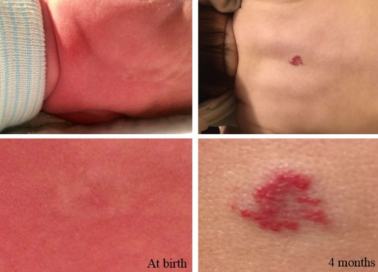
Soft Tissue Sarcoma—Health Professional Version

Soft Tissue Sarcoma—Health Professional Version

Causes & Prevention

NCI does not have PDQ evidence-based information about prevention of soft tissue sarcoma.

Screening

NCI does not have PDQ evidence-based information about screening for soft tissue sarcoma.

Supportive & Palliative Care

We offer evidence-based supportive and palliative care information for health professionals on the assessment and management of cancer-related symptoms and conditions.

Cancer Pain Nausea and Vomiting Nutrition in Cancer Care Transition to End-of-Life Care Last Days of Life View all Supportive and Palliative Care Summaries

Gardasil 9 Vaccine Protects against Additional HPV Types

Summary

In a large randomized clinical trial, a new human papillomavirus (HPV) vaccine effectively prevented infection and disease caused by nine HPV types, including seven types that cause cervical and other cancers—five of which were not covered by the previously available HPV vaccines—and two types that cause genital warts.

Source

New England Journal of Medicine, February 18, 2015 (See the abstract.)

Background

HPV infections are the most common sexually transmitted infections in the United States. More than 40 HPV types can be spread through direct sexual contact. Of these, about a dozen, including HPV types 16, 18, 31, 33, 45, 52, and 58, are high-risk—that is, persistent infection with these HPV types can cause cellular changes that may progress to cancer, including cervical, anal, penile, vaginal, vulvar, and oropharyngeal cancers. HPV types 16 and 18 are responsible for approximately 70 percent of all cervical cancers and HPV types 31, 33, 45, 52, and 58 are responsible for another 20 percent of cervical cancers.

Until December 2014, the FDA had approved two vaccines to protect against HPV infection: Cervarix, a bivalent vaccine that protects against infection with HPV types 16 and 18, and Gardasil, a quadrivalent vaccine that protects against infection with HPV types 6, 11, 16, and 18. Types 6 and 11 are low-risk types that do not cause cancer but can cause warts on or around the genitals, anus, mouth, or throat.

Researchers have been working on developing next-generation HPV vaccines that protect against additional high-risk HPV types. In December 2014, based on the results of this clinical trial, the FDA approved the 9-valent vaccine Gardasil 9, which protects against infection with HPV types 6, 11, 16, 18, 31, 33, 45, 52 and 58.

The Study

In a phase II/III international double-blind clinical trial, researchers randomly assigned 14,215 women between the ages of 16 and 26 to receive Gardasil or Gardasil 9. As part of the clinical trial, a small dose-finding study was first done to determine the dose that would be used in the larger efficacy study. Participants were eligible if they had no history of an abnormal Pap test, no more than four lifetime sexual partners, and no previous abnormal finding on a cervical biopsy.

Women in each group received three intramuscular injections of the vaccine over 6 months.

The study’s primary endpoint was the incidence of high-grade cervical, vulvar, or vaginal disease, including high-grade cervical epithelial neoplasia, adenocarcinoma in situ, cervical cancer, high-grade vulvar intraepithelial neoplasia, high-grade vaginal intraepithelial neoplasia, vulvar cancer, and vaginal cancer. Secondary endpoints included safety and the ability of the vaccine to provoke an immune response (vaccine immunogenicity), as measured by antibody responses to HPV types 6, 11, 16, and 18.

Elmar A. Joura, M.D., of the Medical University of Vienna, Comprehensive Cancer Center in Austria, led the trial, which was sponsored by Merck Sharp & Dohme Corp., a subsidiary of Merck & Co., Inc., the maker of Gardasil and Gardasil 9.

Results

Among study participants who did not have a documented HPV infection at the time of their first vaccine injection and who received all three doses, the number of cases of cervical, vulvar, or vaginal disease related to HPV types 31, 33, 45, 52, and 58 was much lower among patients who received the 9-valent vaccine compared with those who received the quadrivalent vaccine (1 versus 30 cases). The efficacy rate of the 9-valent vaccine was 96.7 percent.

Gardasil 9 was as effective as Gardasil at generating an antibody response against the four HPV types targeted by both vaccines. All cases of high-grade disease detected in the Gardasil 9 group occurred in participants who were HPV-infected when they received their first vaccine dose, which underscores the importance of vaccinating women and girls before they are exposed to HPV, the authors noted.

The safety of Gardasil 9 was evaluated in more than 7,000 women. Adverse events related to the injection site, including mild or moderate pain, swelling, redness, and itching, were more common in the Gardasil 9 group than in the Gardasil group. The rate of adverse systemic events—headache, fever, nausea, dizziness, and fatigue—were similar in the two groups.

Limitations

The researchers compared Gardasil 9 to Gardasil and did not include a placebo control group. Consequently, the researchers were unable to directly determine the efficacy of Gardasil 9 in preventing diseases associated with the HPV types the vaccines have in common (HPV types 6, 11, 16, and 18). However, because Gardasil 9 generated an antibody response similar to that generated by Gardasil and because the incidence of disease related to these four HPV types were similar, the authors concluded that Gardasil 9 would have similar efficacy to Gardasil in preventing diseases associated with these four HPV types.

The study authors noted that longer-term follow-up of participants vaccinated with Gardasil 9 is needed to provide information on the durability of protection.

Comment

This study represents “a milestone in expanding the coverage of cancers associated with the human papillomavirus,” wrote Anne Schuchat, M.D., of the Centers for Disease Control and Prevention, in an accompanying editorial in NEJM.

But even with a vaccine that provides protection against more high-risk HPV types, Dr. Schuchat noted that “vaccination of a much higher proportion of preteens is needed. Otherwise, decades from now oncologists will still be talking about HPV-associated cancers with thousands of new patients every year.”

Aimée Kreimer, Ph.D., of NCI’s Division of Cancer Epidemiology and Genetics, said she is hopeful that Gardasil 9 will protect against HPV infections for the same duration as the earlier HPV vaccines have demonstrated thus far.

Dr. Kreimer also noted that “it will be important to know if currently approved HPV vaccines, including Gardasil 9, may also be given in a reduced dosage schedule without diminishing their efficacy.” This would help “to maximize their benefit in regions of the world where HPV-associated cancers are the leading cause of cancer death in women and among segments of the U.S. population where coverage has been low.”

Cervical Cancer Screening (PDQ®)–Health Professional Version

Cervical Cancer Screening (PDQ®)–Health Professional Version

Overview

Note: The Overview section summarizes the published evidence on this topic. The rest of the summary describes the evidence in more detail.

Other PDQ summaries on Cervical Cancer Prevention, Cervical Cancer Treatment, and Levels of Evidence for Cancer Screening and Prevention Studies are also available.

Screening With the Papanicolaou (Pap) Test: Benefits

Based on solid evidence, regular screening for cervical cancer with the Pap test in an appropriate population of women reduces mortality from cervical cancer. The benefits of screening women younger than 21 years are small because of the low prevalence of lesions that will progress to invasive cancer. Screening is not beneficial in women older than 65 years if they have had a recent history of negative test results.[13]

Magnitude of Effect: Regular Pap screening decreases cervix cancer incidence and mortality by at least 80%.

  • Study Design: Population-based and cohort studies.
  • Internal Validity: Good.
  • Consistency: Good.
  • External Validity: Good.

Screening With the Pap Test: Harms

Based on solid evidence, regular screening with the Pap test leads to additional diagnostic procedures (e.g., colposcopy) and possible overtreatment for low-grade squamous intraepithelial lesions (LSILs). These harms are greatest for younger women, who have a higher prevalence of LSILs, lesions that often regress without treatment. Harms are also increased in younger women because they have a higher rate of false-positive results. Excisional procedures to treat preinvasive disease has been associated with increased risk of long-term consequences for fertility and pregnancy.[4]

Magnitude of Effect: Additional diagnostic procedures were performed in 50% of women undergoing regular Pap testing. Approximately 5% were treated for LSILs. The number of women with impaired fertility and pregnancy complications is unknown.

  • Study Design: Evidence obtained from cohort or case-control studies.
  • Internal Validity: Good.
  • Consistency: Good.
  • External Validity: Good.

Screening With the Human Papillomavirus (HPV) DNA Test: Benefits

Based on solid evidence, screening with an HPV DNA or HPV RNA test detects high-grade cervical dysplasia, a precursor lesion for cervical cancer. Additional clinical trials show that HPV testing is superior to other cervical cancer screening strategies. In April 2014, the U.S. Food and Drug Administration approved an HPV DNA test that can be used alone for the primary screening of cervical cancer risk in women aged 25 years and older.[5]

Magnitude of Effect: In one prospective, clustered, randomized trial, HPV testing was superior to other strategies for preventing cervical cancer mortality.[6,7]

  • Study Design: Clustered randomized controlled trial (RCT).
  • Internal Validity: Good.
  • Consistency: Good.
  • External Validity: Good.

Screening With the HPV DNA Test: Harms

Based on solid evidence, HPV testing identifies numerous infections that will not lead to cervical dysplasia or cervical cancer. This is especially true in women younger than 30 years, in whom rates of HPV infection may be higher.

Magnitude of Effect: In one study, 86.7% of women with a positive HPV test did not develop cervical cancer or related premalignant disease after more than a decade of follow-up.[8]

  • Study Design: Long-term observational trials.
  • Internal Validity: Good.
  • Consistency: Good.
  • External Validity: Good.

Screening With the Pap Test and the HPV DNA Test (Cotesting): Benefits

Based on solid evidence, screening every 5 years with the Pap test and the HPV DNA test (cotesting) in women aged 30 years and older is more sensitive in detecting cervical abnormalities, compared with the Pap test alone. Screening with the Pap test and HPV DNA test reduces the incidence of cervical cancer.[3]

Magnitude of Effect: HPV-based screening provides 60% to 70% greater protection against invasive cervical carcinoma, compared with cytology.[9]

  • Study Design: RCTs.
  • Internal Validity: Good.
  • Consistency: Good.
  • External Validity: Good.

Screening With the Pap Test and the HPV DNA Test (Cotesting): Harms

Based on solid evidence, HPV and Pap cotesting is associated with more false-positives than is the Pap test alone. Abnormal test results can lead to more frequent testing and invasive diagnostic procedures.[3]

Magnitude of Effect: The percentage of U.S. women undergoing cotesting who will have a normal cytology test result and a positive HPV test result (and who will therefore require additional testing) ranges from 11% among women aged 30 to 34 years to 2.6% among women aged 60 to 65 years.[3]

  • Study Design: RCTs.
  • Internal Validity: Good.
  • Consistency: Good.
  • External Validity: Good.

Screening Women Without a Cervix

Based on solid evidence, screening is not helpful in women who do not have a cervix as a result of a hysterectomy for a benign condition.

Magnitude of Effect: Among women without cervices, fewer than 1 per 1,000 had abnormal Pap test results.

  • Study Design: Evidence obtained from a single cohort study.
  • Internal Validity: Good.
  • Consistency: Good.
  • External Validity: Good.
References
  1. Sasieni P, Castanon A, Cuzick J: Effectiveness of cervical screening with age: population based case-control study of prospectively recorded data. BMJ 339: b2968, 2009. [PUBMED Abstract]
  2. Sawaya GF, McConnell KJ, Kulasingam SL, et al.: Risk of cervical cancer associated with extending the interval between cervical-cancer screenings. N Engl J Med 349 (16): 1501-9, 2003. [PUBMED Abstract]
  3. Moyer VA; U.S. Preventive Services Task Force: Screening for cervical cancer: U.S. Preventive Services Task Force recommendation statement. Ann Intern Med 156 (12): 880-91, W312, 2012. [PUBMED Abstract]
  4. Kyrgiou M, Athanasiou A, Paraskevaidi M, et al.: Adverse obstetric outcomes after local treatment for cervical preinvasive and early invasive disease according to cone depth: systematic review and meta-analysis. BMJ 354: i3633, 2016. [PUBMED Abstract]
  5. Wright TC, Stoler MH, Behrens CM, et al.: Primary cervical cancer screening with human papillomavirus: end of study results from the ATHENA study using HPV as the first-line screening test. Gynecol Oncol 136 (2): 189-97, 2015. [PUBMED Abstract]
  6. Sankaranarayanan R, Nene BM, Shastri SS, et al.: HPV screening for cervical cancer in rural India. N Engl J Med 360 (14): 1385-94, 2009. [PUBMED Abstract]
  7. Szarewski A: Cervical screening by visual inspection with acetic acid. Lancet 370 (9585): 365-6, 2007. [PUBMED Abstract]
  8. Chen HC, Schiffman M, Lin CY, et al.: Persistence of type-specific human papillomavirus infection and increased long-term risk of cervical cancer. J Natl Cancer Inst 103 (18): 1387-96, 2011. [PUBMED Abstract]
  9. Ronco G, Dillner J, Elfström KM, et al.: Efficacy of HPV-based screening for prevention of invasive cervical cancer: follow-up of four European randomised controlled trials. Lancet 383 (9916): 524-32, 2014. [PUBMED Abstract]

Natural History, Incidence, and Mortality

It is estimated that 13,360 cases of invasive cervical cancer will be diagnosed and that 4,320 women will die of the disease in the United States in 2025.[1] Cervical cancer incidence and mortality rates have steadily improved over time and have stabilized in recent years, attributed largely to screening with Papanicolaou (Pap) test. However, the mortality rate in Black women is 50% higher and in Native American women is 70% higher than in White women.[1] When corrected for the prevalence of hysterectomy, the mortality rate for Black women is nearly twice the mortality rate for White women.[2]

Invasive squamous carcinoma of the cervix results from the progression of preinvasive precursor lesions called cervical intraepithelial neoplasia (CIN), or dysplasia. CIN is histologically graded into mild dysplasia (CIN 1), moderate dysplasia (CIN 2), or severe dysplasia (CIN 3). Not all of these lesions progress to invasive cancer; many mild and moderate lesions regress. A further categorization, the Bethesda System, is based on cytologic findings: atypical squamous cells of undetermined significance (ASCUS) or cannot rule out low-grade squamous intraepithelial lesions (LSILs), LSILs (consisting of cytologic atypia and CIN 1), and high-grade squamous intraepithelial lesions (HSILs), primarily CIN 2–3 plus carcinoma in situ.[3]

The rate at which invasive cancer develops from CIN is usually slow, measured in years and perhaps decades.[4] This long natural history provides the opportunity for screening to effectively detect this process during the preinvasive phase, thus allowing early treatment and cure. Because many of these preinvasive lesions (especially LSILs) may never progress to invasive cancer,[57] screening also runs the risk of leading to treatment for women who may not need it.

Human papillomavirus (HPV) is an oncogenic virus and the etiologic agent of cervical cancer and related premalignant disease. HPV is transmitted by sexual contact. Sexually inactive women rarely develop cervical cancer, while sexual activity at an early age with multiple sexual partners is a strong risk factor.[8] Nearly all women with invasive cervical cancer have evidence of HPV infection.[912] Most women with HPV infection, however, never develop cervical cancer; thus, this infection is necessary but not sufficient for the development of cancer.[13]

Although cervical cancer mortality increases with age,[14] the prevalence of CIN is highest among women in their 20s and 30s. Mortality is rare among women younger than 30 years; HSILs are rare among women older than 65 years who have been previously screened. About 70% of atypical squamous cells of undetermined significance and CIN 1 lesions regress within 6 years, while about 6% of CIN 1 lesions progress to CIN 3 or worse. In about 10% to 20% of women with CIN 3 lesions, the lesions progress to invasive cancer.[4,7,15]

Historically, cervical cancer mortality rates were substantially higher (twice as high or more) in Black women than in White women younger than 50 years. However, recent rates (2018–2022) have been only modestly higher (17%) in Black women than in White women in this age group. Among women older than 60 years, cervical cancer mortality rates have historically been up to three times as high in Black women than in White women; recent rates (2018–2022) are still almost twice as high in Black women than in White women.[14] In either case, mortality is rare among women of any age who have regular screenings.

References
  1. American Cancer Society: Cancer Facts and Figures 2025. American Cancer Society, 2025. Available online. Last accessed January 16, 2025.
  2. Beavis AL, Gravitt PE, Rositch AF: Hysterectomy-corrected cervical cancer mortality rates reveal a larger racial disparity in the United States. Cancer 123 (6): 1044-1050, 2017. [PUBMED Abstract]
  3. Solomon D, Davey D, Kurman R, et al.: The 2001 Bethesda System: terminology for reporting results of cervical cytology. JAMA 287 (16): 2114-9, 2002. [PUBMED Abstract]
  4. Holowaty P, Miller AB, Rohan T, et al.: Natural history of dysplasia of the uterine cervix. J Natl Cancer Inst 91 (3): 252-8, 1999. [PUBMED Abstract]
  5. Nasiell K, Roger V, Nasiell M: Behavior of mild cervical dysplasia during long-term follow-up. Obstet Gynecol 67 (5): 665-9, 1986. [PUBMED Abstract]
  6. Nash JD, Burke TW, Hoskins WJ: Biologic course of cervical human papillomavirus infection. Obstet Gynecol 69 (2): 160-2, 1987. [PUBMED Abstract]
  7. Melnikow J, Nuovo J, Willan AR, et al.: Natural history of cervical squamous intraepithelial lesions: a meta-analysis. Obstet Gynecol 92 (4 Pt 2): 727-35, 1998. [PUBMED Abstract]
  8. Berrington de González A, Green J; International Collaboration of Epidemiological Studies of Cervical Cancer: Comparison of risk factors for invasive squamous cell carcinoma and adenocarcinoma of the cervix: collaborative reanalysis of individual data on 8,097 women with squamous cell carcinoma and 1,374 women with adenocarcinoma from 12 epidemiological studies. Int J Cancer 120 (4): 885-91, 2007. [PUBMED Abstract]
  9. Bosch FX, Manos MM, Muñoz N, et al.: Prevalence of human papillomavirus in cervical cancer: a worldwide perspective. International biological study on cervical cancer (IBSCC) Study Group. J Natl Cancer Inst 87 (11): 796-802, 1995. [PUBMED Abstract]
  10. Wallin KL, Wiklund F, Angström T, et al.: Type-specific persistence of human papillomavirus DNA before the development of invasive cervical cancer. N Engl J Med 341 (22): 1633-8, 1999. [PUBMED Abstract]
  11. Alani RM, Münger K: Human papillomaviruses and associated malignancies. J Clin Oncol 16 (1): 330-7, 1998. [PUBMED Abstract]
  12. Walboomers JM, Jacobs MV, Manos MM, et al.: Human papillomavirus is a necessary cause of invasive cervical cancer worldwide. J Pathol 189 (1): 12-9, 1999. [PUBMED Abstract]
  13. Ho GY, Bierman R, Beardsley L, et al.: Natural history of cervicovaginal papillomavirus infection in young women. N Engl J Med 338 (7): 423-8, 1998. [PUBMED Abstract]
  14. Surveillance Research Program, National Cancer Institute: SEER*Explorer: An interactive website for SEER cancer statistics. Bethesda, MD: National Cancer Institute. Available online. Last accessed December 30, 2024.
  15. Arends MJ, Buckley CH, Wells M: Aetiology, pathogenesis, and pathology of cervical neoplasia. J Clin Pathol 51 (2): 96-103, 1998. [PUBMED Abstract]

The Pap Test

The Papanicolaou (Pap) test has never been examined in a randomized controlled trial. A large body of consistent observational data, however, supports its effectiveness in reducing mortality from cervical cancer. Both incidence and mortality from cervical cancer have sharply decreased in a number of large populations after the introduction of well-run screening programs.[14] In Iceland, the mortality rate declined by 80% for more than 20 years, and in Finland and Sweden by 50% and 34%, respectively.[1,5] Similar reductions have been observed in large populations in the United States and Canada. Reductions in cervical cancer incidence and mortality were proportional to the intensity of screening.[1,5] Mortality in the Canadian provinces was reduced most remarkably in British Columbia, which had screening rates two to five times those of the other provinces.[6]

Case-control studies have found that the risk of developing invasive cervical cancer is three to ten times higher in women who have not been screened.[710] Risk also increases with long duration after the last normal Pap test, or similarly, with decreasing frequency of screening.[11,12] Screening every 2 to 3 years, however, has not been found to significantly increase the risk of finding invasive cervical cancer above the risk expected with annual screening.[1214]

References
  1. Lăără E, Day NE, Hakama M: Trends in mortality from cervical cancer in the Nordic countries: association with organised screening programmes. Lancet 1 (8544): 1247-9, 1987. [PUBMED Abstract]
  2. Christopherson WM, Lundin FE, Mendez WM, et al.: Cervical cancer control: a study of morbidity and mortality trends over a twenty-one-year period. Cancer 38 (3): 1357-66, 1976. [PUBMED Abstract]
  3. Miller AB, Lindsay J, Hill GB: Mortality from cancer of the uterus in Canada and its relationship to screening for cancer of the cervix. Int J Cancer 17 (5): 602-12, 1976. [PUBMED Abstract]
  4. Johannesson G, Geirsson G, Day N: The effect of mass screening in Iceland, 1965-74, on the incidence and mortality of cervical carcinoma. Int J Cancer 21 (4): 418-25, 1978. [PUBMED Abstract]
  5. Sigurdsson K: Effect of organized screening on the risk of cervical cancer. Evaluation of screening activity in Iceland, 1964-1991. Int J Cancer 54 (4): 563-70, 1993. [PUBMED Abstract]
  6. Benedet JL, Anderson GH, Matisic JP: A comprehensive program for cervical cancer detection and management. Am J Obstet Gynecol 166 (4): 1254-9, 1992. [PUBMED Abstract]
  7. Aristizabal N, Cuello C, Correa P, et al.: The impact of vaginal cytology on cervical cancer risks in Cali, Colombia. Int J Cancer 34 (1): 5-9, 1984. [PUBMED Abstract]
  8. Clarke EA, Anderson TW: Does screening by “Pap” smears help prevent cervical cancer? A case-control study. Lancet 2 (8132): 1-4, 1979. [PUBMED Abstract]
  9. La Vecchia C, Franceschi S, Decarli A, et al.: “Pap” smear and the risk of cervical neoplasia: quantitative estimates from a case-control study. Lancet 2 (8406): 779-82, 1984. [PUBMED Abstract]
  10. Herrero R, Brinton LA, Reeves WC, et al.: Screening for cervical cancer in Latin America: a case-control study. Int J Epidemiol 21 (6): 1050-6, 1992. [PUBMED Abstract]
  11. Celentano DD, Klassen AC, Weisman CS, et al.: Duration of relative protection of screening for cervical cancer. Prev Med 18 (4): 411-22, 1989. [PUBMED Abstract]
  12. Screening for squamous cervical cancer: duration of low risk after negative results of cervical cytology and its implication for screening policies. IARC Working Group on evaluation of cervical cancer screening programmes. Br Med J (Clin Res Ed) 293 (6548): 659-64, 1986. [PUBMED Abstract]
  13. Kleinman JC, Kopstein A: Who is being screened for cervical cancer? Am J Public Health 71 (1): 73-6, 1981. [PUBMED Abstract]
  14. Qin J, Saraiya M, Martinez G, et al.: Prevalence of Potentially Unnecessary Bimanual Pelvic Examinations and Papanicolaou Tests Among Adolescent Girls and Young Women Aged 15-20 Years in the United States. JAMA Intern Med 180 (2): 274-280, 2020. [PUBMED Abstract]

Accuracy of the Pap Test

Ideally, determining the sensitivity and specificity of a screening test would involve a study that applies a gold standard test (such as colposcopy with appropriate biopsy) to all participants (whether the screening test results are positive or negative). Sensitivity (the percentage of true-positive cases that are detected by the screening test) and specificity (the percentage of true-negative cases that are negative by the screening test) could be calculated. Such studies have rarely been done for any screening test for cervical cancer. Studies that compare the Pap test with repeat Pap testing have found that the sensitivity of any abnormality on a single test for detecting high-grade lesions is 55% to 80%.[1,2] Because of the usual slow-growing nature of cervical cancer, the sensitivity of a program of regular Pap testing is likely higher.

To determine the sensitivity and specificity of the Pap smear, both a test threshold (i.e., the point at which the test will be considered to be positive) and a reference-standard threshold (i.e., the point at which the reference standard is considered to be positive) must be defined. In practice, atypical squamous cells of undetermined significance (ASCUS) are often used as the test threshold, and CIN 1 is often used as the reference threshold. This combination gives a sensitivity of about 68% and a specificity of about 75%. A more appropriate test threshold may be LSIL, with a reference threshold of CIN 2–3. This combination gives a sensitivity of 70% to 80%, with a specificity of about 95%.[3]

One important factor in the accuracy of the Pap test is the adequacy of the specimen obtained. Adequate training and using techniques such as the cytobrush may improve sensitivity.[4]

References
  1. Soost HJ, Lange HJ, Lehmacher W, et al.: The validation of cervical cytology. Sensitivity, specificity and predictive values. Acta Cytol 35 (1): 8-14, 1991 Jan-Feb. [PUBMED Abstract]
  2. Benoit AG, Krepart GV, Lotocki RJ: Results of prior cytologic screening in patients with a diagnosis of Stage I carcinoma of the cervix. Am J Obstet Gynecol 148 (5): 690-4, 1984. [PUBMED Abstract]
  3. Nanda K, McCrory DC, Myers ER, et al.: Accuracy of the Papanicolaou test in screening for and follow-up of cervical cytologic abnormalities: a systematic review. Ann Intern Med 132 (10): 810-9, 2000. [PUBMED Abstract]
  4. Martin-Hirsch P, Lilford R, Jarvis G, et al.: Efficacy of cervical-smear collection devices: a systematic review and meta-analysis. Lancet 354 (9192): 1763-70, 1999. [PUBMED Abstract]

Newer Screening Technologies

Newer techniques that employ liquid-based cytology (LBC) (e.g., ThinPrep) have been developed to improve the sensitivity of screening. In 1996, the ThinPrep® Papanicolaou (Pap) test became the first LBC approved by the U.S. Food and Drug Administration.[1] As with the Pap test, the optimal studies to determine the sensitivity and specificity of these technologies have not been conducted. Some less-than-optimal studies show that sensitivity is modestly higher for detecting any degree of cervical intraepithelial neoplasia, with modestly lower specificity.[2,3] One study, however, showed that conventional Pap testing was slightly more sensitive and specific than LBC.[4]

The evidence is also mixed about whether liquid-based techniques improve rates of test adequacy.[2,3] One advantage of LBC is that HPV testing can be performed on the same preparation; one disadvantage is that liquid-based approaches are more expensive than conventional Pap testing. No study has examined whether LBC actually reduces the number of women dying of cervical cancer compared with conventional Pap testing.

References
  1. Gibb RK, Martens MG: The impact of liquid-based cytology in decreasing the incidence of cervical cancer. Rev Obstet Gynecol 4 (Suppl 1): S2-S11, 2011. [PUBMED Abstract]
  2. Hartmann KE, Hall SA, Nanda K, et al.: Screening for Cervical Cancer. Rockville, Md: Agency for Health Research and Quality, 2002. Available online. Last accessed December 18, 2024.
  3. McCrory DC, Matchar DB, Bastian L, et al.: Evaluation of Cervical Cytology. Rockville, Md: Agency for Health Research and Quality, 1999. Evidence Report/Technology Assessment No. 5. AHCPR Publication No. 99-E010. Also available online. Last accessed December 18, 2024.
  4. Coste J, Cochand-Priollet B, de Cremoux P, et al.: Cross sectional study of conventional cervical smear, monolayer cytology, and human papillomavirus DNA testing for cervical cancer screening. BMJ 326 (7392): 733, 2003. [PUBMED Abstract]

Screening Women Who Have Had a Hysterectomy

Women who have had a hysterectomy with removal of the cervix for benign disease rarely have important abnormalities found on Pap testing. Several studies have shown that the rate of high-grade vaginal lesions or vaginal cancer is less than 1 in 1,000 tests;[1,2] no study has shown that screening for vaginal cancer reduces mortality from this rare condition.

References
  1. Fox J, Remington P, Layde P, et al.: The effect of hysterectomy on the risk of an abnormal screening Papanicolaou test result. Am J Obstet Gynecol 180 (5): 1104-9, 1999. [PUBMED Abstract]
  2. Pearce KF, Haefner HK, Sarwar SF, et al.: Cytopathological findings on vaginal Papanicolaou smears after hysterectomy for benign gynecologic disease. N Engl J Med 335 (21): 1559-62, 1996. [PUBMED Abstract]

Screening Interval

Because cervical cancer is slow growing, there is considerable uncertainty about the optimal screening interval. The most direct evidence about this issue comes from a prospective cohort analysis of a randomized controlled trial.[1] Among 2,561 women (mean age, 66.7 years) with normal Pap tests at baseline, 110 had an abnormal Pap test within the next 2 years. No woman was found to have cervical intraepithelial neoplasia (CIN) 2–3 or invasive cancer, and only one woman had CIN 1–2. Thus, the positive predictive value (PPV) of screening 1 year after a negative Pap test was 0%; after 2 years, the PPV was 0.9%. The authors concluded that Pap tests should not be repeated within 2 years of a negative test. A large (N = 332,000) prospective cohort study of cervical cytology and human papillomavirus DNA cotesting in U.S. women aged 30 years and older found that a negative Pap smear was associated with a low risk of developing CIN 3 or cancer (CIN 3+) for up to 5 years after the test (cumulative incidence of CIN 3+ at 3 and 5 years was 0.17% and 0.36%, respectively).[2]

A large study that included data from the National Breast and Cervical Cancer Early Detection Program together with modeling found little further mortality reduction from cervical cancer for screening every year as compared with screening every 3 years.[3] A similar modeling study from Australia found no differences between screening every 2 years and screening every 3 years.[4]

References
  1. Sawaya GF, Grady D, Kerlikowske K, et al.: The positive predictive value of cervical smears in previously screened postmenopausal women: the Heart and Estrogen/progestin Replacement Study (HERS). Ann Intern Med 133 (12): 942-50, 2000. [PUBMED Abstract]
  2. Katki HA, Kinney WK, Fetterman B, et al.: Cervical cancer risk for women undergoing concurrent testing for human papillomavirus and cervical cytology: a population-based study in routine clinical practice. Lancet Oncol 12 (7): 663-72, 2011. [PUBMED Abstract]
  3. Sawaya GF, McConnell KJ, Kulasingam SL, et al.: Risk of cervical cancer associated with extending the interval between cervical-cancer screenings. N Engl J Med 349 (16): 1501-9, 2003. [PUBMED Abstract]
  4. Creighton P, Lew JB, Clements M, et al.: Cervical cancer screening in Australia: modelled evaluation of the impact of changing the recommended interval from two to three years. BMC Public Health 10: 734, 2010. [PUBMED Abstract]

HPV Testing

Noninvasive cervical squamous cell abnormalities are graded histologically as cervical intraepithelial neoplasia (CIN) mild dysplasia (CIN 1), moderate dysplasia (CIN 2), or severe dysplasia (CIN 3), according to the severity of the cell changes and the percent of the epithelium replaced by abnormal cell growth. CIN 3 is a reasonably reproducible diagnosis and, if untreated, has an approximate 30% risk of developing into invasive cancer over many years.[1] CIN 2 has poor interobserver reproducibility,[2] and the biological behavior is variable.[3] CIN 3 is therefore a more rigorous end point for clinical trials, while CIN 2 represents the threshold for treatment to provide an additional measure of safety.

Approximately 15 cancer-associated (high-risk or carcinogenic) HPV genotypes cause virtually all cases of cervical cancer and precursor lesions of CIN 2 and CIN 3. However, carcinogenic HPV infections are very common, particularly in young women, and most infections clear on their own within 1 to 2 years. Therefore, the challenge of incorporating HPV testing in cervical screening programs is to balance sensitivity for detection of CIN 2 or CIN 2+ and to minimize the over-referral of women with transient HPV infections and cervical changes that are destined to regress.

The U.S. Food and Drug Administration has approved several HPV tests. Most of these tests are based on the detection of DNA from one or more oncogenic types of HPV. One test detects HPV RNA. HPV testing is approved for use in two contexts: (1) as a second (i.e., triage) test after an equivocal cytology result of atypical squamous cells of undetermined significance (ASCUS); and (2) for primary screening in conjunction with cervical cytology for women aged 30 years and older.[4] Testing for low-risk HPV types does not identify women at risk of developing CIN 2 or 3.[5,6]

Triage

A large randomized clinical trial, the ASCUS/low-grade squamous intraepithelial lesion (LSIL) Triage Study (ALTS), demonstrated the cost-effectiveness of using HPV testing to clarify the risk of an ASCUS Pap result.[7] ALTS randomly assigned women with ASCUS to one of three management strategies: immediate colposcopy regardless of enrollment test results, referral to colposcopy if HPV test results were positive or if the enrollment cytology was high-grade squamous intraepithelial lesion (HSIL), and referral to colposcopy only if the cytology was HSIL. The HPV triage strategy was as sensitive as immediate colposcopy to detection CIN 2+, while referring only about half of the women for the procedure. Repeat cytology with referral to colposcopy at the threshold of HSIL was less sensitive for CIN 3+ (60%) compared with HPV triage (92%); however, using a cytologic threshold of ASCUS for referral increased sensitivity but resulted in 72% of women with ASCUS undergoing colposcopy.[8] HPV testing is not recommended for adolescent women with ASCUS because most of these women are HPV positive.[9,10]

HPV DNA testing is generally not appropriate or clinically useful after cytology results of LSIL, which is more severe than ASCUS, and most of these women (84%–96%) are carcinogenic HPV DNA positive.[11] One exception may be to clarify the risk for postmenopausal women with cytologic LSIL, which is an interpretation that can be falsely positive, presumably due to atrophic changes.[12]

Primary HPV Screening

Testing for HPV DNA as a primary screening test is an option for women aged 30 years and older. Women who are negative by cytology and HPV testing are at extremely low risk of CIN 3+ and therefore may be screened less frequently. A prospective cohort study of nearly 332,000 U.S. women aged 30 years and older undergoing HPV DNA and cervical cytology cotesting every 3 years found that the cumulative incidence of CIN 3+ in women with negative results for both tests at baseline was 0.047% at 3 years and 0.16% at 5 years.[13] A second study of more than 43,000 women aged 29 to 61 years, one-half of whom underwent three rounds of HPV DNA and cervical cytology cotesting every 5 years, found that the cumulative incidence of CIN 3+ in women with negative results for both tests at baseline was 0.01% (95% confidence interval [CI], 0.00%–0.05%) at 9 years and 0.07% (95% CI, 0.03%–0.17%) after 14 years of follow-up.[14] Screening more frequently than every 3 years would not improve sensitivity significantly but would increase costs and overtreatment.[15,16]

Numerous studies have demonstrated that, compared with cytology, HPV DNA testing is more sensitive for identifying women who have CIN 2+ (range of sensitivities, 84%–97%).[1724] In one randomized trial using both Pap and HPV testing in random order among women aged 30 to 69 years, sensitivity of HPV was 95% compared with 55% for Pap cytology. The combination of HPV and cytology had 100% sensitivity and a referral rate of 7.9%.[18]

The lower specificity of HPV DNA testing compared with cytology is a consideration. Among women older than 30 years, cytology had a specificity of 97% compared with 94% for HPV testing.[18] The specificity of HPV DNA testing would likely be even lower among women younger than 30 years, who have more transient HPV infection that is of little consequence. Thus, detecting such women would potentially increase the number of follow-up diagnostic workups. Potential approaches to minimize over-referral with HPV DNA testing and improve specificity include: (1) triage HPV-positive results with cytology [23] or another more specific molecular assay;[25] and (2) trigger further workup only after two sequential positive HPV test results because it is the persistence of carcinogenic HPV that confers the greatest risk of CIN 2–3.[26,27]

An Italian population-based, randomized, controlled trial of HPV DNA testing versus cervical cytology performed at 3-year intervals in approximately 94,000 women aged 25 to 60 years found a statistically significant decrease in the number of invasive cervical cancer cases diagnosed in the HPV DNA arm at the second round of screening (0 cases vs. 9 cases; P = .004). However, about 48% of individuals in the HPV DNA arm also received conventional cytology testing at the first screening round, making it impossible to discern whether the observed difference resulted from the use of a combined testing strategy or HPV DNA testing alone. Of note, many more women in the HPV DNA arm than in the cytology-alone arm were referred to colposcopy for abnormal findings (4,436 women vs. 1,416 women), prompting the authors to conclude that if the HPV DNA test is used as a primary screening strategy, women with positive test results should be triaged by cytology before referral.[28] A Canadian study of 19,000 women aged 25 to 65 years that compared HPV DNA testing with cervical cytology found that HPV DNA testing identified most women with CIN 3 at initial screening. Women who initially tested HPV DNA negative were at low risk for cervical dysplasia 48 months later. Additionally, there were not an excessive number of women referred for additional diagnostic testing.[24]

A study using data from a population-based randomized trial of cervical cancer screening among women aged 32 to 38 years compared 11 different screening strategies using HPV DNA testing and cytology. The strategy of initial screening with an HPV DNA test and a triage of HPV-positive results with cytology, and subsequent repeat HPV DNA testing after 1 year for women who were HPV positive but cytology negative, increased the sensitivity for detection of CIN 3+ by 30% compared with cytology alone, and increased the total number of screening tests performed by only 12%.[29] In a review of data from a large integrated health system, the added benefit of cotesting versus HPV testing alone would improve detection of CIN 3 or early-stage cervical cancer in very few women. Only 5.1% of locally advanced invasive cancers and 3.6% of CIN 3 were cytology positive and HPV negative, representing a very small fraction of all screened women.[30]

Cytology can be used to triage after primary HPV screening. Triage with cytology can be improved with concomitant detection of p16 and Ki-67 in the same cell (p16/Ki-67 dual stain [DS]). DS can be assessed manually through immunostaining cervical cytology slides. Additionally, artificial intelligence–based deep learning algorithms are currently being investigated and applied to aid in automated identification of p16/Ki-67 dual-stained slides. This approach has been shown to improve specificity without sacrificing sensitivity over manual DS assessment,[31] but it has not yet been validated in population studies.

References
  1. McCredie MR, Sharples KJ, Paul C, et al.: Natural history of cervical neoplasia and risk of invasive cancer in women with cervical intraepithelial neoplasia 3: a retrospective cohort study. Lancet Oncol 9 (5): 425-34, 2008. [PUBMED Abstract]
  2. Stoler MH, Schiffman M; Atypical Squamous Cells of Undetermined Significance-Low-grade Squamous Intraepithelial Lesion Triage Study (ALTS) Group: Interobserver reproducibility of cervical cytologic and histologic interpretations: realistic estimates from the ASCUS-LSIL Triage Study. JAMA 285 (11): 1500-5, 2001. [PUBMED Abstract]
  3. Castle PE, Schiffman M, Wheeler CM, et al.: Evidence for frequent regression of cervical intraepithelial neoplasia-grade 2. Obstet Gynecol 113 (1): 18-25, 2009. [PUBMED Abstract]
  4. Halfon P, Trepo E, Antoniotti G, et al.: Prospective evaluation of the Hybrid Capture 2 and AMPLICOR human papillomavirus (HPV) tests for detection of 13 high-risk HPV genotypes in atypical squamous cells of uncertain significance. J Clin Microbiol 45 (2): 313-6, 2007. [PUBMED Abstract]
  5. Thomsen LT, Frederiksen K, Munk C, et al.: High-risk and low-risk human papillomavirus and the absolute risk of cervical intraepithelial neoplasia or cancer. Obstet Gynecol 123 (1): 57-64, 2014. [PUBMED Abstract]
  6. Castle PE, Hunt WC, Langsfeld E, et al.: Three-year risk of cervical precancer and cancer after the detection of low-risk human papillomavirus genotypes targeted by a commercial test. Obstet Gynecol 123 (1): 49-56, 2014. [PUBMED Abstract]
  7. Kulasingam SL, Kim JJ, Lawrence WF, et al.: Cost-effectiveness analysis based on the atypical squamous cells of undetermined significance/low-grade squamous intraepithelial lesion Triage Study (ALTS). J Natl Cancer Inst 98 (2): 92-100, 2006. [PUBMED Abstract]
  8. ASCUS-LSIL Traige Study (ALTS) Group: Results of a randomized trial on the management of cytology interpretations of atypical squamous cells of undetermined significance. Am J Obstet Gynecol 188 (6): 1383-92, 2003. [PUBMED Abstract]
  9. Wright TC, Massad LS, Dunton CJ, et al.: 2006 consensus guidelines for the management of women with abnormal cervical cancer screening tests. Am J Obstet Gynecol 197 (4): 346-55, 2007. [PUBMED Abstract]
  10. Sherman ME, Schiffman M, Cox JT, et al.: Effects of age and human papilloma viral load on colposcopy triage: data from the randomized Atypical Squamous Cells of Undetermined Significance/Low-Grade Squamous Intraepithelial Lesion Triage Study (ALTS). J Natl Cancer Inst 94 (2): 102-7, 2002. [PUBMED Abstract]
  11. ASCUS-LSIL Traige Study (ALTS) Group: A randomized trial on the management of low-grade squamous intraepithelial lesion cytology interpretations. Am J Obstet Gynecol 188 (6): 1393-400, 2003. [PUBMED Abstract]
  12. Zuna RE, Wang SS, Rosenthal DL, et al.: Determinants of human papillomavirus-negative, low-grade squamous intraepithelial lesions in the atypical squamous cells of undetermined significance/low-grade squamous intraepithelial lesions triage study (ALTS). Cancer 105 (5): 253-62, 2005. [PUBMED Abstract]
  13. Katki HA, Kinney WK, Fetterman B, et al.: Cervical cancer risk for women undergoing concurrent testing for human papillomavirus and cervical cytology: a population-based study in routine clinical practice. Lancet Oncol 12 (7): 663-72, 2011. [PUBMED Abstract]
  14. Dijkstra MG, van Zummeren M, Rozendaal L, et al.: Safety of extending screening intervals beyond five years in cervical screening programmes with testing for high risk human papillomavirus: 14 year follow-up of population based randomised cohort in the Netherlands. BMJ 355: i4924, 2016. [PUBMED Abstract]
  15. Saslow D, Runowicz CD, Solomon D, et al.: American Cancer Society guideline for the early detection of cervical neoplasia and cancer. CA Cancer J Clin 52 (6): 342-62, 2002 Nov-Dec. [PUBMED Abstract]
  16. Goldie SJ, Kim JJ, Wright TC: Cost-effectiveness of human papillomavirus DNA testing for cervical cancer screening in women aged 30 years or more. Obstet Gynecol 103 (4): 619-31, 2004. [PUBMED Abstract]
  17. Arbyn M, Sasieni P, Meijer CJ, et al.: Chapter 9: Clinical applications of HPV testing: a summary of meta-analyses. Vaccine 24 (Suppl 3): S3/78-89, 2006. [PUBMED Abstract]
  18. Mayrand MH, Duarte-Franco E, Rodrigues I, et al.: Human papillomavirus DNA versus Papanicolaou screening tests for cervical cancer. N Engl J Med 357 (16): 1579-88, 2007. [PUBMED Abstract]
  19. Naucler P, Ryd W, Törnberg S, et al.: Human papillomavirus and Papanicolaou tests to screen for cervical cancer. N Engl J Med 357 (16): 1589-97, 2007. [PUBMED Abstract]
  20. Bulkmans NW, Berkhof J, Rozendaal L, et al.: Human papillomavirus DNA testing for the detection of cervical intraepithelial neoplasia grade 3 and cancer: 5-year follow-up of a randomised controlled implementation trial. Lancet 370 (9601): 1764-72, 2007. [PUBMED Abstract]
  21. Cuzick J, Szarewski A, Cubie H, et al.: Management of women who test positive for high-risk types of human papillomavirus: the HART study. Lancet 362 (9399): 1871-6, 2003. [PUBMED Abstract]
  22. Hartmann KE, Hall SA, Nanda K, et al.: Screening for Cervical Cancer. Rockville, Md: Agency for Health Research and Quality, 2002. Available online. Last accessed December 18, 2024.
  23. Cuzick J, Clavel C, Petry KU, et al.: Overview of the European and North American studies on HPV testing in primary cervical cancer screening. Int J Cancer 119 (5): 1095-101, 2006. [PUBMED Abstract]
  24. Ogilvie GS, van Niekerk D, Krajden M, et al.: Effect of Screening With Primary Cervical HPV Testing vs Cytology Testing on High-grade Cervical Intraepithelial Neoplasia at 48 Months: The HPV FOCAL Randomized Clinical Trial. JAMA 320 (1): 43-52, 2018. [PUBMED Abstract]
  25. Carozzi F, Confortini M, Dalla Palma P, et al.: Use of p16-INK4A overexpression to increase the specificity of human papillomavirus testing: a nested substudy of the NTCC randomised controlled trial. Lancet Oncol 9 (10): 937-45, 2008. [PUBMED Abstract]
  26. Koshiol J, Lindsay L, Pimenta JM, et al.: Persistent human papillomavirus infection and cervical neoplasia: a systematic review and meta-analysis. Am J Epidemiol 168 (2): 123-37, 2008. [PUBMED Abstract]
  27. Castle PE: Invited commentary: is monitoring of human papillomavirus infection for viral persistence ready for use in cervical cancer screening? Am J Epidemiol 168 (2): 138-44; discussion 145-8, 2008. [PUBMED Abstract]
  28. Ronco G, Giorgi-Rossi P, Carozzi F, et al.: Efficacy of human papillomavirus testing for the detection of invasive cervical cancers and cervical intraepithelial neoplasia: a randomised controlled trial. Lancet Oncol 11 (3): 249-57, 2010. [PUBMED Abstract]
  29. Naucler P, Ryd W, Törnberg S, et al.: Efficacy of HPV DNA testing with cytology triage and/or repeat HPV DNA testing in primary cervical cancer screening. J Natl Cancer Inst 101 (2): 88-99, 2009. [PUBMED Abstract]
  30. Schiffman M, Kinney WK, Cheung LC, et al.: Relative Performance of HPV and Cytology Components of Cotesting in Cervical Screening. J Natl Cancer Inst 110 (5): 501-508, 2018. [PUBMED Abstract]
  31. Wentzensen N, Lahrmann B, Clarke MA, et al.: Accuracy and Efficiency of Deep-Learning-Based Automation of Dual Stain Cytology in Cervical Cancer Screening. J Natl Cancer Inst 113 (1): 72-79, 2021. [PUBMED Abstract]

Screening Benefit According to Age

Cervical cancer mortality, usually in unscreened women, increases with age, with the maximum mortality for White women between the ages of 45 years and 70 years, and for Black women in their 70s.[1,2] (Also available online.)

Mortality among women with negative Pap screening is low at all ages.

Screening by Pap testing with associated diagnostic testing and treatment is effective in reducing the incidence of all histologies and stages of invasive cervical cancer.[3] The benefit increases with age. Whereas the odds ratio is 0.79 (95% confidence interval [CI], 0.57–1.1) among women screened at age 30 to 31 years for developing cancer at age 35 to 39 years, it improves to 0.26 (95% CI, 0.19–0.36) among women screened at age 52 to 54 years for developing cancer at age 55 to 59 years.

Women aged 20 years and younger are more likely to have Pap abnormalities leading to further testing and treatment, so forgoing Pap testing in these women may improve the benefit-risk balance for this intervention. For more information, see the Evidence of Harm section. Women in this age group have a very low risk of cervical cancer and a high likelihood that cervical cell abnormalities will go away on their own.[4]

High-grade squamous intraepithelial lesions are rare among women older than 65 years who have been previously screened. For women with a negative Pap test at age 60 years and older, the likelihood of having a new diagnosis of CIN 3+ on repeat screening is less than 1 in 1,000 (in some studies, as few as 2–6 in 10,000).[5]

References
  1. Saslow D, Runowicz CD, Solomon D, et al.: American Cancer Society guideline for the early detection of cervical neoplasia and cancer. CA Cancer J Clin 52 (6): 342-62, 2002 Nov-Dec. [PUBMED Abstract]
  2. National Institutes of Health Consensus Development Conference Statement: cervical cancer, April 1-3, 1996. National Institutes of Health Consensus Development Panel. J Natl Cancer Inst Monogr (21): vii-xix, 1996. [PUBMED Abstract]
  3. Sasieni P, Castanon A, Cuzick J: Effectiveness of cervical screening with age: population based case-control study of prospectively recorded data. BMJ 339: b2968, 2009. [PUBMED Abstract]
  4. Saslow D, Solomon D, Lawson HW, et al.: American Cancer Society, American Society for Colposcopy and Cervical Pathology, and American Society for Clinical Pathology screening guidelines for the prevention and early detection of cervical cancer. CA Cancer J Clin 62 (3): 147-72, 2012 May-Jun. [PUBMED Abstract]
  5. Sawaya GF, Grady D, Kerlikowske K, et al.: The positive predictive value of cervical smears in previously screened postmenopausal women: the Heart and Estrogen/progestin Replacement Study (HERS). Ann Intern Med 133 (12): 942-50, 2000. [PUBMED Abstract]

Alternative Screening and Treatment Strategies Including Low-Resource Settings

Choice in methods of screening for cervical cancer in resource-limited countries or underserved populations has prompted the evaluation of alternative methods, including self-collected human papillomavirus (HPV) tests and one-time screen-and-treat approaches.

Visual Inspection of the Cervix With Acetic Acid (VIA)

A clustered, randomized, controlled trial in rural India evaluated the impact of one-time visual VIA and immediate colposcopy, directed biopsy, and cryotherapy (where indicated) on cervical cancer incidence and mortality in healthy women aged 30 to 59 years.[1] Fifty-seven clusters (n = 31,343 women) received the intervention, while 56 control clusters (n = 30,958 women) received counseling and education about cervical cancer screening. After 7 years of follow-up, with adjustments for age, education, marital status, parity, and cluster design, there was a 25% relative reduction in cervical cancer incidence in the intervention arm compared with the control group (hazard ratio [HR], 0.75; 95% confidence interval [CI], 0.55–0.95). Using the same adjustments, cervical cancer mortality rates had a 35% relative reduction in the intervention arm compared with the control group (HR, 0.65; 95% CI, 0.47–0.89); the age-standardized rate of death caused by cervical cancer was 39.6 per 100,000 person-years for the intervention group versus 56.7 per 100,000 person-years for the control group. However, using the same cohort, the same authors subsequently reported that HPV testing is superior at reducing cervical cancer mortality.[2] This population was essentially screen naive at study entry and demonstrated a much higher overall risk of cervical cancer death (11% in the control group) than that observed in the U.S. population; therefore, these findings are not applicable to U.S. and similar Western health care. Histological diagnosis of cervical lesions happened after treatment had already taken place, and approximately 27% of patients in this trial received cryotherapy for lesions later determined to be nonmalignant.[3]

A second cluster-randomized trial of VIA screening in low socioeconomic areas of urban Mumbai, India, similarly demonstrated its efficacy in reducing cervical cancer mortality. In this trial, primary community health workers (as opposed to medical personnel) were trained to provide biennial VIA screening to 75,360 women aged 35 to 64 years. Women with positive screening results were referred to a central hospital for free diagnostic confirmation (including Pap smear, colposcopy, and biopsy, if indicated) and treatment—where warranted—according to hospital protocol. A control group (n = 76,178) received general cancer education. After 12 years, the relative risk (RR) of dying from cervical cancer was reduced by 31% in the screening arm (rate ratio, 0.69; 95% CI, 0.54–0.88), corresponding to about 5 fewer deaths per 100,000 woman-years. Compliance with treatment was about 15% lower for those in the control arm, which may have inflated the observed mortality benefit somewhat.[4]

A demonstration project in Kolkata, India, enrolled 39,740 women aged 30 to 60 years who underwent screening with VIA and Hybrid Capture II HPV DNA testing with colposcopy referral for a positive test, followed by biopsy and treatment if indicated. Estimated test performance for detection of cervical intraepithelial neoplasia (CIN) severe dysplasia (CIN 3+), corrected for verification bias, demonstrated that VIA achieved a sensitivity of 59.9% (95% CI, 49.9%–69.1%) and a specificity of 93.2% (95% CI, 92.9%–93.4%) compared with HPV testing, which resulted in a sensitivity of 91.2% (95% CI, 85.4%–95.7%) and a specificity of 96.9% (95% CI, 96.7%–97.0%). HPV testing identified an additional 32 CIN 3+ cases and 7 invasive cancer cases missed by VIA.[5]

A randomized trial in South Africa evaluated the impact on diagnosis of CIN moderate dysplasia (CIN 2+) at 6 months with a screen-and-treat approach with VIA and HPV versus delayed evaluation.[6] Women underwent HPV DNA testing and VIA testing (N = 6,555) and then returned in 2 to 6 days and were randomly assigned to one of three groups to receive (1) cryotherapy if the HPV DNA test result was positive (n = 2,163; 473 HPV+ and 467 treated); (2) cryotherapy if the VIA test result was positive (n = 2,227; 492 VIA+ and 482 treated); or (3) delayed evaluation (n = 2,165). At 6 months, CIN 2+ was diagnosed in 0.80% of women in the HPV+/cryotherapy group, in 2.23% of the VIA+/cryotherapy group, and in 3.55% of the delayed evaluation group. Differences in the prevalence of CIN 2+ persisted among the subset of women evaluated at 12 months. For the secondary outcome of CIN 3+, the prevalence of CIN 3+ lesions was low among the three groups but followed the same pattern (two cases in the HPV DNA group, three cases in the VIA group, and eight cases in the delayed evaluation group).

While VIA is practical in resource-limited settings, the accuracy and reproducibility are low. Advances in machine deep learning may help improve these metrics. A supervised, deep learning–based approach to predicting cervical precancers and cancers was investigated in a retrospective data set of 9,406 women who underwent cervical cancer screening using photographic images of the cervix. The archived digitized cervical images, taken with a fixed-focus camera (cervicography), were used for training and validation of the deep learning–based algorithm. The automated algorithm achieved better accuracy in predicting precancer and cancer compared with the original physician readers who interpreted the cervicography; it also compared favorably to conventional Pap smear cytology. This automated visual evaluation method needs to be transferred from digitized cervigrams (now obsolete) to contemporary digital cameras.[7]

A study of the feasibility of single-visit management of high-grade cervical lesions was conducted among a predominantly Latina population in California.[8] Women were randomly assigned to a single-visit group (n = 1,716) in which the Pap test was evaluated immediately and treatment administered the same day for women with HSILs or atypical glandular cells of undetermined significance (AGUS); or to usual care (n = 1,805), with results of the Pap test provided within 2 to 4 weeks and referrals for treatment based on results. The program was feasible, with a high degree of acceptability: 14 of 16 women (88%) with abnormal test results completed treatment by 6 months, while 10 of 19 women (53%) in the usual-care arm completed treatment by 6 months. Follow-up at 12 months was also higher among women in the single-visit group with HSILs/AGUS than among those in the usual-care arm; among all women, only 36% in each group had a follow-up Pap test at 1 year.

Self-Collection of HPV Tests

Self-collected HPV testing may be an alternative method for primary cervical screening. Incorporating self-collection of samples for HPV testing may improve access to cervical cancer screening, especially in communities with limited access to health care providers. A pooled analysis of cervical screening studies conducted in China compared the sensitivity and specificity of self-collected cervical specimens for HPV DNA testing, physician-collected specimens for HPV testing, liquid-based cytology (LBC), and VIA. The study included 13,004 participants in the analysis. Women underwent screening with all three sampling methods; in one study included in the pooled analysis, all women had colposcopy and biopsy. The women were instructed in the self-collection methodology by physicians, which likely affected the quality of specimen collection and thus the accuracy of the test in these studies. HPV DNA testing on physician-collected specimens had the highest sensitivity, 97.0% for CIN 2+ (95% CI, 95.2%–98.3%) and 97.8% for CIN 3+ (95% CI, 95.3%–99.2%). The results of HPV DNA testing on self-collected specimens had moderate agreement with that of physician-collected specimens (kappa statistic, 0.67). Pooled sensitivity for self-collected HPV testing was 86.2% for CIN 2+ (95% CI, 82.9%–89.1%) and 86.1% for CIN 3+ (95% CI, 81.4%–90.0%). Pooled specificity for self-collected HPV DNA testing was 80.7% (95% CI, 75.6%–85.8%) for CIN 2+ and 79.5% (95% CI, 74.1%–84.8%) for CIN 3+. The specificity of HPV testing was lowest of all screening modalities. Whereas pooled sensitivity was highest for physician-collected HPV testing, it was lowest for the VIA screening methods—50.3% for CIN 2+ and 55.7% for CIN 3+. Pooled specificity was highest for LBC—94.0% for CIN 2+ and 92.8% for CIN 3+.[9]

A randomized noninferiority trial conducted in the Netherlands found that there was no difference in the CIN 2+ sensitivity or specificity of HPV testing between self-sampling based on written instructions and clinician-based sampling (relative sensitivity, 0.96 [95% CI, 0.90–1.03]; relative specificity, 1.00 [95% CI, 0.99–1.01]).[10] A population-based cluster-randomized trial in Argentina, comparing screening uptake using self-collection of samples for HPV DNA testing with that of clinic-based cervical sample collection with cytology and HPV triage, found that self-collection was associated with increased screening (RR, 4.02; 95% CI, 3.44–4.71), which translated into higher detection of CIN 2+ and treatment.[11] A Dutch study among women who participated in the national cervical cancer screening program found that vaginal self-sampling was highly concordant (96.8%; 95% CI, 96.0%–97.5%) with high-risk HPV prevalence in physician-collected samples and was both convenient and user friendly. Vaginal self-sampling will be offered in the Dutch national screening program for those who do not participate in their routine screening.[12]

A randomized trial within the U.S. Kaiser Permanente health care system evaluated the effectiveness of mailed HPV self-sampling kits versus usual-care reminders for in-clinic screening to increase the uptake of cervical cancer screening and the detection of CIN 2+. A total of 19,851 women who were overdue for screening were randomly assigned to either the self-sampling intervention or the usual-care control group. Screening uptake was higher in the intervention group (26.3%) than in the control group (17.4%) (RR, 1.51; 95% CI, 1.43–1.60). In the intervention group, 12 participants with CIN 2+ were detected compared with 8 participants in the control group (RR, 1.49; 95% CI, 0.61–3.64), and 12 patients were treated compared with 7 of those in the control group (RR, 1.70; 95% CI, 0.67–4.32).[13] As a follow-up to this study, the authors conducted the STEP study (self-testing options in the era for primary HPV screening for cervical cancer), a pragmatic, parallel, single-blinded, randomized clinical trial that compared cervical cancer screening completion across strata of individuals due for screening (screening adherent), overdue for screening, or with unknown screening histories.[14] Overall, 31,355 English-speaking individuals enrolled in Kaiser Permanente Washington were included and randomly assigned to receive usual care (patient reminders and clinical electronic health record alerts), education (usual care plus educational material about screening), direct mail (usual care plus educational materials plus a mailed HPV self-sampling kit), or to opt in (usual care plus educational materials plus the option to request a self-sampling kit). Direct mailing (of HPV self-sampling kits to individuals) increased cervical cancer screening by more than 14% in individuals who were due or overdue for cervical cancer screening, compared with education alone (attention control). Compared with the education group, time-to-screening completion was shorter for the direct-mail and opt-in groups. Time-to-screening completion for the education and the usual-care groups was similar across all screening-history strata. Furthermore, the education and usual-care groups had similar screening rates in this study population. Strengthened by the pragmatic design, this trial was highly inclusive of a diverse patient population with regards to age, race, ethnicity, health care utilization, and household income. Nevertheless, the generalizability of these study results may be limited given that participants were English-speaking and enrolled in a mixed-model managed care system, with both access to health care and insurance coverage. Notably, the HPV self-sampling test used in this study is now approved by the U.S. Food and Drug Administration for use in a health care setting.

A study including underscreened ethnic minority groups and immigrant populations in South Florida evaluated the effectiveness of HPV self-sampling by randomizing women to self-collection via a mailed self-sampling kit or through an in-person visit by a community health worker.[15] The participants self-identified as Hispanic, Haitian, or non-Hispanic Black women between the ages of 30 years and 65 years. After adjusting for study site, age, income, insurance education, Pap smear history, marital status, and citizenship status, women who received the self-sampling intervention via an in-person visit from a community health worker were more likely to complete the self-sampling (odds ratio, 1.81; 95% CI, 1.22–2.69). Completion of HPV self-sampling was high in both study arms, with 81.0% (n = 243) among the in-person visit group and 71.6% (n = 214) among those who received the self-sampling HPV kit via mail.

References
  1. Sankaranarayanan R, Esmy PO, Rajkumar R, et al.: Effect of visual screening on cervical cancer incidence and mortality in Tamil Nadu, India: a cluster-randomised trial. Lancet 370 (9585): 398-406, 2007. [PUBMED Abstract]
  2. Sankaranarayanan R, Nene BM, Shastri SS, et al.: HPV screening for cervical cancer in rural India. N Engl J Med 360 (14): 1385-94, 2009. [PUBMED Abstract]
  3. Szarewski A: Cervical screening by visual inspection with acetic acid. Lancet 370 (9585): 365-6, 2007. [PUBMED Abstract]
  4. Shastri SS, Mittra I, Mishra GA, et al.: Effect of VIA screening by primary health workers: randomized controlled study in Mumbai, India. J Natl Cancer Inst 106 (3): dju009, 2014. [PUBMED Abstract]
  5. Basu P, Mittal S, Banerjee D, et al.: Diagnostic accuracy of VIA and HPV detection as primary and sequential screening tests in a cervical cancer screening demonstration project in India. Int J Cancer 137 (4): 859-67, 2015. [PUBMED Abstract]
  6. Denny L, Kuhn L, De Souza M, et al.: Screen-and-treat approaches for cervical cancer prevention in low-resource settings: a randomized controlled trial. JAMA 294 (17): 2173-81, 2005. [PUBMED Abstract]
  7. Hu L, Bell D, Antani S, et al.: An Observational Study of Deep Learning and Automated Evaluation of Cervical Images for Cancer Screening. J Natl Cancer Inst 111 (9): 923-932, 2019. [PUBMED Abstract]
  8. Brewster WR, Hubbell FA, Largent J, et al.: Feasibility of management of high-grade cervical lesions in a single visit: a randomized controlled trial. JAMA 294 (17): 2182-7, 2005. [PUBMED Abstract]
  9. Zhao FH, Lewkowitz AK, Chen F, et al.: Pooled analysis of a self-sampling HPV DNA Test as a cervical cancer primary screening method. J Natl Cancer Inst 104 (3): 178-88, 2012. [PUBMED Abstract]
  10. Polman NJ, Ebisch RMF, Heideman DAM, et al.: Performance of human papillomavirus testing on self-collected versus clinician-collected samples for the detection of cervical intraepithelial neoplasia of grade 2 or worse: a randomised, paired screen-positive, non-inferiority trial. Lancet Oncol 20 (2): 229-238, 2019. [PUBMED Abstract]
  11. Arrossi S, Thouyaret L, Herrero R, et al.: Effect of self-collection of HPV DNA offered by community health workers at home visits on uptake of screening for cervical cancer (the EMA study): a population-based cluster-randomised trial. Lancet Glob Health 3 (2): e85-94, 2015. [PUBMED Abstract]
  12. Ketelaars PJW, Bosgraaf RP, Siebers AG, et al.: High-risk human papillomavirus detection in self-sampling compared to physician-taken smear in a responder population of the Dutch cervical screening: Results of the VERA study. Prev Med 101: 96-101, 2017. [PUBMED Abstract]
  13. Winer RL, Lin J, Tiro JA, et al.: Effect of Mailed Human Papillomavirus Test Kits vs Usual Care Reminders on Cervical Cancer Screening Uptake, Precancer Detection, and Treatment: A Randomized Clinical Trial. JAMA Netw Open 2 (11): e1914729, 2019. [PUBMED Abstract]
  14. Winer RL, Lin J, Anderson ML, et al.: Strategies to Increase Cervical Cancer Screening With Mailed Human Papillomavirus Self-Sampling Kits: A Randomized Clinical Trial. JAMA 330 (20): 1971-1981, 2023. [PUBMED Abstract]
  15. Kobetz E, Seay J, Koru-Sengul T, et al.: A randomized trial of mailed HPV self-sampling for cervical cancer screening among ethnic minority women in South Florida. Cancer Causes Control 29 (9): 793-801, 2018. [PUBMED Abstract]

Evidence of Harm

Annually in the United States, an estimated 65 million women undergo cervical cancer screening;[1] about 3.9 million (6%) will be referred for further evaluation.[2] About 11,000 cases of invasive cervical cancer were diagnosed in 2008. Thus, Papanicolaou (Pap) test screening results in a large number of colposcopies for benign conditions.

The major potential harm of screening for cervical cancer lies in the screening detection of many cytologic abnormalities such as atypical squamous cells of undetermined significance (ASCUS) and low-grade squamous intraepithelial lesions (LSILs), the majority of which would never progress to cervical cancer. Women with human papillomavirus (HPV)-positive ASCUS or LSILs on Pap testing are usually referred for colposcopy. Histological CIN 2+ is treated with cryotherapy or loop electrosurgical excision procedure. These procedures permanently alter the cervix and have consequences on fertility and pregnancy.[3] Younger women are more likely to acquire HPV infections and be referred for diagnostic workup, and they are more likely to suffer harms from interventions for a condition that often resolves spontaneously.

On the basis of an analysis of screening records from nearly 350,000 women in Bristol, England, investigators projected that 1,000 women would need to be screened for cervical cancer for 35 years to prevent one death from the disease. For each death prevented, the authors estimated that more than 150 women have an abnormal result, more than 80 women are referred for investigation, and more than 50 women have treatment.[4]

References
  1. Solomon D, Breen N, McNeel T: Cervical cancer screening rates in the United States and the potential impact of implementation of screening guidelines. CA Cancer J Clin 57 (2): 105-11, 2007 Mar-Apr. [PUBMED Abstract]
  2. Davey DD, Woodhouse S, Styer P, et al.: Atypical epithelial cells and specimen adequacy: current laboratory practices of participants in the college of American pathologists interlaboratory comparison program in cervicovaginal cytology. Arch Pathol Lab Med 124 (2): 203-11, 2000. [PUBMED Abstract]
  3. Sadler L, Saftlas A, Wang W, et al.: Treatment for cervical intraepithelial neoplasia and risk of preterm delivery. JAMA 291 (17): 2100-6, 2004. [PUBMED Abstract]
  4. Raffle AE, Alden B, Quinn M, et al.: Outcomes of screening to prevent cancer: analysis of cumulative incidence of cervical abnormality and modelling of cases and deaths prevented. BMJ 326 (7395): 901, 2003. [PUBMED Abstract]

Latest Updates to This Summary (04/18/2025)

The PDQ cancer information summaries are reviewed regularly and updated as new information becomes available. This section describes the latest changes made to this summary as of the date above.

Natural History, Incidence, and Mortality

Updated statistics with estimated new cases and deaths for 2025 (cited American Cancer Society as reference 1).

This summary is written and maintained by the PDQ Screening and Prevention Editorial Board, which is editorially independent of NCI. The summary reflects an independent review of the literature and does not represent a policy statement of NCI or NIH. More information about summary policies and the role of the PDQ Editorial Boards in maintaining the PDQ summaries can be found on the About This PDQ Summary and PDQ® Cancer Information for Health Professionals pages.

About This PDQ Summary

Purpose of This Summary

This PDQ cancer information summary for health professionals provides comprehensive, peer-reviewed, evidence-based information about cervical cancer screening. It is intended as a resource to inform and assist clinicians in the care of their patients. It does not provide formal guidelines or recommendations for making health care decisions.

Reviewers and Updates

This summary is reviewed regularly and updated as necessary by the PDQ Screening and Prevention Editorial Board, which is editorially independent of the National Cancer Institute (NCI). The summary reflects an independent review of the literature and does not represent a policy statement of NCI or the National Institutes of Health (NIH).

Board members review recently published articles each month to determine whether an article should:

  • be discussed at a meeting,
  • be cited with text, or
  • replace or update an existing article that is already cited.

Changes to the summaries are made through a consensus process in which Board members evaluate the strength of the evidence in the published articles and determine how the article should be included in the summary.

Any comments or questions about the summary content should be submitted to Cancer.gov through the NCI website’s Email Us. Do not contact the individual Board Members with questions or comments about the summaries. Board members will not respond to individual inquiries.

Levels of Evidence

Some of the reference citations in this summary are accompanied by a level-of-evidence designation. These designations are intended to help readers assess the strength of the evidence supporting the use of specific interventions or approaches. The PDQ Screening and Prevention Editorial Board uses a formal evidence ranking system in developing its level-of-evidence designations.

Permission to Use This Summary

PDQ is a registered trademark. Although the content of PDQ documents can be used freely as text, it cannot be identified as an NCI PDQ cancer information summary unless it is presented in its entirety and is regularly updated. However, an author would be permitted to write a sentence such as “NCI’s PDQ cancer information summary about breast cancer prevention states the risks succinctly: [include excerpt from the summary].”

The preferred citation for this PDQ summary is:

PDQ® Screening and Prevention Editorial Board. PDQ Cervical Cancer Screening. Bethesda, MD: National Cancer Institute. Updated <MM/DD/YYYY>. Available at: /types/cervical/hp/cervical-screening-pdq. Accessed <MM/DD/YYYY>. [PMID: 26389177]

Images in this summary are used with permission of the author(s), artist, and/or publisher for use within the PDQ summaries only. Permission to use images outside the context of PDQ information must be obtained from the owner(s) and cannot be granted by the National Cancer Institute. Information about using the illustrations in this summary, along with many other cancer-related images, is available in Visuals Online, a collection of over 2,000 scientific images.

Disclaimer

The information in these summaries should not be used as a basis for insurance reimbursement determinations. More information on insurance coverage is available on Cancer.gov on the Managing Cancer Care page.

Contact Us

More information about contacting us or receiving help with the Cancer.gov website can be found on our Contact Us for Help page. Questions can also be submitted to Cancer.gov through the website’s Email Us.
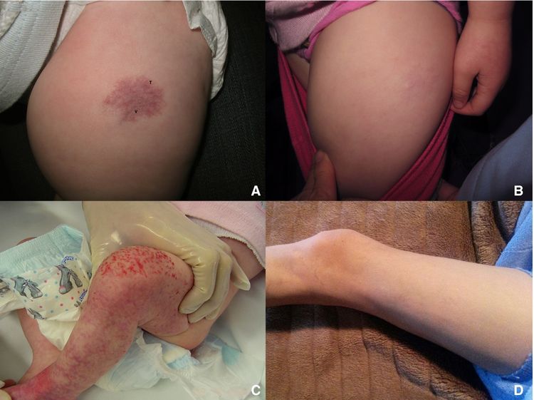
Cervical Cancer Prevention (PDQ®)–Health Professional Version

Cervical Cancer Prevention (PDQ®)–Health Professional Version

Overview

Note: The Overview section summarizes the published evidence on this topic. The rest of the summary describes the evidence in more detail.

Other PDQ summaries on Cervical Cancer Screening and Cervical Cancer Treatment are also available.

Who Is at Risk?

Carcinogenic types of human papillomavirus (HPV) are the primary, etiologic, infectious agents that cause virtually all cases of cervical cancer. HPV type 16 (HPV-16) and HPV type 18 (HPV-18) are most often associated with invasive disease.[1,2] Because HPV can be transmitted during sexual activity, there is an association between beginning sexual activity at a younger age, as well as having a greater number of lifetime sexual partners and an increased risk of developing cervical cancer.[3] Immunosuppression is another risk factor for cervical cancer; for example, coinfection with HIV may lead to long-term persistence of viral infection (i.e., failure to clear).[4,5] Once HPV infection occurs, several additional risk factors are associated with a higher risk of the eventual development of cervical cancer. These risk factors include high parity, long-term use of oral contraceptives, and active and passive cigarette smoking.[68] The risk increases with longer duration and intensity of smoking. Diethylstilbestrol (DES) exposure in utero is also associated with an increased risk of developing cervical dysplasia.[9]

Factors With Adequate Evidence of an Increased Risk of Cervical Cancer

Human papillomavirus (HPV)

Based on solid evidence from observational studies, HPV infection is associated with the development of cervical cancer.

Magnitude of Effect: HPV has been implicated as the primary etiologic infectious agent causing virtually all cases of cervical cancer.

  • Study Design: Evidence obtained from cohort and case-control studies.
  • Internal Validity: Good.
  • Consistency: Good.
  • External Validity: Good.

Immunosuppression

Based on solid evidence, being immunosuppressed is associated with an increased risk of cervical cancer.

  • Study Design: Evidence obtained from cohort and case-control studies.
  • Internal Validity: Good.
  • Consistency: Good.
  • External Validity: Good.

Sexual activity at an early age and with a greater number of partners

Based on solid evidence, sexual activity at a younger age and an increasing number of sexual partners are both associated with an increased risk of HPV infection and subsequent development of cervical cancer.

Magnitude of Effect: Women who experience first sexual intercourse at age 17 years or younger or women who have had six or more lifetime sexual partners have approximately two to three times the risk of squamous cell carcinoma or adenocarcinoma of the cervix, compared with women aged 21 years or older or who have a single sexual partner.[3]

  • Study Design: Evidence obtained from cohort and case-control studies.
  • Internal Validity: Good.
  • Consistency: Good.
  • External Validity: Good.

High parity

Based on solid evidence, high parity is associated with increased risk of cervical cancer in HPV-infected women.

Magnitude of Effect: Among HPV-infected women, those who have had seven or more full-term pregnancies have approximately four times the risk of squamous cell cancer compared with nulliparous women, and HPV-infected women also have two to three times the risk of women who have had one or two full-term pregnancies.[6]

  • Study Design: Evidence obtained from cohort or case-control studies.
  • Internal Validity: Good.
  • Consistency: Good.
  • External Validity: Good.

Long-term use of oral contraceptives

Based on solid evidence, long-term use of oral contraceptives is associated with increased risk of cervical cancer in HPV-infected women.

Magnitude of Effect: Among HPV-infected women, those who used oral contraceptives for 5 to 9 years have approximately three times the incidence of invasive cancer, and those who used them for 10 years or longer have approximately four times the risk.[7]

  • Study Design: Evidence obtained from cohort or case-control studies.
  • Internal Validity: Good.
  • Consistency: Good.
  • External Validity: Good.

Cigarette smoke exposure

Based on solid evidence, cigarette smoking, both active and passive, is associated with an increased risk of cervical cancer in HPV-infected women.

Magnitude of Effect: Among HPV-infected women, current and former smokers have approximately two to three times the incidence of high-grade cervical intraepithelial neoplasia or invasive cancer. Passive smoking is also associated with increased risk but to a lesser extent.

  • Study Design: Evidence obtained from cohort or case-control studies.
  • Internal Validity: Good.
  • Consistency: Good.
  • External Validity: Good.

Diethylstilbestrol (DES) exposure

Based on solid evidence, DES exposure is associated with an increased risk of developing clear cell adenocarcinoma of the cervix.

Magnitude of Effect: About one in 1,000 women exposed to DES in utero will develop a clear cell adenocarcinoma of the cervix.

  • Study Design: Evidence obtained from cohort studies.
  • Internal Validity: Good.
  • Consistency: Good.
  • External Validity: Good.

Factors With Adequate Evidence of a Decreased Risk of Cervical Cancer

Sexual abstinence

Based on solid evidence, abstinence from sexual activity is associated with a near-total reduction in the risk of developing cervical cancer.

Magnitude of Effect: Sexual abstinence essentially precludes HPV transmission.

  • Study Design: Evidence obtained from cohort or case-control studies.
  • Internal Validity: Good.
  • Consistency: Good.
  • External Validity: Good.

Interventions With Adequate Evidence of a Decreased Risk of Cervical Cancer

Note: Based on solid evidence, screening with the Papanicolaou (Pap) test and screening with the HPV DNA test reduces cervical cancer incidence. For more information on these screening tests, see Cervical Cancer Screening.

HPV vaccination: Benefits

Based on solid evidence, vaccination against HPV-16/HPV-18 is effective in preventing HPV infection in HPV-naive individuals and is associated with a reduced incidence of cervical intraepithelial neoplasia 2 and 3. By extrapolation, these vaccines should also be associated with a reduced incidence of cervical cancer.

Magnitude of Effect: Vaccination against HPV-16 and HPV-18 reduces incident and persistent infections with efficacy of 91.6% (95% confidence interval [CI], 64.5%–98.0%) and 100% (95% CI, 45%–100%), respectively.

  • Study Design: Evidence obtained from randomized controlled trials (for intraepithelial precursor lesions) and cohort study analyses (for invasive cervical cancers).
  • Internal Validity: Good.
  • Consistency: Good.
  • External Validity: Good.

HPV vaccination: Harms

Based on solid evidence, harms of HPV vaccines include injection-site reactions, dizziness and syncope, headache, and fever. Vaccination during pregnancy has not been associated with adverse pregnancy outcomes.[10] Allergic reactions occur rarely.

  • Study Design: Evidence obtained from randomized controlled trials.
  • Internal Validity: Good.
  • Consistency: Good.
  • External Validity: Good.

Use of barrier protection during sexual intercourse: Benefits

Based on solid evidence, the use of barrier methods (e.g., condoms) during sexual intercourse is associated with a decreased risk of cervical cancer.

Magnitude of Effect: Total use of barrier protection decreases cervical cancer incidence (relative risk, 0.4; 95% CI, 0.2–0.9).

  • Study Design: Evidence obtained from cohort and case-control studies.
  • Internal Validity: Good.
  • Consistency: Good.
  • External Validity: Good.

Use of barrier protection during sexual intercourse: Harms

Based on fair evidence, the use of barrier methods during sexual intercourse is associated with few serious harms. Barrier methods can break, potentially resulting in unintended pregnancy. Allergic reactions to the barrier material (e.g., natural latex) can occur.

  • Study Design: Evidence obtained from cohort and case-control studies.
  • Internal Validity: Good.
  • Consistency: Good.
  • External Validity: Good.
References
  1. Schiffman M, Castle PE, Jeronimo J, et al.: Human papillomavirus and cervical cancer. Lancet 370 (9590): 890-907, 2007. [PUBMED Abstract]
  2. Trottier H, Franco EL: The epidemiology of genital human papillomavirus infection. Vaccine 24 (Suppl 1): S1-15, 2006. [PUBMED Abstract]
  3. Berrington de González A, Green J; International Collaboration of Epidemiological Studies of Cervical Cancer: Comparison of risk factors for invasive squamous cell carcinoma and adenocarcinoma of the cervix: collaborative reanalysis of individual data on 8,097 women with squamous cell carcinoma and 1,374 women with adenocarcinoma from 12 epidemiological studies. Int J Cancer 120 (4): 885-91, 2007. [PUBMED Abstract]
  4. Abraham AG, D’Souza G, Jing Y, et al.: Invasive cervical cancer risk among HIV-infected women: a North American multicohort collaboration prospective study. J Acquir Immune Defic Syndr 62 (4): 405-13, 2013. [PUBMED Abstract]
  5. Grulich AE, van Leeuwen MT, Falster MO, et al.: Incidence of cancers in people with HIV/AIDS compared with immunosuppressed transplant recipients: a meta-analysis. Lancet 370 (9581): 59-67, 2007. [PUBMED Abstract]
  6. Muñoz N, Franceschi S, Bosetti C, et al.: Role of parity and human papillomavirus in cervical cancer: the IARC multicentric case-control study. Lancet 359 (9312): 1093-101, 2002. [PUBMED Abstract]
  7. Moreno V, Bosch FX, Muñoz N, et al.: Effect of oral contraceptives on risk of cervical cancer in women with human papillomavirus infection: the IARC multicentric case-control study. Lancet 359 (9312): 1085-92, 2002. [PUBMED Abstract]
  8. Appleby P, Beral V, Berrington de González A, et al.: Carcinoma of the cervix and tobacco smoking: collaborative reanalysis of individual data on 13,541 women with carcinoma of the cervix and 23,017 women without carcinoma of the cervix from 23 epidemiological studies. Int J Cancer 118 (6): 1481-95, 2006. [PUBMED Abstract]
  9. Hoover RN, Hyer M, Pfeiffer RM, et al.: Adverse health outcomes in women exposed in utero to diethylstilbestrol. N Engl J Med 365 (14): 1304-14, 2011. [PUBMED Abstract]
  10. Scheller NM, Pasternak B, Mølgaard-Nielsen D, et al.: Quadrivalent HPV Vaccination and the Risk of Adverse Pregnancy Outcomes. N Engl J Med 376 (13): 1223-1233, 2017. [PUBMED Abstract]

Incidence and Mortality

An estimated 13,360 new cervical cancers and 4,320 cervical cancer deaths will occur in the United States in 2025.[1] When corrected for the prevalence of hysterectomy, the mortality rate for Black women is nearly twice the mortality rate for White women.[2] Also, approximately 1,250,000 women will be diagnosed with precancers annually by cytology using the Papanicolaou (Pap) smear. A continuum of pathological changes may be diagnosed, ranging from atypical squamous cells of undetermined significance to low-grade squamous intraepithelial lesions (LSIL) to high-grade squamous intraepithelial lesions (HSIL) to invasive cancer. The precancerous conditions LSIL and HSIL are also referred to as cervical intraepithelial neoplasia (CIN) 1, 2, and 3. Lesions can regress, persist, or progress to an invasive malignancy, with LSIL (CIN 1) more likely to regress spontaneously and HSIL (CIN 2/CIN 3) more likely to persist or progress. The average time for progression of CIN 3 to invasive cancer has been estimated to be 10 to 15 years.[3]

References
  1. American Cancer Society: Cancer Facts and Figures 2025. American Cancer Society, 2025. Available online. Last accessed January 16, 2025.
  2. Beavis AL, Gravitt PE, Rositch AF: Hysterectomy-corrected cervical cancer mortality rates reveal a larger racial disparity in the United States. Cancer 123 (6): 1044-1050, 2017. [PUBMED Abstract]
  3. Holowaty P, Miller AB, Rohan T, et al.: Natural history of dysplasia of the uterine cervix. J Natl Cancer Inst 91 (3): 252-8, 1999. [PUBMED Abstract]

Factors With Adequate Evidence of an Increased Risk of Cervical Cancer

HPV

Epidemiological studies to evaluate risk factors for the development of squamous intraepithelial lesions and cervical malignancy demonstrate conclusively a sexual mode of transmission of a carcinogen.[1] It is now widely accepted that human papillomavirus (HPV) is the primary etiologic infectious agent that causes virtually all cases of cervical cancer.[2,3] Other sexually transmitted factors, including herpes simplex virus 2 and Chlamydia trachomatis, may play a co-causative role.[4] More than 80 distinct types of HPV have been identified, approximately 30 of which infect the human genital tract. HPV type 16 (HPV-16) and HPV type 18 (HPV-18) are most often associated with invasive disease. Characterization of carcinogenic risk associated with HPV types is an important step in the process of developing a combination HPV vaccine for the prevention of cervical neoplasia. In a population-based study of HPV infection and cervical neoplasia in Costa Rica, 80% of high-grade squamous intraepithelial lesions (HSIL) and invasive lesions were associated with HPV infection by one or more of 13 cancer-associated types.[5] In this study, the risk of about 50% of HSIL and invasive cervical cancer was attributable to HPV-16. HPV-18 was associated with 15% of invasive disease but only 5% of HSIL, suggesting that HPV-18 may have a role in more aggressive cases of cervical malignancy. There may be differences in the prevalence and carcinogenic risk of individual high-risk HPV genotypes by race and geographical regions, and additional studies are ongoing.[6]

Immunosuppression

Most cases of HPV infection are resolved by the host immune system. Immunosuppression leads to persistence of viral infection with a subsequent increased risk of cervical neoplasia. Women with immunosuppression resulting from HIV infection have been studied over the past three decades of the AIDS epidemic. In one North American study, a group of 13,690 HIV-infected women were studied for a median of 5 years. The rate of invasive cervical cancer in the HIV-infected women was 26 cases per 100,000 women, and this was approximately four times greater than an HIV-uninfected control group.[7] HIV-infected women with the lowest CD4 lymphocyte counts were at the highest risk of invasive cancer. Women who are immunosuppressed resulting from organ transplantation are also at risk of invasive cervical cancer, and one meta-analysis found a twofold increased risk.[8]

Sexual Activity at an Early Age and With a Greater Number of Partners

HPV infection has been established as a necessary cause of almost all cases of cervical cancer, and the primary mode of transmission is sexual contact. This provides context for the findings that younger age at first intercourse and an increasing number of lifetime sexual partners are both associated with an increased risk of developing cervical cancer. Pooled, individual, patient-level data from 12 cohort and case-control studies demonstrated statistically significantly increased risks of developing cervical cancer in women who were aged 17 years or younger at first intercourse, compared with women who were aged 21 years or older at first intercourse (relative risk [RR] for squamous cell cancer, 2.24; 95% confidence interval [CI], 2.11–2.38 and RR for adenocarcinoma, 2.06; 95% CI, 1.83–2.33). Similar findings were observed in women who had six or more lifetime sexual partners, compared with women who had one lifetime sexual partner (RR for squamous cell cancer, 2.98; 95% CI, 2.62–3.40 and RR for adenocarcinoma, 2.64; 95% CI, 2.07–3.36).[9]

High Parity

High parity has long been recognized as a risk factor for cervical cancer, but the relation of parity to HPV infection was uncertain. A meta-analysis of 25 epidemiological studies, including 16,563 women with cervical cancer and 33,542 women without cervical cancer, showed that the number of full-term pregnancies was associated with increased risk, regardless of age at first pregnancy. This finding was also true if analyses were limited to patients with high-risk HPV infections (RR, 4.99; 95% CI, 3.49–7.13 for seven or more pregnancies vs. no pregnancies; linear trend test X2 = 30.69; P < .001).[10]

Long-Term Use of Oral Contraceptives

Long-term use of oral contraceptives has also been known to be associated with cervical cancer, but its relation to HPV infection was also uncertain. A pooled analysis of HPV-positive women from the studies described above was undertaken. Compared with women who have never used oral contraceptives, those who have used them for fewer than 5 years did not have an increased risk of cervical cancer (odds ratio [OR], 0.73; 95% CI, 0.52–1.03). The OR for women who used oral contraceptives for 5 to 9 years was 2.82 (95% CI, 1.46–5.42), and for 10 or more years, the OR was 4.03 (95% CI, 2.09–8.02).[11] A meta-analysis of 24 epidemiological studies confirmed the increased risk associated with oral contraceptives, which is proportionate to the duration of use. Risk decreases after cessation and returns to normal risk levels in 10 years.[12]

Cigarette Smoke Exposure

Cigarette smoking by women is associated with an increased risk of squamous cell carcinoma.[1,13,14] This risk increases with longer duration and intensity of smoking. The risk among smokers may be present with exposure to environmental tobacco smoke and may be as high as four times that of women who are nonsmokers who are not exposed to environmental smoking.[1] Case-control studies of women infected with HPV have examined the effect of various types and levels of tobacco exposure and found similar results.[1416]

DES Exposure

Diethylstilbestrol (DES) is a synthetic form of estrogen that was prescribed to pregnant women in the United States between 1940 and 1971 to prevent miscarriage and premature labor. DES is associated with a substantially increased risk of developing clear cell adenocarcinoma of the vagina and cervix among the daughters of women who used the drug during pregnancy (standardized incidence ratio, 24.23; 95% CI, 8.89–52.74); the risk persists as these women age into their 40s.[17] Despite the greatly elevated risk relative to the general population, this type of cancer is still rare; about one in 1,000 daughters exposed to DES will develop a clear cell adenocarcinoma.

DES exposure in utero is also associated with an increased risk of developing cervical dysplasia. An evaluation of three cohorts, including the Diethylstilbestrol Adenosis study, the Dieckmann study, and the Women’s Health Study, with long-term follow-up of more than 4,500 women exposed in utero to DES, found that 6.9% of exposed women developed grade II or higher cervical intraepithelial neoplasia, compared with 3.4% of nonexposed women (hazard ratio, 2.28; 95% CI, 1.59–3.27).[18]

References
  1. Brinton LA: Epidemiology of cervical cancer–overview. IARC Sci Publ (119): 3-23, 1992. [PUBMED Abstract]
  2. Schiffman M, Castle PE, Jeronimo J, et al.: Human papillomavirus and cervical cancer. Lancet 370 (9590): 890-907, 2007. [PUBMED Abstract]
  3. Trottier H, Franco EL: The epidemiology of genital human papillomavirus infection. Vaccine 24 (Suppl 1): S1-15, 2006. [PUBMED Abstract]
  4. Ault KA: Epidemiology and natural history of human papillomavirus infections in the female genital tract. Infect Dis Obstet Gynecol 2006 (Suppl): 40470, 2006. [PUBMED Abstract]
  5. Herrero R, Hildesheim A, Bratti C, et al.: Population-based study of human papillomavirus infection and cervical neoplasia in rural Costa Rica. J Natl Cancer Inst 92 (6): 464-74, 2000. [PUBMED Abstract]
  6. Risley C, Clarke MA, Geisinger KR, et al.: Racial differences in HPV type 16 prevalence in women with ASCUS of the uterine cervix. Cancer Cytopathol 128 (8): 528-534, 2020. [PUBMED Abstract]
  7. Abraham AG, D’Souza G, Jing Y, et al.: Invasive cervical cancer risk among HIV-infected women: a North American multicohort collaboration prospective study. J Acquir Immune Defic Syndr 62 (4): 405-13, 2013. [PUBMED Abstract]
  8. Grulich AE, van Leeuwen MT, Falster MO, et al.: Incidence of cancers in people with HIV/AIDS compared with immunosuppressed transplant recipients: a meta-analysis. Lancet 370 (9581): 59-67, 2007. [PUBMED Abstract]
  9. Berrington de González A, Green J; International Collaboration of Epidemiological Studies of Cervical Cancer: Comparison of risk factors for invasive squamous cell carcinoma and adenocarcinoma of the cervix: collaborative reanalysis of individual data on 8,097 women with squamous cell carcinoma and 1,374 women with adenocarcinoma from 12 epidemiological studies. Int J Cancer 120 (4): 885-91, 2007. [PUBMED Abstract]
  10. International Collaboration of Epidemiological Studies of Cervical Cancer: Cervical carcinoma and reproductive factors: collaborative reanalysis of individual data on 16,563 women with cervical carcinoma and 33,542 women without cervical carcinoma from 25 epidemiological studies. Int J Cancer 119 (5): 1108-24, 2006. [PUBMED Abstract]
  11. Moreno V, Bosch FX, Muñoz N, et al.: Effect of oral contraceptives on risk of cervical cancer in women with human papillomavirus infection: the IARC multicentric case-control study. Lancet 359 (9312): 1085-92, 2002. [PUBMED Abstract]
  12. Appleby P, Beral V, Berrington de González A, et al.: Cervical cancer and hormonal contraceptives: collaborative reanalysis of individual data for 16,573 women with cervical cancer and 35,509 women without cervical cancer from 24 epidemiological studies. Lancet 370 (9599): 1609-21, 2007. [PUBMED Abstract]
  13. Hellberg D, Nilsson S, Haley NJ, et al.: Smoking and cervical intraepithelial neoplasia: nicotine and cotinine in serum and cervical mucus in smokers and nonsmokers. Am J Obstet Gynecol 158 (4): 910-3, 1988. [PUBMED Abstract]
  14. Brock KE, MacLennan R, Brinton LA, et al.: Smoking and infectious agents and risk of in situ cervical cancer in Sydney, Australia. Cancer Res 49 (17): 4925-8, 1989. [PUBMED Abstract]
  15. Ho GY, Kadish AS, Burk RD, et al.: HPV 16 and cigarette smoking as risk factors for high-grade cervical intra-epithelial neoplasia. Int J Cancer 78 (3): 281-5, 1998. [PUBMED Abstract]
  16. Plummer M, Herrero R, Franceschi S, et al.: Smoking and cervical cancer: pooled analysis of the IARC multi-centric case–control study. Cancer Causes Control 14 (9): 805-14, 2003. [PUBMED Abstract]
  17. Verloop J, van Leeuwen FE, Helmerhorst TJ, et al.: Cancer risk in DES daughters. Cancer Causes Control 21 (7): 999-1007, 2010. [PUBMED Abstract]
  18. Hoover RN, Hyer M, Pfeiffer RM, et al.: Adverse health outcomes in women exposed in utero to diethylstilbestrol. N Engl J Med 365 (14): 1304-14, 2011. [PUBMED Abstract]

Factors With Adequate Evidence of a Decreased Risk of Cervical Cancer

Sexual Abstinence

Nearly all cases of cervical cancer are associated with human papillomavirus (HPV) infection, which is transmitted during sexual activity. Therefore, cervical cancer is seen more frequently in women with sexual activity at an early age and with multiple partners.[1] Lifetime abstinence from sexual activity is associated with a near-total reduction in the risk of developing cervical cancer. For more information, see the HPV section.

References
  1. IARC Working Group on the Evaluation of Carcinogenic Risks to Humans: Human papillomaviruses. IARC Monogr Eval Carcinog Risks Hum 100 (Pt B), 255-296, 2012. Available online. Last accessed January 31, 2025.

Interventions With Adequate Evidence of a Decreased Risk of Cervical Cancer

HPV Vaccination

Given the etiological role of human papillomavirus (HPV) in the pathogenesis of cervical neoplasia, vaccines to immunize individuals against HPV infection offer a primary prevention strategy for cervical cancer. A quadrivalent (HPV type 6 [HPV-6], type 11 [HPV-11], type 16 [HPV-16], and type 18 [HPV-18]) vaccine using a late protein L1 construct to induce antibody-mediated immunity was approved for use by the U.S. Food and Drug Administration in 2006; a bivalent (HPV-16, -18) vaccine was approved in 2009; and a vaccine targeting nine HPV types was approved in 2014. Vaccination during pregnancy has not been associated with adverse pregnancy outcomes.[1]

Persistent infection with oncogenic types of HPV, such as HPV-16 and HPV-18, is associated with the development of cervical cancer.[2] A vaccine to prevent HPV infection with oncogenic-type viruses has the potential to reduce the incidence of cervical cancer. A vaccine against HPV-16 using empty-viral capsids called virus-like particles (VLP) was developed and tested for efficacy in preventing persistent infection with HPV-16.

A multicenter, double-blind, placebo-controlled trial enrolled 2,391 women aged 16 to 23 years and randomly assigned them to receive either 40 µg of HPV-16 L1 VLP vaccine or placebo on day 1, at 2 months, and at 6 months. Papanicolaou (Pap) tests and genital samples for HPV-16 DNA were obtained on day 1, at 7 months, and every 6 months for 48 months. Colposcopy and cervical biopsies were obtained when clinically indicated at study exit. Serum HPV-16 antibody titers were obtained at study entry, at 7 months, and then every 6 months. A total of 1,505 women (755 receiving vaccine and 750 receiving placebo) completed all three vaccinations and had follow-up after month 7. After immunization, HPV titers peaked at month 7, declined through month 18, and then stabilized in months 30 through 48. There were no cases of cervical intraepithelial neoplasia (CIN) in the vaccine-treated women, but there were 12 cases in the placebo group (six CIN 2 and six CIN 3). HPV-16 infection that persisted for at least 4 months was seen in seven vaccine-treated women, compared with 111 placebo-treated women.[3]

An international, double-blind, placebo-controlled trial of a bivalent HPV-16/HPV-18 VLP vaccine was performed in 1,113 women aged 15 to 25 years with normal cervical cytology who were seronegative for HPV-16, HPV-18, and 12 other oncogenic HPV types at enrollment. Women received either vaccine or placebo at 0, 1, and 6 months and were assessed by cervical cytology and self-obtained cervicovaginal samples for at least 18 months. A masked treatment-allocation follow-up study was performed for an additional 3 years, for a combined analysis of up to 6.4 years of follow-up. The 12-month persistent infection rate of HPV-16 or HPV-18 in an according-to-protocol cohort (i.e., women who received all three doses of vaccine or placebo on the correct schedule) was 0 of 401 women in the vaccine arm, compared with 20 of 372 women in the placebo arm, with a vaccine efficacy rate of 100% (95% confidence interval [CI], 81.8%–100%). Diagnoses of CIN 2 or higher in a total vaccinated cohort (i.e., women who received at least one dose of vaccine or placebo) were 0 of 481 women in the vaccine arm compared with 9 of 470 women in the placebo arm, with a vaccine efficacy of 100% (95% CI, 51.3%–100%). Adverse events were similar in vaccinated and placebo-treated women. Neither analysis was intention-to-treat (ITT), making it difficult to know what the true vaccine efficacy for either virological or cytohistological end points would be in the routine clinical setting. Furthermore, cytohistological outcomes were reported only as composite end points (CIN 2+), making it impossible to distinguish the vaccine’s efficacy against invasive cervical cancer alone and potentially inflating the observed efficacy by including lesions with a relatively high probability (approximately 50% for CIN 2 [4]) of spontaneous regression.[5] A register-based observational study in England reported the impact of a national bivalent vaccination program on cervical cancer and CIN 3.[6] Routine vaccinations were offered to girls aged 12 to 13 years with a catch-up program for those aged 14 to 18 years. Data from 13.7 million years of follow-up in women aged 20 years to younger than 30 years showed a substantial reduction in cervical cancer and CIN 3 incidence after a national HPV program was introduced. This was especially true in individuals who were offered the vaccine between ages 12 and 13 years (see Table 1).

Table 1. Estimated Relative Reduction in Cervical Cancer or CIN 3 by Age When Vaccine Was Offered Compared With the Reference Unvaccinated Cohort
Estimated Relative Reduction (95% CI)
Age Vaccine Was Offered Cervical Cancer CIN 3
CI = confidence interval; CIN = cervical intraepithelial neoplasia.
16–18 y 34% (25–41) 39% (36–41)
14–16 y 62% (52–71) 75% (72–77)
12–13 y 87% (72–94) 97% (96–78)

A quadrivalent vaccine (HPV types-6, -11, -16, and -18) was evaluated in a multinational, double-blind, randomized controlled trial of 17,622 women aged 15 to 26 years (FUTURE I and II).[7] Women received either the HPV vaccine or placebo at 0, 2, and 6 months; participants were assessed by clinical exam, Pap test, and HPV DNA testing for 4 or more years. Two analyses were reported. One group was considered to be HPV naive: negative to 14 HPV types. The second group was an ITT analysis, which approximates a sexually active population. The composite end point for cervical disease included the incidence of HPV-16/HPV-18–related, CIN 2, CIN 3, adenocarcinoma in situ, or invasive carcinoma. Outcomes were reported as follows:

Table 2. Vaccine Efficacy of the Quadrivalent HPV Vaccine
Population Point Estimate and 95% CI
CI = confidence interval; CIN = cervical intraepithelial neoplasia; HPV = human papillomavirus; ITT = intention-to-treat.
HPV-naive population for HPV-CIN 3 100% (90.5%–100%) for lesions associated with HPV-6, -11, -16, or -18
ITT CIN 3 45.3% (29.8%–57.6%) for lesions associated with HPV-6, -11, -16, or -18

This study also demonstrated decreased rates of abnormal Pap tests and subsequent diagnostic procedures. No cases of invasive cervical cancer were identified during the trial.

A 9-valent HPV (9vHPV) vaccine was studied in another international randomized trial, which included 14,215 women. This new vaccine, 9vHPV, includes the four HPV types in the quadrivalent vaccine, qHPV (6, 11, 16, 18) and also 5 more oncogenic types (31, 33, 45, 52, 58). Sexually active women aged 16 to 26 years with fewer than five lifetime sexual partners received three intramuscular injections (day 1, month 2, and month 6) of either the qHPV vaccine or the 9vHPV vaccine. Women were evaluated every 6 months for up to 5 years. The rate of high-grade cervical, vulvar, or vaginal disease was the same in both groups (14.0 per 1,000 person-years) because of preexisting HPV infection, but the rate of disease related to HPV-31, -35, -45, -52, and -58 was lower in the 9vHPV vaccine group (0.1 vs. 1.6 per 1,000 person-years). Injection-site reactions were more common in the 9vHPV group.[8] Although not addressed in this study, the benefit of HPV vaccination is optimal in younger females before the onset of sexual activity.

All forms of the HPV vaccine are currently recommended by the Centers for Disease Control and Prevention (CDC) in the United States as a two-dose schedule at least 6 months apart for adolescents younger than 15 years. The current CDC recommendation for older individuals is to receive the original three-dose series. Recently, given issues of cost and adherence, there has been published data investigating whether similar vaccine efficacy could be obtainable using a reduced-dose schedule. A post hoc combined analysis of two phase III randomized controlled trials of the bivalent HPV vaccine (the Costa Rica Vaccine Trial and the PApilloma TRIal against Cancer In young Adults [PATRICIA] Trial) found that among women who were not HPV positive at enrollment for the specific virus type being studied, vaccine efficacy against either one-time incident detection of HPV 16/18 or incident infection that persisted at least 6 months was not statistically significantly different for those who received all three, two, or only one of the scheduled HPV vaccine doses (resulting from nonadherence or other factors) for up to 4 years of follow-up. Vaccine efficacy rates for persistent HPV 16/18 infection ranged from 89.1% (95% CI, 86.8%–91.0%) for three doses, to 89.7% (95% CI, 73.3%–99.8%) for two doses, to 96.6% (95% CI, 81.7%–99.8%) for one dose. To date, there are no randomized controlled trials that directly assess this clinical question.[9] A recent international study compared a two-dose schedule with a three-dose schedule in adolescents younger than 15 years who received the 9vHPV vaccine. The antibody response was noninferior in the two-dose schedule, leading to the current recommendation that two doses are sufficient for this age group.[10] Long-term follow-up data from the Costa Rica Vaccine Trial suggested that all HPV-vaccinated women aged 18 to 25 years at the time of initial vaccination remained HPV-16/HPV-18 seropositive more than a decade after initial vaccination, regardless of the number of doses received. The antibody levels were lower in the women who received only one dose than in the women who received two or three doses of the bivalent vaccine, but the levels remained higher than estimated levels achieved via natural immunity. The long-term vaccine efficacy rates against prevalent HPV-16 or HPV-18 infection were 80.2% (95% CI, 70.7%–87.0%) among women who received three doses, 83.8% (95% CI, 19.5%–99.2%) among those who received two doses, and 82.1% (95% CI, 40.2%–97.0%) among those who received one dose.[11] Additionally, there was prolonged efficacy against CIN 2 and CIN 3 after 7 to 11 years of follow-up.[12] The women in this long-term follow-up study were not randomly assigned to one, two, or three doses, and the number of women who received only one dose is low. However, the promising findings of the long-term stability of HPV-antibody levels and vaccine efficacy in women who were older than the recommended age at the time of initial vaccination has influenced the design of a currently ongoing trial, which will answer the question of the efficacy of a single dose more definitively. The ESCUDDO study (NCT03180034) is a trial enrolling adolescent girls who will be randomly assigned to either one dose or two doses of the bivalent or nonavalent vaccines. A concurrent population survey of comparable, unvaccinated, age-matched females in the same region will be used for comparison. Results are anticipated in 2025.

On the basis of their mechanism of action, L1/2 HPV vaccines do not appear to impact preexisting infections. The FUTURE II trial demonstrated a markedly lower vaccine efficacy rate in the total randomized study population, which included individuals who were positive for HPV at baseline, compared with the per-protocol population (44% for lesions associated with HPV-16 or HPV-18, and 17% for lesions associated with any HPV type vs. 98%; see Table 2 above).[7] Additionally, an intermediate analysis of a randomized controlled trial primarily evaluating the efficacy of the HPV-16/18 vaccine in preventing infection found no effect on viral clearance rates in women aged 18 to 25 years who were positive at the time of study enrollment.[13]

The type-specific vaccines, if successful in preventing invasive cancer, will offer protection for only a subset of cases, the proportion of which will vary worldwide.[14] Using data from a multicenter case-control study conducted in 25 countries, it was estimated that a vaccine containing the seven most common HPV types could prevent 87% of cervical cancers worldwide. A vaccine with the two most common strains, HPV-16 and HPV-18, would prevent 71% of cervical cancers worldwide.[14]

There is growing evidence of population-level impacts and herd immunity with HPV vaccination. There are data that explore the impact of national HPV vaccination programs and report on vaccine effectiveness. These data come from studies conducted in different countries throughout the world including England, Denmark, Australia, Costa Rica, and the United States. In England, 15,459 residual genital specimens from women aged 16 to 24 years, collected for Chlamydia trachomatis screening between 2010 and 2016, were utilized for national HPV surveillance.[15] In this study, vaccine effectiveness for HPV-16/HPV-18 was 82% (95% CI, 60.6%–91.8%) for women who were vaccinated before age 15 years. Within the younger age groups, the prevalence of HPV-16/HPV-18 significantly decreased within the postvaccination period between 2010 and 2011 to 2016 from 8.2% to 1.6% in 16 to 18 year olds and from 14.0% to 1.6% in 19 to 21 year olds (compared with 17.6% and 16.9% in the prevaccination era).[15] Data from a Danish nationwide cohort study reported the dose-related effectiveness of the quadrivalent HPV vaccine.[16] In this cohort of 590,083 women aged 17 to 25 years, 215,309 (36%) women were vaccinated at age 16 years or younger, and 40,742 (19%) women received less than three doses. Cervical cancer screening rates were similar in the vaccinated and unvaccinated cohorts. In the overall cohort, there were 5,561 cases of CIN 3+ during the follow-up period. Only 5% of cases were in vaccinated women. The incidence rate was 355 cases per 100,000 person-years in unvaccinated women compared with 41 cases per 100,000 person-years in vaccinated women. The incidence rate was independent of the number of vaccine doses administered (incidence rates 40, 41, 40 cases per 100,000 person-years for 1, 2 and 3 doses, respectively).[16]

A study of cervical HPV DNA among 202 Australian women aged 18 to 24 years who were sampled between 2005 and 2007, before implementation of a national quadrivalent prophylactic HPV vaccine program, compared the results with a matched group of 1,058 women who were sampled in the postvaccination era (2010–2012). This study found an adjusted prevalence ratio (PR) among fully vaccinated women of 0.07 (95% CI, 0.04–0.14; P < .0001) for vaccine-related HPV types and a smaller but statistically significant magnitude of protection of 0.65 (95% CI, 0.43–0.96; P < .03) among unvaccinated women, suggesting herd immunity (protection of unvaccinated individuals).[17] These data strengthen previous results that suggest herd immunity in this population, manifested as a reduction in genital warts among heterosexual men, a group that includes sexual partners of vaccinated women.[18] Data also suggest cross protection against carcinogenic types that are not directly targeted by the quadrivalent vaccine but are included in the new nonvalent HPV vaccine.[17] Pooled data from the Costa Rica Vaccine Trial and PATRICIA Study showed that the AS04-adjuvanted HPV-16/HPV-18 vaccine provides additional cross protection beyond established protected types (e.g., 34/35/39/40/42/43/44/51/52/53/54/56/58/59/66/68/73/70/74; efficacy 9/9%; 95% CI, 1.7%–1.4%). This may partially explain the high efficacy of the AS04-adjuvanted HPV-16/HPV-18 vaccine against CIN 3+ (87.8%; 95% CI, 71.1%–95.7%).[19] A meta-analysis that included data from 14 high-income countries with cumulated data from more than 60 million individuals over 8 years reported an 83% decrease in prevalence of HPV-16 and HPV-18 (RR, 0.17; 95% CI, 0.11–0.25) among girls aged 13 to 19 years. There was also evidence of benefit to a more proximal cancer end point. After 5 to 9 years of HPV vaccination, decreased risk of CIN 2+ was also identified among screened girls aged 15 to 19 years (RR, 0.49; 95% CI, 0.42–0.58), and among women aged 20 to 24 years (RR, 0.69; 95% CI, 0.57–0.84) in comparison to an increase seen among screened and mostly unvaccinated women aged 25 to 29 years (RR, 1.19; 95% CI, 1.06–1.32) and aged 30 to 39 years (RR, 1.23; 95% CI, 1.13–1.34).[20]

Data from the National Health and Nutrition Examination Survey (NHANES) from 2003 to 2018 demonstrated an increasing impact of the HPV vaccination program and herd protection in the United States.[21] Overall, the data demonstrated an increase in HPV vaccination coverage among sexually experienced females and males. Importantly, vaccination before age 15 years also increased from 2011 to 2014 and from 2015 to 2018 in both females (27.2%, 48.6%, respectively) and males (18.6%, 48.7%, respectively). From 2015 to 2018, the 4-valent HPV (4vHPV)-type prevalence among sexually experience females aged 14 to 24 years was 85% overall, 90% in vaccinated females, and 74% in unvaccinated females. Estimates of the vaccine’s effectiveness and its impact among vaccinated females were similar from 2007 to 2010 (64% and 61%, respectively) and from 2011 to 2014 (84% and 89%, respectively). However, in 2015 to 2018, these statistics diverged (60% and 90%, respectively). This indicates that as herd protection increases and prevalence among unvaccinated individuals decreases, vaccine effectiveness can be difficult to estimate (1-prevalence ratio between vaccinated and unvaccinated individuals x 100). In 2013 to 2016, the prevalence of 4vHPV types was 1.8% in sexually experienced males who were vaccinated and 3.5% in sexually experienced males who were unvaccinated (PR, 0.49; 95% CI, 0.11–2.20), resulting in an estimated vaccine effectiveness of 51%. Significant declines were not observed in non–4vHPV-type prevalence for females or males. Although notable limitations of this survey study included self-report of HPV vaccine and dose, small sample sizes, and estimates of the impact and effectiveness based on history of at least one vaccine dose, this nationally representative data reflects an increasing impact of the U.S. vaccination program and herd protection.

Association of HPV vaccination with reduced incidence of invasive cervical cancer

In a nationwide population-based cohort study of the impact of the national vaccination program in Sweden using quadrivalent vaccine, more than 1.67 million women aged 10 to 30 years with no previous history of HPV vaccination were followed through the national registry using individual person linkage.[22] The cumulative risk of cervical cancer by age 30 years was 47 cases per 100,000 in vaccinated women, compared with 94 cases per 100,000 in unvaccinated women (incidence rate ratio [IRR], 0.51; 95% CI, 0.32–0.82, adjusting only for age at follow-up). After adjusting for all collected potential confounding factors, the IRR for women vaccinated before age 17 years was 0.12 (95% CI, 0.00–0.34).

Use of Barrier Protection During Sexual Intercourse

Barrier methods of contraception are associated with a reduced incidence of squamous intraepithelial lesions (SIL) presumptively secondary to protection from sexually transmitted disease.[23,24] The effectiveness of condom use for the prevention of HPV infections has been evaluated in a prospective study of women aged 18 to 22 years who were virgins.[25] The number of vulvovaginal HPV infections was reduced with consistent condom use, and the HPV infection rate was 37.8 infections per 100 patient-years among women whose partners used condoms 100% of the time in the 8 months before testing, compared with 89.3 infections per 100 patient-years among women whose partners used condoms less than 5% of the time (P trend = .005). No cervical SIL were detected among women reporting 100% condom use by their partner.[25]

References
  1. Scheller NM, Pasternak B, Mølgaard-Nielsen D, et al.: Quadrivalent HPV Vaccination and the Risk of Adverse Pregnancy Outcomes. N Engl J Med 376 (13): 1223-1233, 2017. [PUBMED Abstract]
  2. Wallin KL, Wiklund F, Angström T, et al.: Type-specific persistence of human papillomavirus DNA before the development of invasive cervical cancer. N Engl J Med 341 (22): 1633-8, 1999. [PUBMED Abstract]
  3. Mao C, Koutsky LA, Ault KA, et al.: Efficacy of human papillomavirus-16 vaccine to prevent cervical intraepithelial neoplasia: a randomized controlled trial. Obstet Gynecol 107 (1): 18-27, 2006. [PUBMED Abstract]
  4. Castle PE, Schiffman M, Wheeler CM, et al.: Evidence for frequent regression of cervical intraepithelial neoplasia-grade 2. Obstet Gynecol 113 (1): 18-25, 2009. [PUBMED Abstract]
  5. Romanowski B, de Borba PC, Naud PS, et al.: Sustained efficacy and immunogenicity of the human papillomavirus (HPV)-16/18 AS04-adjuvanted vaccine: analysis of a randomised placebo-controlled trial up to 6.4 years. Lancet 374 (9706): 1975-85, 2009. [PUBMED Abstract]
  6. Falcaro M, Castañon A, Ndlela B, et al.: The effects of the national HPV vaccination programme in England, UK, on cervical cancer and grade 3 cervical intraepithelial neoplasia incidence: a register-based observational study. Lancet 398 (10316): 2084-2092, 2021. [PUBMED Abstract]
  7. FUTURE II Study Group: Quadrivalent vaccine against human papillomavirus to prevent high-grade cervical lesions. N Engl J Med 356 (19): 1915-27, 2007. [PUBMED Abstract]
  8. Joura EA, Giuliano AR, Iversen OE, et al.: A 9-valent HPV vaccine against infection and intraepithelial neoplasia in women. N Engl J Med 372 (8): 711-23, 2015. [PUBMED Abstract]
  9. Kreimer AR, Struyf F, Del Rosario-Raymundo MR, et al.: Efficacy of fewer than three doses of an HPV-16/18 AS04-adjuvanted vaccine: combined analysis of data from the Costa Rica Vaccine and PATRICIA trials. Lancet Oncol 16 (7): 775-86, 2015. [PUBMED Abstract]
  10. Iversen OE, Miranda MJ, Ulied A, et al.: Immunogenicity of the 9-Valent HPV Vaccine Using 2-Dose Regimens in Girls and Boys vs a 3-Dose Regimen in Women. JAMA 316 (22): 2411-2421, 2016. [PUBMED Abstract]
  11. Kreimer AR, Sampson JN, Porras C, et al.: Evaluation of Durability of a Single Dose of the Bivalent HPV Vaccine: The CVT Trial. J Natl Cancer Inst 112 (10): 1038-1046, 2020. [PUBMED Abstract]
  12. Porras C, Tsang SH, Herrero R, et al.: Efficacy of the bivalent HPV vaccine against HPV 16/18-associated precancer: long-term follow-up results from the Costa Rica Vaccine Trial. Lancet Oncol 21 (12): 1643-1652, 2020. [PUBMED Abstract]
  13. Hildesheim A, Herrero R, Wacholder S, et al.: Effect of human papillomavirus 16/18 L1 viruslike particle vaccine among young women with preexisting infection: a randomized trial. JAMA 298 (7): 743-53, 2007. [PUBMED Abstract]
  14. Muñoz N, Bosch FX, Castellsagué X, et al.: Against which human papillomavirus types shall we vaccinate and screen? The international perspective. Int J Cancer 111 (2): 278-85, 2004. [PUBMED Abstract]
  15. Mesher D, Panwar K, Thomas SL, et al.: The Impact of the National HPV Vaccination Program in England Using the Bivalent HPV Vaccine: Surveillance of Type-Specific HPV in Young Females, 2010-2016. J Infect Dis 218 (6): 911-921, 2018. [PUBMED Abstract]
  16. Verdoodt F, Dehlendorff C, Kjaer SK: Dose-related Effectiveness of Quadrivalent Human Papillomavirus Vaccine Against Cervical Intraepithelial Neoplasia: A Danish Nationwide Cohort Study. Clin Infect Dis 70 (4): 608-614, 2020. [PUBMED Abstract]
  17. Tabrizi SN, Brotherton JM, Kaldor JM, et al.: Assessment of herd immunity and cross-protection after a human papillomavirus vaccination programme in Australia: a repeat cross-sectional study. Lancet Infect Dis 14 (10): 958-66, 2014. [PUBMED Abstract]
  18. Donovan B, Franklin N, Guy R, et al.: Quadrivalent human papillomavirus vaccination and trends in genital warts in Australia: analysis of national sentinel surveillance data. Lancet Infect Dis 11 (1): 39-44, 2011. [PUBMED Abstract]
  19. Tota JE, Struyf F, Hildesheim A, et al.: Efficacy of AS04-Adjuvanted Vaccine Against Human Papillomavirus (HPV) Types 16 and 18 in Clearing Incident HPV Infections: Pooled Analysis of Data From the Costa Rica Vaccine Trial and the PATRICIA Study. J Infect Dis 223 (9): 1576-1581, 2021. [PUBMED Abstract]
  20. Drolet M, Bénard É, Pérez N, et al.: Population-level impact and herd effects following the introduction of human papillomavirus vaccination programmes: updated systematic review and meta-analysis. Lancet 394 (10197): 497-509, 2019. [PUBMED Abstract]
  21. Rosenblum HG, Lewis RM, Gargano JW, et al.: Human Papillomavirus Vaccine Impact and Effectiveness Through 12 Years After Vaccine Introduction in the United States, 2003 to 2018. Ann Intern Med 175 (7): 918-926, 2022. [PUBMED Abstract]
  22. Lei J, Ploner A, Elfström KM, et al.: HPV Vaccination and the Risk of Invasive Cervical Cancer. N Engl J Med 383 (14): 1340-1348, 2020. [PUBMED Abstract]
  23. Parazzini F, Negri E, La Vecchia C, et al.: Barrier methods of contraception and the risk of cervical neoplasia. Contraception 40 (5): 519-30, 1989. [PUBMED Abstract]
  24. Hildesheim A, Brinton LA, Mallin K, et al.: Barrier and spermicidal contraceptive methods and risk of invasive cervical cancer. Epidemiology 1 (4): 266-72, 1990. [PUBMED Abstract]
  25. Winer RL, Hughes JP, Feng Q, et al.: Condom use and the risk of genital human papillomavirus infection in young women. N Engl J Med 354 (25): 2645-54, 2006. [PUBMED Abstract]

Latest Updates to This Summary (04/18/2025)

The PDQ cancer information summaries are reviewed regularly and updated as new information becomes available. This section describes the latest changes made to this summary as of the date above.

Incidence and Mortality

Updated statistics with estimated new cases and deaths for 2025 (cited American Cancer Society as reference 1).

This summary is written and maintained by the PDQ Screening and Prevention Editorial Board, which is editorially independent of NCI. The summary reflects an independent review of the literature and does not represent a policy statement of NCI or NIH. More information about summary policies and the role of the PDQ Editorial Boards in maintaining the PDQ summaries can be found on the About This PDQ Summary and PDQ® Cancer Information for Health Professionals pages.

About This PDQ Summary

Purpose of This Summary

This PDQ cancer information summary for health professionals provides comprehensive, peer-reviewed, evidence-based information about cervical cancer prevention. It is intended as a resource to inform and assist clinicians in the care of their patients. It does not provide formal guidelines or recommendations for making health care decisions.

Reviewers and Updates

This summary is reviewed regularly and updated as necessary by the PDQ Screening and Prevention Editorial Board, which is editorially independent of the National Cancer Institute (NCI). The summary reflects an independent review of the literature and does not represent a policy statement of NCI or the National Institutes of Health (NIH).

Board members review recently published articles each month to determine whether an article should:

  • be discussed at a meeting,
  • be cited with text, or
  • replace or update an existing article that is already cited.

Changes to the summaries are made through a consensus process in which Board members evaluate the strength of the evidence in the published articles and determine how the article should be included in the summary.

Any comments or questions about the summary content should be submitted to Cancer.gov through the NCI website’s Email Us. Do not contact the individual Board Members with questions or comments about the summaries. Board members will not respond to individual inquiries.

Levels of Evidence

Some of the reference citations in this summary are accompanied by a level-of-evidence designation. These designations are intended to help readers assess the strength of the evidence supporting the use of specific interventions or approaches. The PDQ Screening and Prevention Editorial Board uses a formal evidence ranking system in developing its level-of-evidence designations.

Permission to Use This Summary

PDQ is a registered trademark. Although the content of PDQ documents can be used freely as text, it cannot be identified as an NCI PDQ cancer information summary unless it is presented in its entirety and is regularly updated. However, an author would be permitted to write a sentence such as “NCI’s PDQ cancer information summary about breast cancer prevention states the risks succinctly: [include excerpt from the summary].”

The preferred citation for this PDQ summary is:

PDQ® Screening and Prevention Editorial Board. PDQ Cervical Cancer Prevention. Bethesda, MD: National Cancer Institute. Updated <MM/DD/YYYY>. Available at: /types/cervical/hp/cervical-prevention-pdq. Accessed <MM/DD/YYYY>. [PMID: 26389433]

Images in this summary are used with permission of the author(s), artist, and/or publisher for use within the PDQ summaries only. Permission to use images outside the context of PDQ information must be obtained from the owner(s) and cannot be granted by the National Cancer Institute. Information about using the illustrations in this summary, along with many other cancer-related images, is available in Visuals Online, a collection of over 2,000 scientific images.

Disclaimer

The information in these summaries should not be used as a basis for insurance reimbursement determinations. More information on insurance coverage is available on Cancer.gov on the Managing Cancer Care page.

Contact Us

More information about contacting us or receiving help with the Cancer.gov website can be found on our Contact Us for Help page. Questions can also be submitted to Cancer.gov through the website’s Email Us.