Cryosurgery is a treatment that uses extreme cold produced by liquid nitrogen or argon gas to destroy cancer cells and abnormal tissue. It is a local treatment, which means that it is directed toward a specific part of your body. Cryosurgery is used to treat tumors on the skin, as well as certain tumors inside the body.
Cryosurgery freezes tissue, causing cells in the treated area to die.
For tumors on the skin, the doctor applies liquid nitrogen directly to the abnormal area with a cotton swab or spraying device. For tumors inside the body, the doctor may use a device called a cryoprobe to freeze the tumor tissue. Cryoprobes may be put into the body during surgery or through a small cut in the skin. As liquid nitrogen or argon gas flows through the cryoprobe, the doctor places it directly on the tumor. During this procedure, the doctor uses ultrasound or MRI to guide the cryoprobe to the correct spot, which helps limit damage to nearby healthy tissue. Sometimes, more than one cryoprobe is used to freeze different parts of the tumor.
When the frozen tissue thaws, the cells die. Tumors that were frozen inside the body will be absorbed. Tumors that were frozen on the skin will form a scab that will fall off as the damaged skin heals.
Cryosurgery may be used with other cancer treatments such as hormone therapy, chemotherapy, immunotherapy, radiation therapy, or surgery. For example, the tissue remaining after a primary bone tumor has been removed by surgery may be treated with cryotherapy to help reduce the risk that the tumor will come back.
Benefits of cryosurgery
Cryosurgery has many benefits.
For tumors inside the body, only a small cut or puncture is usually needed to insert the cryoprobe through the skin. As a result, pain, bleeding, and other problems that come with surgery are reduced.
Cryosurgery can often be done with local anesthesia and may not require a hospital stay.
Since cryosurgery is a local treatment and doctors can focus treatment on a precise area, damage to nearby healthy tissue can be reduced.
Cryosurgery can be repeated safely and may be used with other cancer treatments.
Cryosurgery may be used when tumors can’t be removed with surgery or when people can’t have surgery because of their age or other medical problems.
Cryosurgery may be an option when the cancer does not respond to standard treatments.
Drawbacks of cryosurgery
For some uses of cryosurgery, doctors do not know how well it controls cancer or improves how long people live over the long term. Also, cryosurgery can only be used to treat tumors that can be seen by using imaging tests.
Because the long-term value of cryosurgery for some cancers and precancers is still being tested, its use may not be covered by insurance.
Side effects from cryosurgery
Cryosurgery can cause side effects, although they are likely to be less severe than those from other local treatments, such as surgery or radiation therapy. The side effects that you might have depend mostly on the part of your body that is treated. For instance:
Cryosurgery to treat abnormal cervical cells can cause cramping, pain, or bleeding.
Cryosurgery for skin tumors may cause scarring and swelling. If nerves are damaged, you may have a loss of feeling. Rarely, it may cause a loss of skin pigment or hair in the treated area.
Cryosurgery to treat tumors in bone may lead to the damage of nearby bone tissue that in time will cause broken bones.
Cryosurgery to treat tumors in the liver may cause damage to the bile ducts and major blood vessels, which can lead to heavy bleeding or infection.
Cryosurgery to treat prostate cancer can cause urine flow to be blocked, incontinence, impotence, and damage to the rectum. You are more likely to have side effects from cryosurgery if you have also had radiation therapy to the prostate.
Where to go for cryosurgery
For simple procedures, you will have cryosurgery in a doctor’s office as an outpatient. For more complex ones, you may need to stay in the hospital.
A small number of hospitals and cancer centers throughout the country have skilled doctors and machines needed to perform more complex procedures. Talk with your doctor or contact hospitals and cancer centers in your area to find out if they are using cryosurgery.
Cryosurgery research
Researchers are studying cryosurgery as a possible treatment for:
oral cancer and conditions of the mouth that can turn into cancer
breast cancer
colon cancer
pancreatic cancer
kidney cancer
They are also studying the use of cryotherapy with other cancer treatments, such as hormone therapy, chemotherapy, immunotherapy, radiation therapy, and surgery.
Donating Blood Stem Cells for Stem Cell Transplants
Donations of blood stem cells are used in stem cell transplants, which help people recover from treatments with high doses of cancer treatment that destroy their own stem cells.
Credit: iStock
Blood-forming stem cells are immature cells that grow into the three different types of blood cells that we all have in our bodies. These blood cells are white blood cells, red blood cells, and platelets.
Blood stem cells are used in stem cell transplants, which help people recover from treatments with high doses of chemotherapy and radiation that destroy their own blood-forming stem cells. A blood stem cell transplant is a medical procedure used to treat patients with certain life-threatening diseases.
The blood stem cells used in transplants can come from yourself or a donor. If the stem cell comes from you, the transplant is called autologous. If they come from a donor, the transplant is called allogeneic. Whoever the donor is, the cells need to match yours as closely as possible. Rarely, stem cells may come from an identical twin, known as a syngeneic transplant.
How are blood stem cells collected for a stem cell transplant?
Blood stem cells used in a stem cell transplant can be collected from the bone marrow or blood. Donor blood stem cells can also come from the umbilical cord of a newborn baby.
If you are having an autologous stem cell transplant, your stem cells will be collected before you start treatment.
Bone marrow is the liquid center of the bone and is rich in blood stem cells. Cells from the bone marrow are collected with a thick needle that is inserted through your skin and into your hip bone. For this procedure, you will need general anesthesia, which puts you to sleep.
It takes an hour to 90 minutes to harvest enough stem cells for a transplant.
Stem cells from bone marrow can be frozen and preserved for many years.
Collecting stem cells from blood
Stem cells from the blood are collected from your bloodstream through a central venous catheter or a large vein in your arm. During this procedure, the blood flows through a machine that removes the stem cells. Then it returns your blood back to you. It takes 4 to 6 hours to complete this process, which is called apheresis or leukapheresis. Once the stem cells are collected, they can be frozen until you are ready for them. You may need to have a second collection if enough cells are not collected on the first day.
Four to five days before your stem cells are collected, you will receive medicine to increase the number of stem cells flowing through your bloodstream.
Collecting stem cells from an umbilical cord
To donate umbilical cord blood, parents must contact a cord blood bank before the baby is born to arrange for the collection of the blood.
After the baby is born and the umbilical cord has been cut, blood is retrieved from the umbilical cord and placenta. The blood is then processed and frozen for future use.
About cord blood banks
Cord blood banks may be public or commercial. Public cord blood banks accept donations of cord blood. They store the cord blood until it is a match for someone who needs a stem cell transplant. Commercial cord blood banks charge money to store the cord blood for the family of the baby in case it is needed later for the baby or another family member.
Only a small amount of blood can be retrieved from the umbilical cord and placenta, so the collected stem cells are most often used for children or small adults.
What are the risks of donating blood stem cells?
Risks of donating bone marrow
The most serious risk of donating bone marrow involves the use of anesthesia during the procedure. General anesthesia is safe but is the type of anesthesia most likely to cause problems. The most common problems are nausea, vomiting, chills, and confusion. If you do have problems like this, you should be better in a day or two. You might also have a sore throat caused by a breathing tube.
Though rare, there is also a risk of a very serious allergic reaction to the anesthesia. Other side effects include reduced blood pressure, headache, and pain at the site of where the needle was inserted..
The area where the bone marrow was taken out may feel stiff or sore for a few days.
You may feel weak and tired until your body replaces the bone marrow that you donated. Some people are back to normal within a few days. Others may take several weeks to fully recover their strength.
Risks of donating stem cells from the blood
Apheresis might cause a small amount of discomfort. During apheresis, you may feel lightheaded and have chills, numbness around the lips, and cramping in the hands.
The medicine that you receive to increase the number of stem cells in the bloodstream may cause flu- like symptoms, bone and muscle aches, headaches, fatigue, nausea, vomiting, and difficulty sleeping. These side effects should stop 2 to 3 days after the last dose. You can take acetaminophen (such as Tylenol®) for these symptoms and report to your doctor if they get worse.
Risks of donating umbilical cord blood
Donating cord blood involves no risks to the mother or the baby.
What are the costs of donating blood stem cells?
All medical costs for stem cell donation are covered by the nonprofit Be The Match® program, or by the patient or patient’s health plan. Travel expenses and other costs are also covered. The only costs to you, the donor, might be time taken off from work.
You can donate your baby’s umbilical cord blood to public cord blood banks at no charge. But commercial cord blood banks charge to store the cord blood for your family’s private use.
How to become a blood stem cell donor
The National Marrow Donor Program, a nonprofit organization, manages the Be The Match® Registry, which helps connect patients with matching donors.
Surgery is used to treat many types of cancer. It works best for solid tumors that are contained in one area.
Credit: National Cancer Institute
Surgery, when used to treat cancer, is a procedure in which a surgeon removes cancer from your body. Surgeons are medical doctors with special training in surgery.
How surgery is performed
Surgeons often use small, thin knives, called scalpels, and other sharp tools to cut your body during surgery. Surgery often requires cuts through skin, muscles, and sometimes bone. After surgery, these cuts can be painful and take some time to heal.
Anesthesia keeps you from feeling pain during surgery. Anesthesia refers to drugs or other substances that cause you to lose feeling or awareness. There are three types of anesthesia.
Local anesthesia causes loss of feeling in one small area of the body.
Regional anesthesia causes loss of feeling in a part of the body, such as an arm or leg.
General anesthesia causes loss of feeling and a complete loss of awareness that seems like a very deep sleep.
There are other ways of performing surgery that do not involve cuts with scalpels. Some of these include:
Cryosurgery Cryosurgery is a type of treatment in which extreme cold produced by liquid nitrogen or argon gas is used to destroy abnormal tissue. Cryosurgery may be used to treat early-stage skin cancer, retinoblastoma, and precancerous growths on the skin and cervix. Cryosurgery is also called cryotherapy.
Lasers This is a type of treatment in which powerful beams of light are used to cut through tissue. Lasers can focus very accurately on tiny areas, so they can be used for precise surgeries. Lasers can also be used to shrink or destroy tumors or growths that might turn into cancer.
Lasers are most often used to treat tumors on the surface of the body or on the inside lining of internal organs. Examples include basal cell carcinoma, cervical changes that might turn into cancer, and cervical, vaginal, esophageal, and non-small cell lung cancer.
Hyperthermia Hyperthermia is a type of treatment in which small areas of body tissue are exposed to high temperatures. The high heat can damage and kill cancer cells or make them more sensitive to radiation and certain chemotherapy drugs. Radiofrequency ablation is one type of hyperthermia that uses high-energy radio waves to generate heat. Hyperthermia is not widely available and is being studied in clinical trials.
Photodynamic Therapy Photodynamic therapy is a type of treatment that uses drugs which react to a certain type of light. When the tumor is exposed to this light, these drugs become active and kill nearby cancer cells. Photodynamic therapy is used most often to treat or relieve symptoms caused by skin cancer, mycosis fungoides, and non-small cell lung cancer.
There are many types of surgery. The types differ based on the purpose of the surgery, the part of the body that requires surgery, the amount of tissue to be removed, and, in some cases, what the patient prefers.
Surgery may be open or minimally invasive.
In open surgery, the surgeon makes one large cut to remove the tumor, some healthy tissue, and maybe some nearby lymph nodes.
In minimally invasive surgery, the surgeon makes a few small cuts instead of one large one. They insert a long, thin tube with a tiny camera into one of the small cuts. This tube is called a laparoscope. The camera projects images from the inside of the body onto a monitor, which allows the surgeon to see what they are doing. They use special surgery tools that are inserted through the other small cuts to remove the tumor and some healthy tissue.
Because minimally invasive surgery requires smaller cuts, it takes less time to recover from than open surgery.
To learn about the type of surgery that may be used to treat your type of cancer, see the cancer treatment summaries for adult and childhood cancers.
Types of cancer treated with surgery
Many types of cancer are treated with surgery. Surgery works best for solid tumors that are contained in one area. It is a local treatment, meaning that it treats only the part of your body with the cancer. It is not used for leukemia (a type of blood cancer) or for cancers that have spread.
Sometimes surgery will be the only treatment you need. But most often, you will also have other cancer treatments.
How surgery works against cancer
Depending on your type of cancer and how advanced it is, surgery can be used to:
Remove the entire tumor Surgery removes cancer that is contained in one area.
Debulk a tumor Surgery removes some, but not all, of a cancer tumor. Debulking is used when removing an entire tumor might damage an organ or the body. Removing part of a tumor can help other treatments work better.
Ease cancer symptoms Surgery is used to remove tumors that are causing pain or pressure.
Risks of surgery
Surgeons are highly trained and will do everything they can to prevent problems during surgery. Even so, sometimes problems do occur. Common problems are:
Pain After surgery, most people will have pain in the part of the body that was operated on. How much pain you feel will depend on the extent of the surgery, the part of your body where you had surgery, and how you feel pain.
Your doctor or nurse can help you manage pain after surgery. Talk with your doctor or nurse before surgery about ways to control pain. After surgery, tell them if your pain is not controlled.
Infection Infection is another problem that can happen after surgery. To help prevent infection, follow your nurse’s instructions about caring for the area where you had surgery. If you do develop an infection, your doctor can prescribe a medicine (called an antibiotic) to treat it.
Other risks of surgery include bleeding, damage to nearby tissues, and reactions to the anesthesia. Talk to your doctor about possible risks for the type of surgery you will have.
How much surgery costs
The cost of surgery depends on many factors, including:
the type of surgery you have
how many specialists are involved in your surgery
if you need local, regional, or general anesthesia
where you have surgery—at an outpatient clinic, a doctor’s office, or the hospital
if you need to stay in the hospital, and for how long
the part of the country where you live
Talk with your health insurance company about what services it will pay for. Most insurance plans pay for surgery to treat cancer. To learn more, talk with the business office of the clinic or hospital where you go for treatment. If you need financial assistance, there are organizations that may be able to help. To find such organizations, go to the NCI database, Organizations that Offer Support Services and search for “financial assistance.” Or call toll-free 1-800-4-CANCER (1-800-422-6237) to ask for information on organizations that may help.
Where you have surgery
Where you have surgery depends on:
the type of surgery
how extensive it is
where the surgeon practices
the type of facility your insurance will cover
You can have outpatient surgery in a doctor’s office, surgery center, or hospital. Outpatient means that you do not spend the night. Or, you may have surgery in the hospital and stay the night. How many nights you stay will depend on the type of surgery you have and how quickly you recover.
What to expect before, during, and after surgery
Before surgery
Before surgery, a nurse may call you to tell you how to prepare. They may tell you about tests and exams you need to have before the surgery. Common tests that you may need, if you have not had them lately, are:
You may not be able to eat or drink for a certain period of time before the surgery. It is important to follow the instructions about eating and drinking. If you don’t, your surgery may need to be rescheduled.
You may also be asked to have supplies on hand for taking care of your wounds after surgery. Supplies might include antiseptic ointment and bandages.
During surgery
Once you are under anesthesia, the surgeon removes the cancer, usually along with some healthy tissue around it. Removing this healthy tissue helps improve the chances that all the cancer has been removed.
Sometimes, the surgeon might also remove lymph nodes or other tissues near the tumor. These tissues will be checked under a microscope to see if the cancer has spread. Knowing if the nearby tissue contains cancer will help your doctors suggest the best treatment plan for you after surgery.
After surgery
Once you are ready to go home after surgery, the nurse will tell you how to take care of yourself. They will explain:
how to control pain
activities you should and should not do
how to take care of your wound
how to spot signs of infection and steps to take if you do
when you can return to work
You will have at least one more visit with the surgeon a week or two after you go home. For more complex surgeries, you may need to see the surgeon more often. You may have stitches removed, and the surgeon will check to make sure you are healing as you should.
Special diet needs before and after surgery
Surgery increases your need for good nutrition. If you are weak or underweight, you may need to eat a high-protein, high-calorie diet before surgery.
Some types of surgery may change how your body uses food. Surgery can also affect eating if you have surgery of the mouth, stomach, intestines, or throat. If you have trouble eating after surgery, you may be given nutrients through a feeding tube or IV (through a needle directly into a vein).
You will need to take time off from work to have and recover from surgery. You may need only 1 day or many weeks. How long you need to recover depends on many factors, such as:
The type of anesthesia you have. If you have local or regional anesthesia, you will probably return to work more quickly than if you have general anesthesia.
The type of surgery you have, and how extensive it is
The type of work you do. If you have an active job, you may need to take off more time than if you sit at a desk. If your job allows, you may want to see if you can work at home, or start back part time, to help you ease back into a full day.
Ask your doctor how long you will need to recover from your surgery. If you expect a longer recovery time, talk with your employer to find out if you can take medical leave. Check to make sure your health insurance will cover costs if you are on medical leave and not working for a time.
Laser therapy uses an intense, narrow beam of light to remove or destroy cancer and abnormal cells that can turn into cancer.
Credit: iStock
What is laser therapy?
Laser therapy uses an intense, narrow beam of light to remove or destroy cancer and abnormal cells that can turn into cancer. Tumor cells absorb light of different wavelengths (or colors) than normal cells do. So, tumor cells can be targeted by selecting the proper wavelength of the laser. Laser therapy is a type of local treatment, which means it treats a specific part of your body.
Lasers can also be used in other types of local treatment, including photodynamic therapy and a treatment that is like hyperthermia, called laser interstitial thermal therapy, or LITT.
Laser therapy can also be used with surgery. Doctors can use lasers to seal:
Lasers may also be used to ease certain symptoms of advanced cancer, such as bleeding or blockages. For example, lasers can be used to destroy parts of a tumor that is blocking the windpipe, throat, colon, or stomach.
How laser therapy is given
Laser therapy is often given through an endoscope, a narrow, lighted tube used to look at tissues inside the body. Flexible endoscopes use optical fibers, which are thin fibers, used singly or in bundles to transmit light to the therapy site. It is inserted through an opening in the body, such as the mouth, nose, anus, or vagina. Laser light is then precisely aimed to cut or destroy a tumor.
Types of lasers used in cancer treatment
Three types of lasers are used to treat cancer:
carbon dioxide (CO2) lasers
argon lasers
neodymium:yttrium-aluminum-garnet (Nd:YAG) lasers
CO2 and argon lasers can cut the skin’s surface without going into deeper layers. So, they can be used to remove cancers on the surface of the body, such as skin cancer.
The Nd:YAG laser is more often used through an endoscope to treat internal organs, such as the uterus, esophagus, and colon.
Nd:YAG laser light can also travel through optical fibers into specific areas of the body during LITT.
Argon lasers are often used in photodynamic therapy.
Benefits of laser therapy
When cutting tissue, lasers seal the cut from bleeding, so they may cause less damage to normal tissues when used in surgery. As a result, you usually have less pain, bleeding, swelling, and scarring. With laser therapy, the time in surgery is usually shorter. In fact, laser therapy can often be done on an outpatient basis. It takes less time to heal after laser surgery, and you are less likely to get an infection.
Drawbacks of laser therapy
Surgeons must have special training before they can do laser therapy and strict safety measures must be followed. Laser therapy is expensive and requires specialized equipment. Also, the effects of laser therapy may not last long, so doctors may have to repeat the treatment for you to get the full benefit.
Not every hospital or cancer center in the country has skilled doctors and the machines needed to use lasers in cancer treatment. Talk with your doctor or contact hospitals and cancer centers in your area to find out if they are using lasers.
Laser therapy research
Doctors are testing lasers to treat cancers and precancers. If you are interested in finding a clinical trial that uses lasers, use the advanced clinical trials search form or call NCI’s Cancer Information Service at 1–800–4–CANCER (1–800–422–6237).
Radiation not only kills or slows the growth of cancer cells, it can also affect nearby healthy cells. Damage to healthy cells can cause side effects.
Many people who get radiation therapy have fatigue. Fatigue is feeling exhausted and worn out. It can happen all at once or come on slowly. People feel fatigue in different ways and you may feel more or less fatigue than someone else who is getting the same amount of radiation therapy to the same part of the body.
Other radiation therapy side effects you may have depend on the part of the body that is treated. To see which side effects you might expect, find the part of your body being treated in the following chart. Many of the side effects in the list link to more information in the Side Effects section.
Discuss this chart with your doctor or nurse. Ask them about the side effects that you might expect.
Healthy cells that are damaged during radiation treatment usually recover within a few months after treatment is over. But sometimes people may have side effects that do not improve. Other side effects may show up months or years after radiation therapy is over. These are called late effects. Whether you might have late effects, and what they might be, depends on the part of your body that was treated, other cancer treatments you’ve had, genetics, and other factors, such as smoking.
Ask your doctor or nurse which late effects you should watch for. See the section on Late Effects to learn more.
External beam radiation therapy comes from a machine that aims radiation at your cancer.
Credit: National Cancer Institute
External beam radiation therapy comes from a machine that aims radiation at your cancer. It is a local treatment, which means it treats a specific part of your body. For example, if you have cancer in your lung, you will have radiation only to your chest, not to your whole body.
External beam radiation therapy is used to treat many types of cancer.
Types of beams used in radiation therapy
Radiation beams used in external radiation therapy come from three types of particles:
photons
protons
electrons
Photons
Most radiation therapy machines use photon beams. Photons are also used in x-rays, but x-rays use lower doses. Photon beams can reach tumors deep in the body. As they travel through the body, photon beams scatter little bits of radiation along their path. These beams do not stop once they reach the tumor but go into normal tissue past it.
Protons are particles with a positive charge. Like photon beams, proton beams can also reach tumors deep in the body. However, proton beams do not scatter radiation on their path through the body and they stop once they reach the tumor. Doctors think that proton beams might reduce the amount of normal tissue that is exposed to radiation. Clinical trials are underway to compare radiation therapy using proton beams with that using photons beams. Some cancer centers are using proton beams in radiation therapy, but the high cost and size of the machines are limiting their use.
Electrons
Electrons are particles with a negative charge. Electron beams cannot travel very far through body tissues. Therefore, their use is limited to tumors on the skin or near the surface of the body.
Types of external beam radiation therapy
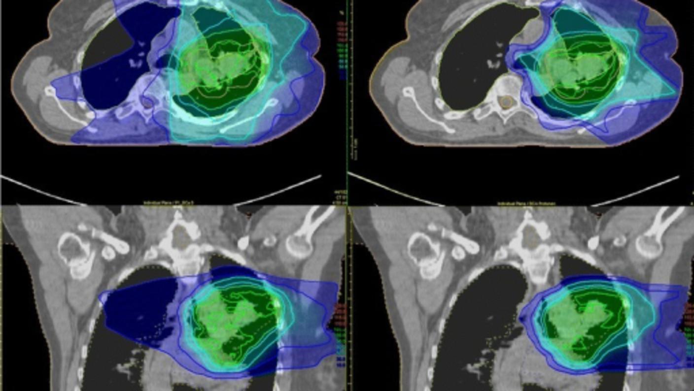
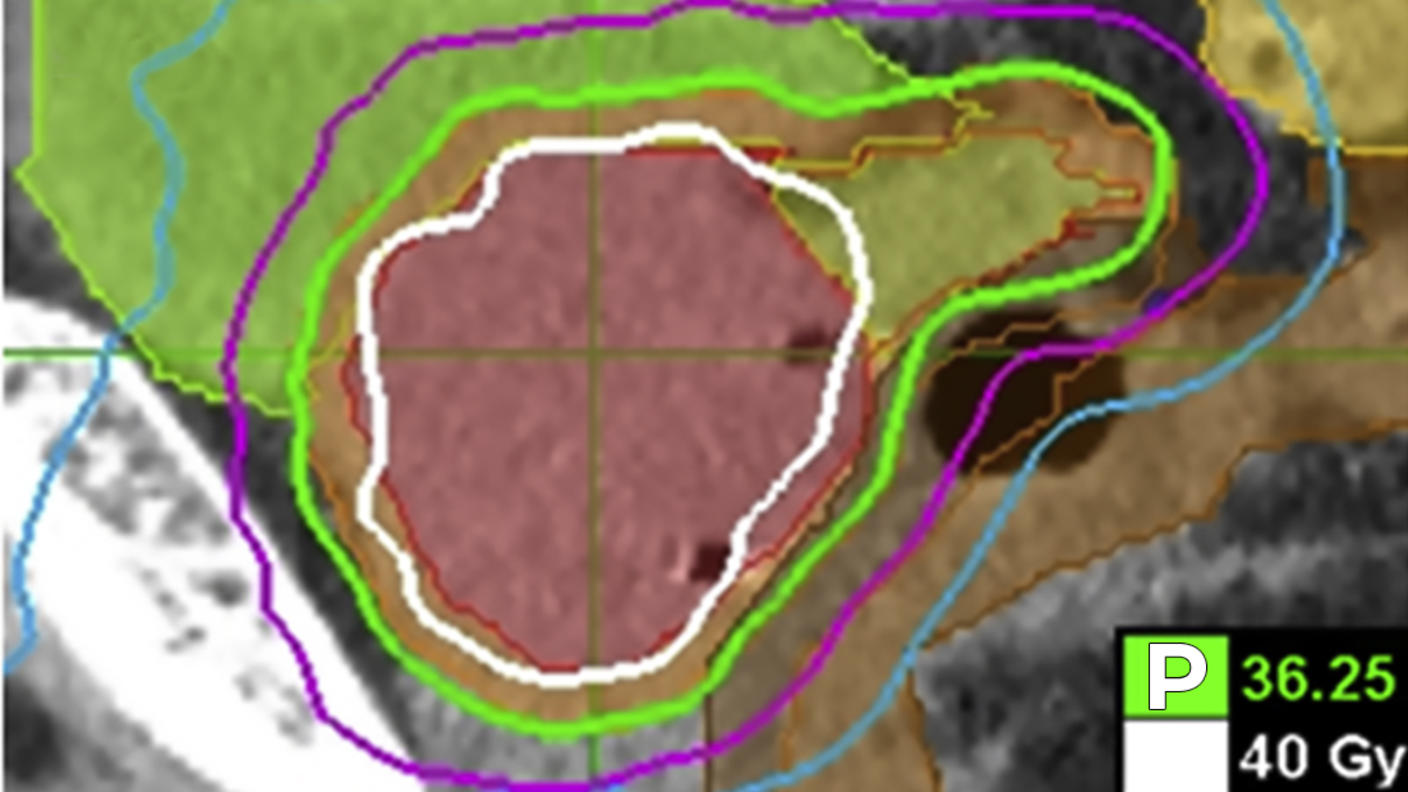
There are many types of external beam radiation therapy, all of which share the goal of delivering the highest prescribed dose of radiation to the tumor while sparing the normal tissue around it. Each type relies on a computer to analyze images of the tumor in order to calculate the most precise dose and treatment path possible.
Types of external beam radiation therapy include:
3-D conformal radiation therapy
What it is
3-D conformal radiation therapy is a common type of external beam radiation therapy. It uses images from CT, MRI, and PET scans to precisely plan the treatment area, a process called simulation. A computer program is used to analyze the images and to design radiation beams that conform to the shape of the tumor.
How it works
3-D conformal radiation conforms to the shape of the tumor by delivering beams from many directions. The precise shaping makes it possible to use higher doses of radiation to the tumor while sparing normal tissue.
Treatment schedule
Most people have treatment once a day, Monday through Friday. The number of treatments vary from person to person based on details about your cancer, such as the type and stage of the cancer and the size and location of the tumor.
Intensity-modulated radiation therapy (IMRT)
What it is
IMRT is a type of 3-D conformal radiation therapy.
How it works
Like 3-D conformal radiation, radiation beams are aimed at the tumor from several directions.
IMRT uses many more smaller beams than 3-D conformal and the strength of the beams in some areas can be changed to give higher doses to certain parts of the tumor.
Treatment schedule
Most people have treatment once a day, Monday through Friday. The number of treatments varies from person to person based on details about your cancer, such as the type and stage of the cancer and the size and location of the tumor.
Image-guided radiation therapy (IGRT)
What it is
IGRT is a type of IMRT. However, it uses imaging scans not only for treatment planning before radiation therapy sessions but also during radiation therapy sessions.
How it works
During treatment, you will have repeated scans, such as CT, MRI, or PET scans. These scans are processed by computers to detect changes in the tumor’s size and location. The repeated imaging allows for your position or the radiation dose to be adjusted during treatment if needed. These adjustments can improve the accuracy of treatment and help spare normal tissue.
Treatment schedule
Most people have treatment once a day, Monday through Friday. The number of treatments varies from person to person based on details about your cancer, such as the type and stage of the cancer and the size and location of the tumor.
Tomotherapy®
What it is
Tomotherapy® is a type of IMRT that uses a machine that is a combination of a CT scanner and an external-beam radiation machine.
How it works
Tomotherapy® machines take images of the tumor right before treatment sessions to allow for very precise tumor targeting and sparing of normal tissues. It rotates around you during treatment, delivering radiation in a spiral pattern, slice by slice. Tomotherapy® might be better at sparing normal tissue than 3-D conformal radiation therapy, but it has not been tested in clinical trials to be sure.
Treatment schedule
Most people have treatment once a day, Monday through Friday. The number of treatments varies from person to person based on details about your cancer, such as the type and stage of the cancer and the size and location of the tumor.
Stereotactic radiosurgery
What it is
Stereotactic radiosurgery is the use of focused, high-energy beams to treat small tumors with well-defined edges in the brain and central nervous system. It may be an option if surgery is too risky due to your age or other health problems or if the tumor cannot safely be reached with surgery. GammaKnife is a type of stereotactic radiosurgery.
How it works
You will be placed in a head frame or some other device to make sure you do not move during treatment. In stereotactic radiosurgery, many small beams of radiation are aimed at the tumor from different directions. Each beam has very little effect on the tissue it passes through, but a precisely targeted dose of radiation is delivered to the site where all the beams come together.
Treatment schedule
Treatment schedules can vary, but treatment is usually given in one dose. In some cases, you may receive up to five doses, given once per day.
Stereotactic body radiation therapy is similar to stereotactic radiosurgery, but it is used for small, isolated tumors outside the brain and spinal cord, often in the liver or lung. It may be an option when you cannot have surgery due to age, health problems, or the location of the tumor.
How it works
As in stereotactic radiosurgery, stereotactic body radiation therapy uses special equipment to hold you still during treatment. It delivers a highly precise beam to a limited area.
Treatment schedule
Tumors outside of the brain are more likely to move with the normal motion of the body, such as with breathing or digesting. Therefore, the radiation beams cannot be targeted as precisely as they are in stereotactic radiosurgery. For this reason, stereotactic body radiation is usually given in more than one dose. You may have up to five doses, given once per day.
What to expect when having external beam radiation therapy
How often you will have external beam radiation therapy
Most people have external beam radiation therapy once a day, five days a week, Monday through Friday. Radiation is given in a series of treatments to allow healthy cells to recover and to make radiation more effective. How many weeks you have treatment depends on the type of cancer you have, the goal of your treatment, the radiation dose, and the radiation schedule.
The span of time from your first radiation treatment to the last is called a course of treatment.
Researchers are looking at different ways to adjust the radiation dose or schedule in order to reach the total dose of radiation more quickly or to limit damage to healthy cells. Different ways of delivering the total radiation dose include:
Accelerated fractionation, which is treatment given in larger daily or weekly doses to reduce the number of weeks of treatment.
Hyperfractionation, which is a smaller dose than the usual daily dose of radiation given more than once a day.
Hypofractionation, which is larger doses given once a day or less often to reduce the number of treatments.
Researchers hope these different schedules for delivering radiation may be more effective and cause fewer side effects than the usual way of doing it or be as effective but more convenient.
Where you go for external beam radiation therapy
Most of the time, you will get external beam radiation therapy as an outpatient. This means that you will have treatment at a clinic or radiation therapy center and will not stay the night in the hospital.
What happens before your first external beam radiation therapy treatment
You will have a 1- to 2-hour meeting with your doctor or nurse before you begin radiation therapy. At this time, you will have a physical exam, talk about your medical history, and maybe have imaging tests. Your doctor or nurse will discuss external beam radiation therapy, its benefits and side effects, and ways you can care for yourself during and after treatment. You can then choose whether to have external beam radiation therapy.
If you decide to have external beam radiation therapy, you will be scheduled for a treatment planning session called a simulation. At this time:
A radiation oncologist (a doctor who specializes in using radiation to treat cancer) and radiation therapist will figure out your treatment area. You may also hear the treatment area referred to as the treatment port or treatment field. These terms refer to the places in your body that will get radiation. You will be asked to lie very still while x-rays or scans are taken.
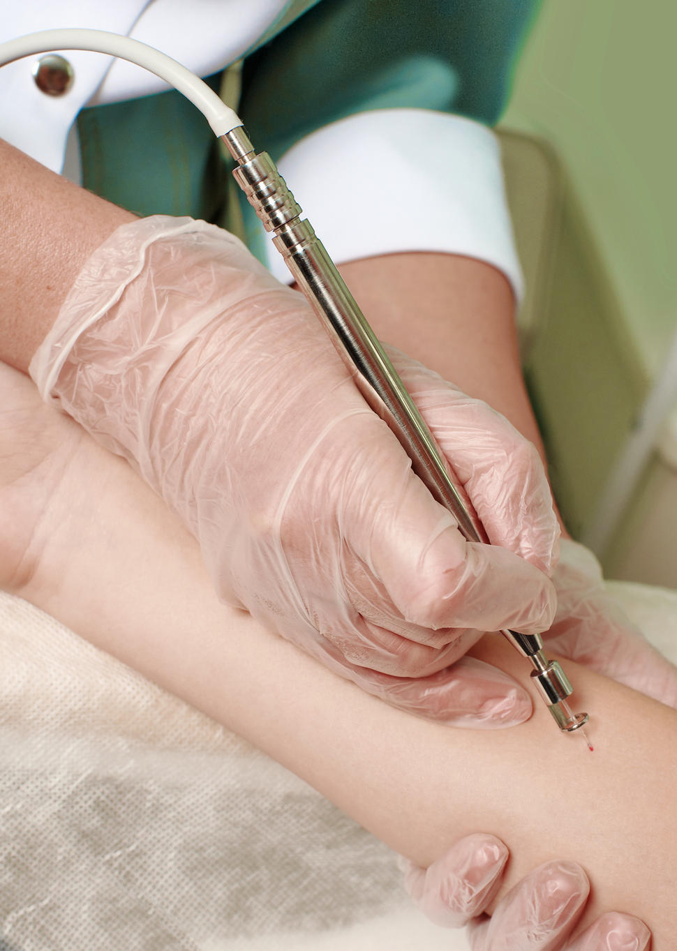
The radiation therapist will tattoo or draw small dots of colored ink on your skin to mark the treatment area. These dots will be needed throughout your course of radiation therapy. The radiation therapist will use them to make sure you are in exactly the same position for every treatment. The dots are about the size of a freckle. If the dots are tattooed, they will remain on your skin for the rest of your life. Ink markings will fade over time. Be careful not to remove them and tell the radiation therapist if they fade or lose color.
A body mold may be made of the part of the body that is being treated. This is a plastic or plaster form that keeps you from moving during treatment. It also helps make sure that you are in exactly the same position for each treatment
A mask fitted to your face helps make sure that you are in exactly the same position for each treatment.
Credit: National Cancer Institute
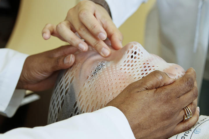
If you are getting radiation to the head and neck area you may be fitted for a mask. The mask has many air holes. It attaches to the table where you will lie for your treatments. The mask helps keep your head from moving so that you are in exactly the same position for each treatment.
What to wear for your treatments
Wear clothes that are comfortable and made of soft fabric, such as fleece or cotton. Choose clothes that are easy to take off, since you may need to expose the treatment area or change into a hospital gown. Do not wear clothes that are tight, such as close-fitting collars or waistbands, near your treatment area. Also, do not wear jewelry, adhesive bandages, or powder in the treatment area.
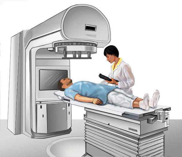
What happens during a treatment session
You may be asked to change into a hospital gown or robe.
You will go to the treatment room where you will receive radiation. The temperature in this room will be very cool.
Depending on where your cancer is, you will either lie down on a treatment table or sit in a special chair. The radiation therapist will use the dots on your skin and body mold or face mask, if you have one, to help place you in the right position.
You may see colored lights pointed at your skin marks. These lights are harmless and help the therapist position you for treatment.
You will need to stay very still so the radiation goes to the exact same place each time. You will get radiation for 1 to 5 minutes. During this time, you can breathe normally.
The radiation therapist will leave the room just before your treatment begins. He or she will go to a nearby room to control the radiation machine. The therapist watches you on a TV screen or through a window and talks with you through a speaker in the treatment room. Make sure to tell the therapist if you feel sick or are uncomfortable. He or she can stop the radiation machine at any time. You will hear the radiation machine and see it moving around, but you won’t be able to feel, hear, see, or smell the radiation.
Most visits last from 30 minutes to an hour, with most of that time spent placing you in the correct position.
How to relax for treatment sessions
Keep yourself busy while you wait.
Read a book or magazine.
Work on crossword puzzles or needlework.
Use headphones to listen to music or recorded books.
Meditate, breathe deeply, pray, use imagery, or find other ways to relax.
See Learning to Relax for exercises and other ideas on how to relax.
External beam radiation therapy will not make you radioactive
People often wonder if they will be radioactive when they are having treatment with radiation. External beam radiation therapy will not make you radioactive. You may safely be around other people, even pregnant women, babies, and young children.
What To Know About External Beam Radiation Therapy
Narrator: What to know about external beam radiation therapy.
Let’s listen in on a visit between Cara and her oncologist, Dr. Ross.
Cara: Dr. Ross, my sister and I have been learning more about external beam radiation therapy. We made a list of questions that I brought with me today. Can we go over them together?
Dr. Ross: Of course! I’d be happy to, Cara. And I’m glad you brought along your sister for support and to take notes. Feel free to tape record our conversation, too, if that would be helpful.
Cara: Thank you. Would you tell us how external beam radiation therapy works?
Dr. Ross: Sure, Cara. It’s a common cancer treatment that uses high doses of radiation to destroy cancer cells and shrink tumors. A large machine aims radiation at the cancer. The machine moves around you, without touching you. We can visit the treatment room later, if you’re interested.
Cara: Okay, thanks. I also wondered if this treatment would make me radioactive, and does it hurt?
Dr. Ross: No, it won’t make you radioactive, so it’s safe to be around people. And the treatment itself doesn’t hurt.
Cara: That’s good to know. How long does the treatment take?
Dr. Ross: The length of treatment for each person depends on the type and stage of the cancer. Most courses of treatment take 2 to 10 weeks. And it’s usually done in an outpatient center, so no overnight hospital stays.
Cara: How often will I come in for visits during those weeks?
Dr. Ross: People generally come in for treatment once a day for 5 days in a row.
Cara: Will I have side effects?
Dr. Ross: You probably will have some side effects. The side effects you may have depend on the part of your body being treated.
Side effects happen because radiation can injure healthy cells that are near the cancer cells it is destroying. We’ll talk in a lot more detail during your next visit about specific side effects and how to manage them.
Cara: What are the next steps if I decide to get external beam radiation therapy?
Dr. Ross: We’ll make another, longer appointment for a checkup and to go through your complete medical history.
Cara: That sounds good. Thanks for answering my questions, Dr. Ross. I’m gonna go home and talk things over with the rest of my family.
Dr. Ross: That sounds good. Now that you have a basic understanding of what external beam radiation therapy is, let’s talk more about your treatment plan.
Narrator Summary: External beam radiation therapy uses high doses of radiation to destroy cancer cells and shrink tumors. A large machine aims radiation at the cancer. The machine moves around you, without touching you.
Most treatments last from 2 to 10 weeks. People usually receive treatment once a day for 5 days in a row.
People often have some side effects from treatment. They usually happen because radiation therapy can injure healthy cells that are near the cancer cells it is destroying. Side effects depend on the part of your body being treated. It’s important to keep in mind that they differ in degree from person to person.
During a follow-up appointment with your doctor, you will have a checkup and go through your complete medical history.
Talk with your doctor to learn more about your treatment goals and plan and how to manage any side effects you may have.
To collect stem cells for a stem cell transplant, the donor is connected to an apheresis machine. After the machine collects blood stem cells from the donor, it returns the rest of the blood to their body.
Credit: Terese Winslow
Stem cell transplants are procedures that restore blood stem cells in people who have had theirs destroyed by the high doses of chemotherapy or radiation therapy that are used to treat certain cancers, blood disorders, and autoimmune disorders. Blood-forming stem cells are vital because they grow into different types of blood cells. The main types of blood cells are:
white blood cells, which are part of your immune system and help your body fight infection
red blood cells, which carry oxygen throughout your body
platelets, which help the blood clot and prevent bleeding
Types of cancer treated with stem cell transplants
Stem cell transplants for other types of cancer are being studied in clinical trials, which are research studies involving people. To find a study that may be an option for you, see Find a Clinical Trial.
How stem cell transplants work against cancer
Stem cell transplants do not usually work against cancer directly. Instead, they restore your body’s ability to produce new blood cells after treatment with the very high doses of chemotherapy and maybe other treatments, such as radiation therapy, that are used to destroy cancer cells.
But in leukemia, the stem cell transplant may work against cancer directly. This happens because of an effect called graft-versus-tumor or graft-versus-leukemia, which can occur after transplants that use stem cells from a donor. This effect occurs when white blood cells from your donor (the graft) attack any cancer cells that remain in your body (the tumor or leukemia cells). This effect improves the chances of success of the transplant.
Types of stem cell transplants
In a stem cell transplant, you receive healthy blood-forming stem cells through a needle in your vein. Most of the blood-forming stem cells that are used in transplants come from the bloodstream. When stem cells come from the blood, the transplant may be called a peripheral blood stem cell transplant, or PBSCT. But blood stem cells can also come from the bone marrow or umbilical cord, which is blood collected when a baby is born. When the stem cells come from the bone marrow, the procedure may be called a bone marrow transplant, or BMT. When they come from cord blood, the procedure may be called a cord blood transplant.
Once they enter your bloodstream, the stem cells travel to the bone marrow, where they take the place of the cells that were destroyed by treatment. Transplants can be:
autologous, which means the stem cells come from you, the person with cancer
allogeneic, which means the stem cells come from someone else. The donor may be a blood relative or someone who is not related, if the cells are a close enough match to yours
syngeneic, which means the stem cells come from your identical twin
There are benefits and risks to both autologous and allogeneic stem cell transplants. With autologous transplants, the transplanted cells will match. But there is a small risk that cancer cells will be transplanted.
With allogeneic transplants, it is important that the cells match closely enough that your immune system won’t see the transplanted blood stem cells as foreign and destroy them.
Mini-transplants are a type of allogeneic transplant that use lower doses of cancer treatment than a regular transplant. They do not kill all your blood-forming stem cells, but they still kill some of the cancer cells. This type of allogeneic transplant can prevent rejection of the donor’s stem cells by suppressing your immune system.
Tandem transplants are a type of autologous transplant. During a tandem transplant, you receive a round of high-dose chemotherapy followed by a stem cell transplant. Then after many weeks or months, you have another round of high-dose chemotherapy followed by another stem cell transplant.
Whether a stem cell transplant is right for you and which type you might have depends on many factors, such as:
the type of cancer you have
how advanced your cancer is
if you can use your own stem cells
if matching donor stem cells are available
if there are other treatments that are likely to work for your cancer
if you can tolerate high doses of chemotherapy
if you have other serious health problems
other treatments you’ve had in the past
Your doctor will carefully weigh these issues with the risks and benefits of each type of stem cell transplant and discuss them with you.
How blood-forming stem cells are matched
To decide if the stem cells from a donor are a match for you, they will be tested for their HLAs (which stands for human leukocyte antigens). HLAs are sets of proteins, or markers, that you have on most cells in your body. Each person has a different set of HLAs. The more HLAs that you and the donor have in common, the better the chance that your body will accept the donor’s stem cells.
Most often, the best match for an allogeneic stem cell transplant is a brother or sister.
If you have an allogeneic transplant, you might develop a serious problem called graft-versus-host disease. Graft-versus-host disease can occur when white blood cells from your donor (the graft) see cells in your body (the host) as foreign and attack them. This problem can cause damage to your skin, liver, intestines, and many other organs.
Graft-versus-host disease can be acute or chronic. Acute graft-versus-host disease occurs within the first 3 months after transplant. Chronic graft-versus-host disease occurs 3 months after a transplant or later.
Graft-versus-host disease can be treated with steroids or other drugs that suppress your immune system.
There are a few ways that the risk of graft-versus-host disease can be reduced.
The closer your donor’s stem cells match yours, the less likely you are to have graft-versus-host disease.
Your doctor may give you drugs to suppress your immune system.
Donated stem cells can be treated to remove the white blood cells (called T cells) that cause graft-versus-host disease. This process is called T-cell depletion.
How much stem cell transplants cost
Stem cells transplants are complicated procedures that are very expensive. They require long hospital stays at special treatment centers and require the services of many health care providers. If you do not live nearby, you will need to stay in a hotel or apartment when you are not in the hospital. If you have no problems, you can go home 100 days after you’ve received the donor stem cells. But you will need to be closely followed by a doctor who has experience in taking care of people who have had a stem cell transplant.
Transplants can cause serious side effects that can be expensive to manage.
If you need to travel for treatment, you might have extra costs for transportation, housing, and childcare.
Most insurance plans cover some of the costs of transplants for certain types of cancer. Talk with your health plan about which services it will pay for. The business office of your treatment center may help you understand all the costs involved.
When you need an allogeneic stem cell transplant, you will need to go to a hospital that has a specialized transplant center. The National Marrow Donor Program® maintains a list of transplant centers in the United States.
How long it takes to have a stem cell transplant
A stem cell transplant can take a few months to complete. The process begins with treatment with high doses of chemotherapy and maybe radiation therapy. This treatment goes on for a week or two. Once you have finished, you will have a few days to rest.
Next, you will receive the blood stem cells. The day you receive your stem cells is often called “day zero.” The stem cells will be given to you through an intravenous (IV) catheter. This process is like receiving a blood transfusion. It takes 1 to 5 hours to receive all the stem cells.
After receiving the stem cells, you begin the recovery phase. During this time, doctors will follow the progress of the new blood cells by checking your blood counts often. As the new stem cells produce blood cells, your blood counts will go up.
Even after your blood counts return to normal, it takes much longer for your immune system to fully recover—several months for autologous transplants, and 1 to 2 years for allogeneic or syngeneic transplants.
How you may feel after a stem cell transplant
Stem cell transplants affect people in different ways. How you feel depends on:
the type of transplant that you have
the doses of treatment you have before the transplant
how you respond to the high-dose treatments
your type of cancer
how advanced your cancer is
how healthy you were before the transplant
Since people respond to stem cell transplants in different ways, your doctor or nurses cannot know for sure how the procedure will make you feel.
Whether or not you can work during a stem cell transplant may depend on the type of job you have. The process of a stem cell transplant, with the high-dose treatments, the transplant, and recovery, can take many months. You will be in and out of the hospital during this time. Even when you are not in the hospital, sometimes you will need to stay near it, rather than staying in your own home.
You will be more tired and your ability to concentrate on work may be affected. You will be visiting the hospital two or three times a week after discharge. You may need to spend a few hours in the hospital for blood or platelet transfusions or replacing minerals in your body.
So, if your job allows, you may want to arrange to work remotely part-time. Many employers are required by law to change your work schedule to meet your needs during cancer treatment. Talk with your employer about ways to adjust your work during treatment. You can learn more about these laws by talking with a social worker.
For more information about working with cancer and your legal rights, see Going Back to Work.
Brachytherapy is a type of internal radiation therapy that is often used to treat cancers of the head and neck, breast, cervix, prostate, and eye.
Credit: iStock
Brachytherapy is a type of internal radiation therapy in which seeds, ribbons, or capsules that contain a radiation source are placed in your body, in or near the tumor. Brachytherapy is a local treatment and treats only a specific part of your body. It is often used to treat cancers of the head and neck, breast, cervix, prostate, and eye.
What happens before your first brachytherapy treatment
You will have a 1- to 2-hour meeting with your doctor or nurse to plan your treatment before you begin brachytherapy. At this time, you will have a physical exam, talk about your medical history, and maybe have imaging tests. Your doctor will discuss the type of brachytherapy that is best for you, its benefits and side effects, and ways you can care for yourself during and after treatment. You can then decide whether to have brachytherapy.
Radiation Therapy Audio Transcript
What To Know About Brachytherapy (A Type of Internal Radiation Therapy)
Narrator: What to know about brachytherapy, a type of internal radiation therapy.
Let’s listen in as Ravi and Sona talk with Ravi’s oncologist, Dr. Williams, about brachytherapy.
Ravi: Dr. Williams, my wife, Sona, and I made a list of questions that we hope to talk with you about today. We’d like to know more about brachytherapy as a treatment option for me.
Dr. Williams: Of course. Brachytherapy is a type of internal radiation that uses radiation to destroy cancer cells and shrink tumors. The radiation often comes in the form of seeds, ribbons, or wires. These are put into your body, in or near the cancer.
Ravi: How do you get it in me?
Dr. Williams: A small holder, usually a thin tube called a catheter, is placed into your body. Then the seeds, ribbons, or wires are put inside this small holder so that the radiation can reach and destroy cancer cells.
Sona: Dr. Williams, Ravi and I have heard that there are 3 types of brachytherapy. Would you tell us more about each of them?
Dr. Williams: Sure, there are 3 types of brachytherapy: low-dose rate implants, high-dose rate implants, and permanent implants.
Low-dose rate implants, often called LDR for short, are implants that stay in for 7 days or less before they are taken out. You are likely to stay in a special room in the hospital if you receive this type of implant.
High-dose rate implants, or HDR implants, stay in for place for a few minutes at a time and are then taken out. You’ll probably make daily trips to the hospital for your treatment, or you may stay in the hospital.
I know we’re covering a lot of information. Do you have any questions so far?
Ravi: No, I’m following you.
Dr. Williams: Okay. Then there’s a third type called permanent implants. These implants are put in your body and stay there. Over time, the radiation dose gets weaker.
Sona: How do you know which implant is right for Ravi?
Dr. Williams: We choose a treatment based on the type of cancer and where it’s located, the person’s health, and any treatments they’ve had before. Depending upon the type of treatment selected, we’ll also go over special instructions for Ravi to follow.
Ravi: Will I have side effects?
Dr. Williams: You probably will have some side effects. The side effects you may have depend on the part of your body being treated.
Side effects happen because radiation can injure healthy cells that are near the cancer cells it’s destroying. We’ll talk in a lot more detail during your next visit about specific side effects and how to manage them.
Ravi: Thanks. This was helpful.
Dr. Williams: I’m glad to hear that. I want to make sure I clearly explained brachytherapy. Ravi, can you tell me a little bit about what I went over?
Ravi: Sure. You said that this treatment uses radiation to destroy the cancer cells or shrink the tumor. The radiation is put into the body.
You talked about 3 types of brachytherapy – low-dose rate implants, high-dose rate implants, and permanent implants – and said that these implants can stay in a few minutes, a few days, or the rest of my life.
Hmm, Sona, did I forget anything?
Sona: There was the part where Dr. Williams explained that the radiation is often placed in the body using a small, thin tube. The radiation looks like seeds, ribbons, or wires and is put into the thin tube so it can reach and destroy cancer cells.
Dr. Williams: Exactly. Oh, and before I forget, here’s a fact sheet that covers some of what we talked about today. Now before we talk about your treatment plan, do you have any other questions?
Ravi: Not right now, I think we’re set on the basics. I am eager to hear about which treatment you recommend for me, though.
Dr. Williams: Of course, let’s get started.
How brachytherapy is put in place
Most brachytherapy is put in place through a catheter, which is a small, stretchy tube. Sometimes, brachytherapy is put in place through a larger device called an applicator. The way the brachytherapy is put in place depends on your type of cancer. Your doctor will place the catheter or applicator into your body before you begin treatment.
Techniques for placing brachytherapy:
Interstitial brachytherapy, in which the radiation source is placed within the tumor. This technique is used for prostate cancer, for instance.
Intracavity brachytherapy, in which the radiation source is placed within a body cavity or a cavity created by surgery. For example, radiation can be placed in the vagina to treat cervical or endometrial cancer.
Episcleral brachytherapy, in which the radiation source is attached to the eye. This technique is used to treat melanoma of the eye.
Once the catheter or applicator is in place, the radiation source is placed inside it. The radiation source may be kept in place for a few minutes, for many days, or for the rest of your life. How long it remains in place depends on the type of radiation source, your type of cancer, where the cancer is in your body, your health, and other cancer treatments you have had.
Types of brachytherapy
There are three types of brachytherapy
Low-dose rate (LDR) implants: In this type of brachytherapy, the radiation source stays in place for 1 to 7 days. You are likely to be in the hospital during this time. Once your treatment is finished, your doctor will remove the radiation source and the catheter or applicator.
High-dose rate (HDR) implants: In this type of brachytherapy, the radiation source is left in place for just 10 to 20 minutes at a time and then taken out. You may have treatment twice a day for 2 to 5 days or once a week for 2 to 5 weeks. The schedule depends on your type of cancer. During the course of treatment, your catheter or applicator may stay in place, or it may be put in place before each treatment. You may be in the hospital during this time, or you may make daily trips to the hospital to have the radiation source put in place. As with LDR implants, your doctor will remove the catheter or applicator once you have finished treatment.
Permanent implants: After the radiation source is put in place, the catheter is removed. The implants remain in your body for the rest of your life, but the radiation gets weaker each day. As time goes on, almost all the radiation will go away. When the radiation is first put in place, you may need to limit your time around other people and take other safety measures. Be extra careful not to spend time with children or pregnant women.
What to expect when the catheter is removed
Once you finish treatment with LDR or HDR implants, the catheter will be removed. Here are some things to expect:
You will get medicine for pain before the catheter or applicator is removed.
The area where the catheter or applicator was might be tender for a few months.
There is no radiation in your body after the catheter or applicator is removed. It is safe for people to be near you–even young children and pregnant women.
For a week or two, you may need to limit activities that take a lot of effort. Ask your doctor what kinds of activities are safe for you and which ones you should avoid.
Brachytherapy will make you give off radiation
With brachytherapy, the radiation source in your body will give off radiation for a while. If the radiation you receive is a very high dose, you may need to follow some safety measures.
Staying in a private hospital room to protect others from radiation coming from your body.
Being treated quickly by nurses and other hospital staff. They will provide all the care you need but may stand at a distance, talk with you from the doorway of your room, and wear protective clothing.
Your visitors will also need to follow safety measures, which may include
not being allowed to visit when the radiation is first put in
needing to check with the hospital staff before they go to your room
standing by the doorway rather than going into your hospital room
keeping visits short, about 30 minutes or less each day (the length of visits depends on the type of radiation being used and the part of your body being treated)
not having visits from pregnant women and children younger than a year old
You may also need to follow safety measures once you leave the hospital, such as not spending much time with other people. Your doctor or nurse will talk with you about any safety measures you should follow when you go home.
Hyperthermia to treat cancer is not widely available. But at some centers, it is used for advanced cancers along with other treatments such as radiation therapy and chemotherapy.
Credit: National Cancer Institute
What is hyperthermia treatment?
Hyperthermia is a type of treatment in which body tissue is heated to as high as 113 °F to help damage and kill cancer cells with little or no harm to normal tissue. Hyperthermia to treat cancer is also called thermal therapy, thermal ablation, or thermotherapy.
Different types of techniques may be used to create heat for hyperthermia treatment. These techniques include:
heating fluids such as blood or chemotherapy drugs and putting them into the body (called perfusion)
placing the entire body in a heated chamber or hot water bath or wrapping with heated blankets
Cancers treated with hyperthermia
Hyperthermia to treat cancer is not widely available. But at some centers it is used, along with other treatments such as radiation therapy and chemotherapy, for advanced cancers. It has been used to treat these types of advanced cancers:
Hyperthermia is almost always used with other forms of cancer treatment. Many clinical trials have shown that hyperthermia, when used with treatments such as radiation therapy and chemotherapy, helps shrink tumors and may make it easier for them to kill cancer cells.
How hyperthermia is given
During treatment, the doctor numbs the treatment area and inserts small probes with tiny thermometers into the tumor. Thermometers help the doctor closely watch the temperature of the tumor and nearby tissue during treatment. Imaging techniques, such as CT scans, may be used to make sure the probes are in the proper place.
Types of hyperthermia treatment
Hyperthermia can be used to treat small areas of the body, large areas, or the entire body.
In local hyperthermia, doctors apply heat to a small area. The type of local hyperthermia used depends on where the tumor is located.
External hyperthermia is used to treat tumors that are on or just below the skin. For this type of hyperthermia, doctors place devices that create heat around or near the treatment area.
Intraluminal or endocavitary hyperthermia is used to treat tumors within or near body cavities, such as the esophagus or rectum. In this type of hyperthermia, doctors place probes that create heat inside the cavity and insert them into the tumor.
Interstitial hyperthermia is used to treat tumors deep within the body, such as in the brain. This type of hyperthermia allows the tumor to be heated to higher temperatures than external techniques. The doctor will insert probes or needles into your tumor while you are under anesthesia. Imaging techniques, such as ultrasound, may be used to help make sure the probe is in the right place. The heat source is then inserted into the probe.
Radiofrequency ablation is a type of interstitial hyperthermia that uses radio waves to heat and kill cancer cells.
In regional hyperthermia, doctors apply heat to large areas of the body, such as a cavity, organ, or limb. Techniques used in regional hyperthermia include deep tissue techniques, regional perfusion, and continuous hyperthermic peritoneal perfusion.
Deep tissue techniques treat cancers within the body, such as cervical or bladder cancer. During this procedure, devices that deliver heat are placed around the cavity or organ to be treated and energy is focused on the area to raise its temperature.
Regional perfusion techniques treat cancers in the arms and legs, such as melanoma, or in some organs, such as the liver or lung. During this procedure, some of your blood is removed, heated, and then pumped back into the limb or organ. Chemotherapy is often given during this treatment.
Continuous hyperthermic peritoneal perfusion treats cancer within the peritoneal cavity, which is the space within the abdomen that contains the intestines, stomach, and liver.
This treatment is given during surgery. While you are under anesthesia, heated chemotherapy drugs flow from a warming device through your peritoneal cavity, causing the temperature in the area to reach 106 to 108°F.
Whole-body hyperthermia treats cancer that has spread throughout the body. In this type of hyperthermia, you are placed in a thermal chamber or wrapped in hot water blankets that raise your body temperature to 107 or 108 °F for short periods of time.
Benefits of hyperthermia
Hyperthermia can help other cancer treatments, such as chemotherapy and radiation therapy, work better.
Drawbacks of hyperthermia to treat cancer
Treatment with hyperthermia requires special equipment and expertise and is not widely available. It is also not clear if it helps people live longer.
Hyperthermia side effects
Most healthy tissue is not damaged during hyperthermia if the temperature stays under 111°F. But different features of different tissues may cause higher temperatures to occur in certain spots. This can cause burns, blisters, discomfort, or pain.
Perfusion techniques can cause swelling, blood clots, bleeding, and other damage to the normal tissues in the treated area. But most of these side effects improve after treatment.
Diarrhea, nausea, and vomiting are common after whole-body hyperthermia. It can also cause more serious side effects that are not common, including heart and blood vessel problems.
Where to go for hyperthermia treatment
A small number of hospitals and cancer centers throughout the country have skilled doctors and the machines needed to perform hyperthermia. Talk with your doctor or contact hospitals and cancer centers in your area to find out if they are using hyperthermia.
Hyperthermia treatment research
In clinical trials, doctors are studying how effective hyperthermia is for treating different cancers and when used with other cancer treatments. Other trials focus on improving techniques for delivering hyperthermia. If you are interested in joining a research study that is testing hyperthermia. Search for research studies by using the advanced clinical trials search form or contact NCI’s Cancer Information Service.
Cancer cells that have absorbed a drug called a photosensitizer. When activated by light, the drug kills the cancer cells.
Credit: National Cancer Institute
What is photodynamic therapy?
Photodynamic therapy uses a drug that is activated by light, called a photosensitizer or photosensitizing agent, to kill cancer cells. The light can come from a laser or other source, such as LEDs. Photodynamic therapy is also called PDT.
Photodynamic therapy is most often used as a local treatment, which means it treats a specific part of the body.
Cancer and precancers treated with photodynamic therapy
The FDA has approved photodynamic therapy to treat:
non-small cell lung cancer when it blocks the airways
How photodynamic therapy treats cancer
When cells that have absorbed photosensitizers are exposed to a specific wavelength of light, the photosensitizer produces a form of oxygen, called an oxygen radical, that kills them.
Photodynamic therapy may also damage blood vessels in the tumor, which prevents it from receiving the blood it needs to keep growing. And, it may trigger the immune system to attack tumor cells, even in other areas of the body.
How photodynamic therapy is given
Photodynamic therapy is a two-step process. First, you will first receive a photosensitizer. The drug may be taken by mouth, spread on the skin, or given through an IV, depending on where the tumor is in the body. After 24 to 72 hours, most of the drug will have left normal cells but remain in cancer or precancer cells. Then your tumor will be exposed to the light source.
How the light is applied depends on where the tumor is. For skin tumors, the light is aimed right at the cancer. For tumors in the throat, airways, and lungs, your doctor will insert an endoscope down your throat. An endoscope is a thin, lighted tube that can help the doctor see inside the body. Once the endoscope is in place, the doctor threads a fiber optic cable that transmits light through it to reach the treatment areas.
One type of photodynamic therapy called extracorporeal photopheresis (ECP) is used to treat abnormal white blood cells that can cause skin symptoms in people with cutaneous T-cell lymphoma. In ECP, a machine collects your blood cells, treats them with a photosensitizer, exposes them to light, and then returns them to your body through a needle in a vein.
Most often, you will have photodynamic therapy as an outpatient, which means you go home after treatment and do not spend the night in the hospital. You may have photodynamic therapy by itself, or you may have it along with other cancer treatments.
Benefits of photodynamic therapy
Photodynamic therapy limits damage to healthy cells because the photosensitizers tend to build up in abnormal cells and the light is focused directly on them.
Photodynamic therapy does not cause scarring, which makes it good for people with skin cancers and precancers.
Drawbacks of photodynamic therapy
Photodynamic therapy can harm normal cells in the treatment area and cause side effects.
The light used in photodynamic therapy can’t pass through more than about 1/3-inch of tissue, or 1 centimeter. So, photodynamic therapy can only be used to treat tumors that are on or just under the skin or on the lining of internal organs or cavities.
Because the light can’t reach very far into large tumors, photodynamic therapy is less helpful in treating them.
Side effects of photodynamic therapy
Damage to normal cells is limited but photodynamic therapy can still cause burns, swelling, pain, and scarring in the treatment area. Other side effects may occur depending on the area that is treated, including:
cough
trouble swallowing
stomach pain
painful breathing
shortness of breath
skin problems, such as redness, stinging, swelling, or itching
A type of photosensitizer called porfimer sodium makes the skin and eyes sensitive to light for about 6 weeks. During this time, you should avoid direct sunlight and bright indoor light.
A small number of hospitals and cancer centers throughout the country have skilled doctors and the machines needed to perform photodynamic therapy. Talk with your doctor or contact hospitals and cancer centers in your area to find out if they are using this treatment.
Photodynamic therapy research
Researchers are looking for ways to expand photodynamic therapy to other cancers, including improving the equipment used and the delivery of the light.
Studies are underway to see if ECP may be used for other blood cancers and to help reduce rejection after stem cell transplants.
Researchers are also developing a new type of PDT called photoimmunotherapy, or PIT. In this treatment, a photosensitizer is combined with an immune protein that delivers the photosynthesizer to cancer cells. When light is applied, the photosynthesizer kills the cancer cells. This process also causes an immune response inside the tumor that can cause more cancer cells to die. Studies of PIT are going on with people with head and neck cancers. Other research is focused on finding photosensitizers that:
are more powerful
target cancer cells more precisely
are triggered by light that can go through tissue to treat deep or large tumors