Stomach Cancer Screening

Screening is looking for cancer before a person has any symptoms. This can help detect cancer at an early stage. When abnormal tissue or cancer is found early, it may be easier to treat.  

In the United States, there are no standard or routine screening tests to detect stomach cancer in people at average risk. Researchers are working to develop such tests. If a test reduces the number of deaths from stomach cancer, it may become a standard screening test.

Upper endoscopy; drawing shows an endoscope (a thin, lighted tube) inserted through the mouth and down the throat into the esophagus and stomach. An inset shows a patient on a table having an upper endoscopy.

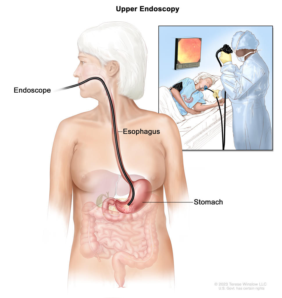
Upper endoscopy. A thin, lighted tube called an endoscope is inserted through the mouth and down the throat to check for abnormal areas in the esophagus, stomach, and first part of the small intestine.

Credit: © Terese Winslow

Tests to screen for stomach cancer

Some people who have a higher risk of stomach cancer may benefit from screening with upper endoscopy, including: 

Learn more about risk factors for stomach cancer

Upper endoscopy

Upper endoscopy is a procedure to look inside the esophagus, stomach, and duodenum (first part of the small intestine) to check for abnormal areas. An endoscope is passed through the mouth and down the throat into the esophagus. An endoscope is a thin, tube-like instrument with a light and a lens for viewing. It may also have a tool to remove tissue, which is checked under a microscope for signs of disease.

Risks of stomach cancer screening

Talk with your doctor about whether stomach cancer screening is right for you.   

Potential risks of stomach cancer screening include:  

  • False-negative test results. Screening test results may appear to be normal even though stomach cancer is present. A person who receives a false-negative test result (one that shows there is no cancer when there really is) may delay seeking medical care even if there are symptoms.
  • False-positive test results. Screening test results may appear to be abnormal even though no cancer is present. A false-positive test result (one that shows there is cancer when there really isn’t) can cause anxiety and is usually followed by more tests and procedures which also have risks.
  • Risks of the screening test itself. Upper endoscopy may cause the following rare, but serious, complications:
    • a small hole (puncture) in the esophagus or stomach
    • heart problems
    • breathing problems
    • lung infection from inhaling food, fluid, or stomach acid into the lung
    • severe bleeding that needs to be treated at a hospital
    • reactions to medicine used during the procedure 
       

Gallbladder Cancer Treatment (PDQ®)–Patient Version

Gallbladder Cancer Treatment (PDQ®)–Patient Version

General Information About Gallbladder Cancer

Key Points

  • Gallbladder cancer is a rare type of cancer that starts in the tissues of the gallbladder.
  • Signs and symptoms of gallbladder cancer include jaundice, fever, and pain.
  • Gallbladder cancer is difficult to detect (find) and diagnose early.
  • Tests that examine the gallbladder and nearby organs are used to detect (find), diagnose, and stage gallbladder cancer.
  • Certain factors affect the prognosis (chance of recovery) and treatment options.

Gallbladder cancer is a rare type of cancer that starts in the tissues of the gallbladder.

Gallbladder cancer occurs when cells in the gallbladder start to grow out of control. The gallbladder is a pear-shaped organ that lies just under the liver in the upper abdomen. The gallbladder stores bile, a fluid made by the liver to digest fat. When food is being broken down in the stomach and intestines, bile is released from the gallbladder through a tube called the common bile duct, which connects the gallbladder and liver to the first part of the small intestine.

EnlargeAnatomy of the gallbladder; shows the liver, common hepatic duct, cystic duct, common bile duct, pancreas, and small intestine. The inset shows the liver, bile ducts, gallbladder, pancreas, and small intestine.
Anatomy of the gallbladder. The gallbladder is just below the liver. Bile is stored in the gallbladder and flows through the cystic duct and the common bile duct into the small intestine when food is being digested.

The wall of the gallbladder has 4 main layers of tissue:

Primary gallbladder cancer starts in the inner layer and spreads through the outer layers as it grows.

Signs and symptoms of gallbladder cancer include jaundice, fever, and pain.

These and other signs and symptoms may be caused by gallbladder cancer or by other conditions. Check with your doctor if you have:

Gallbladder cancer is difficult to detect (find) and diagnose early.

Gallbladder cancer is difficult to detect and diagnose because:

  • People rarely have signs or symptoms in the early stages of gallbladder cancer.
  • The symptoms of gallbladder cancer, when present, are like the symptoms of many other illnesses.
  • The gallbladder is hidden behind the liver.

Gallbladder cancer is sometimes found when the gallbladder is removed for other reasons. Although gallstones are the most common risk factor for developing gallbladder cancer, the overall risk is low. Most people with gallstones do not develop gallbladder cancer.

Tests that examine the gallbladder and nearby organs are used to detect (find), diagnose, and stage gallbladder cancer.

Procedures that make pictures of the gallbladder and the area around it help diagnose gallbladder cancer and show how far the cancer has spread. The process used to find out if cancer cells have spread within and around the gallbladder is called staging.

To plan treatment, it is important to know if the gallbladder cancer can be removed by surgery. Tests and procedures to detect, diagnose, and stage gallbladder cancer are usually done at the same time. In addition to asking about your personal and family health history and doing a physical exam, your doctor may perform the following tests and procedures:

  • Liver function tests: A procedure in which a blood sample is checked to measure the amounts of certain substances released into the blood by the liver. A higher-than-normal amount of a substance can be a sign of liver disease that may be caused by gallbladder cancer.
  • Blood chemistry studies: A procedure in which a blood sample is checked to measure the amounts of certain substances released into the blood by organs and tissues in the body. An unusual (higher or lower than normal) amount of a substance can be a sign of disease.
  • CT scan (CAT scan): A procedure that makes a series of detailed pictures of areas inside the body, such as the chest, abdomen, and pelvis, taken from different angles. The pictures are made by a computer linked to an x-ray machine. A dye may be injected into a vein or swallowed to help the organs or tissues show up more clearly. This procedure is also called computed tomography, computerized tomography, or computerized axial tomography.
  • Ultrasound exam: A procedure in which high-energy sound waves (ultrasound) are bounced off internal tissues or organs and make echoes. The echoes form a picture of body tissues called a sonogram. An abdominal ultrasound is done to diagnose gallbladder cancer.
  • PTC (percutaneous transhepatic cholangiography): A procedure used to x-ray the liver and bile ducts. A thin needle is inserted through the skin below the ribs and into the liver. Dye is injected into the liver or bile ducts and an x-ray is taken. If a blockage is found, a thin, flexible tube called a stent is sometimes left in the liver to drain bile into the small intestine or a collection bag outside the body.
  • ERCP (endoscopic retrograde cholangiopancreatography): A procedure used to x-ray the ducts (tubes) that carry bile from the liver to the gallbladder and from the gallbladder to the small intestine. Sometimes gallbladder cancer causes these ducts to narrow and block or slow the flow of bile, causing jaundice. An endoscope (a thin, lighted tube) is passed through the mouth, esophagus, and stomach into the first part of the small intestine. A catheter (a smaller tube) is then inserted through the endoscope into the bile ducts. A dye is injected through the catheter into the ducts and an x-ray is taken. If the ducts are blocked by a tumor, a fine tube may be inserted into the duct to unblock it. This tube (or stent) may be left in place to keep the duct open. Tissue samples may also be taken.
  • MRI (magnetic resonance imaging) with gadolinium: A procedure that uses a magnet, radio waves, and a computer to make a series of detailed pictures of areas inside the body. A substance called gadolinium is injected into a vein. The gadolinium collects around the cancer cells so they show up brighter in the picture. This procedure is also called nuclear magnetic resonance imaging (NMRI).
  • Endoscopic ultrasound (EUS): A procedure in which an endoscope is inserted into the body, usually through the mouth or rectum. An endoscope is a thin, tube-like instrument with a light and a lens for viewing. A probe at the end of the endoscope is used to bounce high-energy sound waves (ultrasound) off internal tissues or organs and make echoes. The echoes form a picture of body tissues called a sonogram. This procedure is also called endosonography.
  • Laparoscopy: A surgical procedure to look at the organs inside the abdomen to check for signs of disease. Small incisions (cuts) are made in the wall of the abdomen and a laparoscope (a thin, lighted tube) is inserted into one of the incisions. Other instruments may be inserted through the same or other incisions to perform procedures such as removing organs or taking tissue samples for biopsy. Laparoscopy helps to find out if the cancer is within the gallbladder only or has spread to nearby tissues and if it can be removed by surgery.
  • Biopsy: The removal of cells or tissues so they can be viewed under a microscope by a pathologist to check for signs of cancer. The biopsy may be done after surgery to remove the tumor. If the tumor clearly cannot be removed by surgery, the biopsy may be done using a fine needle to remove cells from the tumor.

Certain factors affect the prognosis (chance of recovery) and treatment options.

The prognosis and treatment options depend on:

  • the stage of the cancer (whether the cancer has spread from the gallbladder to other places in the body)
  • whether the cancer can be completely removed by surgery
  • the type of gallbladder cancer (how the cancer cell looks under a microscope)
  • whether the cancer has just been diagnosed or has recurred (come back)

Treatment may also depend on the age and general health of the patient and whether the cancer is causing signs or symptoms.

Gallbladder cancer can be cured only if it is found before it has spread, when it can be removed by surgery. If the cancer has spread, palliative treatment can improve the patient’s quality of life by controlling the symptoms and complications of this disease.

Stages of Gallbladder Cancer

Key Points

  • Tests and procedures to stage gallbladder cancer are usually done at the same time as diagnosis.
  • There are three ways that cancer spreads in the body.
  • Cancer may spread from where it began to other parts of the body.
  • The following stages are used for gallbladder cancer:
    • Stage 0 (Carcinoma in Situ)
    • Stage I
    • Stage II
    • Stage III
    • Stage IV
  • For gallbladder cancer, stages are also grouped according to how the cancer may be treated. There are two treatment groups:
    • Localized (Stage I)
    • Unresectable, recurrent, or metastatic (Stage II, Stage III, and Stage IV)

Tests and procedures to stage gallbladder cancer are usually done at the same time as diagnosis.

Learn more about tests and procedures used to detect, diagnose, and stage gallbladder cancer in the General Information section.

There are three ways that cancer spreads in the body.

Cancer can spread through tissue, the lymph system, and the blood:

  • Tissue. The cancer spreads from where it began by growing into nearby areas.
  • Lymph system. The cancer spreads from where it began by getting into the lymph system. The cancer travels through the lymph vessels to other parts of the body.
  • Blood. The cancer spreads from where it began by getting into the blood. The cancer travels through the blood vessels to other parts of the body.

Cancer may spread from where it began to other parts of the body.

When cancer spreads to another part of the body, it is called metastasis. Cancer cells break away from where they began (the primary tumor) and travel through the lymph system or blood.

  • Lymph system. The cancer gets into the lymph system, travels through the lymph vessels, and forms a tumor (metastatic tumor) in another part of the body.
  • Blood. The cancer gets into the blood, travels through the blood vessels, and forms a tumor (metastatic tumor) in another part of the body.

The metastatic tumor is the same type of cancer as the primary tumor. For example, if gallbladder cancer spreads to the liver, the cancer cells in the liver are actually gallbladder cancer cells. The disease is metastatic gallbladder cancer, not liver cancer.

Many cancer deaths are caused when cancer moves from the original tumor and spreads to other tissues and organs. This is called metastatic cancer. This animation shows how cancer cells travel from the place in the body where they first formed to other parts of the body.

The following stages are used for gallbladder cancer:

Stage 0 (Carcinoma in Situ)

In stage 0, abnormal cells are found in the mucosa (innermost layer) of the gallbladder wall. These abnormal cells may become cancer and spread into nearby normal tissue. Stage 0 is also called carcinoma in situ.

Stage I

In stage I, cancer has formed in the mucosa (innermost layer) of the gallbladder wall and may have spread to the muscle layer of the gallbladder wall.

Stage II

Stage II is divided into stages IIA and IIB, depending on where the cancer has spread in the gallbladder.

  • In stage IIA, cancer has spread through the muscle layer to the connective tissue layer of the gallbladder wall on the side of the gallbladder that is not near the liver.
  • In stage IIB, cancer has spread through the muscle layer to the connective tissue layer of the gallbladder wall on the same side as the liver. Cancer has not spread to the liver.

Stage III

Stage III is divided into stages IIIA and IIIB, depending on where the cancer has spread.

  • In stage IIIA, cancer has spread through the connective tissue layer of the gallbladder wall and one or more of the following is true:
  • In stage IIIB, cancer has formed in the mucosa (innermost layer) of the gallbladder wall and may have spread to the muscle, connective tissue, or serosa (layer of tissue that covers the gallbladder) and may have also spread to the liver or to one nearby organ or structure (such as the stomach, small intestine, colon, pancreas, or the bile ducts outside the liver). Cancer has spread to one to three nearby lymph nodes.

Stage IV

Stage IV is divided into stages IVA and IVB.

  • In stage IVA, cancer has spread to the portal vein or hepatic artery or to two or more organs or structures other than the liver. Cancer may have spread to one to three nearby lymph nodes.
  • In stage IVB, cancer may have spread to nearby organs or structures. Cancer has spread:
    • to four or more nearby lymph nodes; or
    • to other parts of the body, such as the peritoneum and liver.

For gallbladder cancer, stages are also grouped according to how the cancer may be treated. There are two treatment groups:

Localized (Stage I)

Cancer is found in the wall of the gallbladder and can be completely removed by surgery.

Unresectable, recurrent, or metastatic (Stage II, Stage III, and Stage IV)

Unresectable cancer cannot be removed completely by surgery. Most patients with gallbladder cancer have unresectable cancer.

Recurrent cancer is cancer that has recurred (come back) after it has been treated. Gallbladder cancer may come back in the gallbladder or in other parts of the body.

Metastasis is the spread of cancer from the primary site (place where it started) to other places in the body. Metastatic gallbladder cancer may spread to surrounding tissues, organs, throughout the abdominal cavity, or to distant parts of the body.

Treatment Option Overview

Key Points

  • There are different types of treatment for patients with gallbladder cancer.
  • The following types of treatment are used:
    • Surgery
    • Radiation therapy
    • Chemotherapy
  • New types of treatment are being tested in clinical trials.
    • Radiation sensitizers
    • Targeted therapy
    • Immunotherapy
  • Treatment for gallbladder cancer may cause side effects.
  • Patients may want to think about taking part in a clinical trial.
  • Patients can enter clinical trials before, during, or after starting their cancer treatment.
  • Follow-up care may be needed.

There are different types of treatment for patients with gallbladder cancer.

Different types of treatments are available for patients with gallbladder cancer. Some treatments are standard (the currently used treatment), and some are being tested in clinical trials. A treatment clinical trial is a research study meant to help improve current treatments or obtain information on new treatments for patients with cancer. When clinical trials show that a new treatment is better than the standard treatment, the new treatment may become the standard treatment. Patients may want to think about taking part in a clinical trial. Some clinical trials are open only to patients who have not started treatment.

The following types of treatment are used:

Surgery

Gallbladder cancer may be treated with a cholecystectomy, surgery to remove the gallbladder and some of the tissues around it. Nearby lymph nodes may be removed. A laparoscope is sometimes used to guide gallbladder surgery. The laparoscope is attached to a video camera and inserted through an incision (port) in the abdomen. Surgical instruments are inserted through other ports to perform the surgery. Because there is a risk that gallbladder cancer cells may spread to these ports, tissue surrounding the port sites may also be removed.

If the cancer has spread and cannot be removed, the following types of palliative surgery may relieve symptoms:

  • Biliary bypass: If the tumor is blocking the bile duct and bile is building up in the gallbladder, a biliary bypass may be done. During this operation, the doctor will cut the gallbladder or bile duct in the area before the blockage and sew it to the small intestine to create a new pathway around the blocked area.
  • Endoscopic stent placement: If the tumor is blocking the bile duct, surgery may be done to put in a stent (a thin tube) to drain bile that has built up in the area. The doctor may place the stent through a catheter that drains the bile into a bag on the outside of the body or the stent may go around the blocked area and drain the bile into the small intestine.
  • Percutaneous transhepatic biliary drainage: A procedure done to drain bile when there is a blockage and endoscopic stent placement is not possible. An x-ray of the liver and bile ducts is done to locate the blockage. Images made by ultrasound are used to guide placement of a stent, which is left in the liver to drain bile into the small intestine or a collection bag outside the body. This procedure may be done to relieve jaundice before surgery.

Radiation therapy

Radiation therapy is a cancer treatment that uses high-energy x-rays or other types of radiation to kill cancer cells or keep them from growing. External radiation therapy uses a machine outside the body to send radiation toward area of the body with cancer.

Chemotherapy

Chemotherapy is a cancer treatment that uses drugs to stop the growth of cancer cells, either by killing the cells or by stopping the cells from dividing. When chemotherapy is taken by mouth or injected into a vein or muscle, the drugs enter the bloodstream and can reach cancer cells throughout the body (systemic therapy).

New types of treatment are being tested in clinical trials.

This summary section describes treatments that are being studied in clinical trials. It may not mention every new treatment being studied. Information about clinical trials is available from the NCI website.

Radiation sensitizers

Clinical trials are studying ways to improve the effect of radiation therapy on tumor cells, including:

  • Hyperthermia therapy: A treatment in which body tissue is exposed to high temperatures to damage and kill cancer cells or to make cancer cells more sensitive to the effects of radiation therapy and certain anticancer drugs.
  • Radiosensitizers: Drugs that make tumor cells more sensitive to radiation therapy. Giving radiation therapy together with radiosensitizers may kill more tumor cells.

Targeted therapy

Targeted therapy is a type of treatment that uses drugs or other substances to identify and attack specific cancer cells. The following targeted therapies are being studied in patients with gallbladder cancer that is locally advanced and cannot be removed by surgery or has spread to other parts of the body:

  • Ivosidenib is a type of targeted therapy that blocks a specific mutation in a gene called IDH1. It works by slowing or stopping the growth of cancer cells.
  • Pemigatinib is a type of targeted therapy that blocks specific changes in a gene called FGFR2. This may help keep cancer cells from growing and may kill them.

Immunotherapy

Immunotherapy is a treatment that uses the patient’s immune system to fight cancer. Substances made by the body or made in a laboratory are used to boost, direct, or restore the body’s natural defenses against cancer.

Immune checkpoint inhibitor therapy is a type of immunotherapy that may be used to treat gallbladder cancer.

  • PD-1 and PD-L1 inhibitor therapy: PD-1 is a protein on the surface of T cells that helps keep the body’s immune responses in check. PD-L1 is a protein found on some types of cancer cells. When PD-1 attaches to PD-L1, it stops the T cell from killing the cancer cell. PD-1 and PD-L1 inhibitors keep PD-1 and PD-L1 proteins from attaching to each other. This allows the T cells to kill cancer cells.
    • Pembrolizumab is a type of PD-1 inhibitor that may be used in patients whose cancer is locally advanced and cannot be removed by surgery or has spread to other parts of the body.
    • Durvalumab is a type of PD-L1 inhibitor that is being studied in combination with chemotherapy in previously untreated patients whose cancer is locally advanced, recurrent, or has spread to other parts of the body.
EnlargeImmune checkpoint inhibitor; the panel on the left shows the binding of proteins PD-L1 (on the tumor cell) to PD-1 (on the T cell), which keeps T cells from killing tumor cells in the body. Also shown are a tumor cell antigen and T cell receptor. The panel on the right shows immune checkpoint inhibitors (anti-PD-L1 and anti-PD-1) blocking the binding of PD-L1 to PD-1, which allows the T cells to kill tumor cells.
Immune checkpoint inhibitor. Checkpoint proteins, such as PD-L1 on tumor cells and PD-1 on T cells, help keep immune responses in check. The binding of PD-L1 to PD-1 keeps T cells from killing tumor cells in the body (left panel). Blocking the binding of PD-L1 to PD-1 with an immune checkpoint inhibitor (anti-PD-L1 or anti-PD-1) allows the T cells to kill tumor cells (right panel).
Immunotherapy uses the body’s immune system to fight cancer. This animation explains one type of immunotherapy that uses immune checkpoint inhibitors to treat cancer.

Treatment for gallbladder cancer may cause side effects.

For information about side effects caused by treatment for cancer, visit our Side Effects page.

Patients may want to think about taking part in a clinical trial.

For some patients, taking part in a clinical trial may be the best treatment choice. Clinical trials are part of the cancer research process. Clinical trials are done to find out if new cancer treatments are safe and effective or better than the standard treatment.

Many of today’s standard treatments for cancer are based on earlier clinical trials. Patients who take part in a clinical trial may receive the standard treatment or be among the first to receive a new treatment.

Patients who take part in clinical trials also help improve the way cancer will be treated in the future. Even when clinical trials do not lead to effective new treatments, they often answer important questions and help move research forward.

Patients can enter clinical trials before, during, or after starting their cancer treatment.

Some clinical trials only include patients who have not yet received treatment. Other trials test treatments for patients whose cancer has not gotten better. There are also clinical trials that test new ways to stop cancer from recurring (coming back) or reduce the side effects of cancer treatment.

Clinical trials are taking place in many parts of the country. Information about clinical trials supported by NCI can be found on NCI’s clinical trials search webpage. Clinical trials supported by other organizations can be found on the ClinicalTrials.gov website.

Follow-up care may be needed.

As you go through treatment, you will have follow-up tests or check-ups. Some tests that were done to diagnose or stage the cancer may be repeated to see how well the treatment is working. Decisions about whether to continue, change, or stop treatment may be based on the results of these tests.

Some of the tests will continue to be done from time to time after treatment has ended. The results of these tests can show if your condition has changed or if the cancer has recurred (come back).

Treatment of Localized and Locally Advanced Gallbladder Cancer

For information about the treatments listed below, visit the Treatment Option Overview section.

Treatment of localized and locally advanced gallbladder cancer may include:

Use our clinical trial search to find NCI-supported cancer clinical trials that are accepting patients. You can search for trials based on the type of cancer, the age of the patient, and where the trials are being done. General information about clinical trials is also available.

Treatment of Unresectable, Metastatic, or Recurrent Gallbladder Cancer

For information about the treatments listed below, visit the Treatment Option Overview section.

Treatment of unresectable, metastatic, or recurrent gallbladder cancer is usually within a clinical trial. Treatment may include:

Use our clinical trial search to find NCI-supported cancer clinical trials that are accepting patients. You can search for trials based on the type of cancer, the age of the patient, and where the trials are being done. General information about clinical trials is also available.

To Learn More About Gallbladder Cancer

About This PDQ Summary

About PDQ

Physician Data Query (PDQ) is the National Cancer Institute’s (NCI’s) comprehensive cancer information database. The PDQ database contains summaries of the latest published information on cancer prevention, detection, genetics, treatment, supportive care, and complementary and alternative medicine. Most summaries come in two versions. The health professional versions have detailed information written in technical language. The patient versions are written in easy-to-understand, nontechnical language. Both versions have cancer information that is accurate and up to date and most versions are also available in Spanish.

PDQ is a service of the NCI. The NCI is part of the National Institutes of Health (NIH). NIH is the federal government’s center of biomedical research. The PDQ summaries are based on an independent review of the medical literature. They are not policy statements of the NCI or the NIH.

Purpose of This Summary

This PDQ cancer information summary has current information about the treatment of gallbladder cancer. It is meant to inform and help patients, families, and caregivers. It does not give formal guidelines or recommendations for making decisions about health care.

Reviewers and Updates

Editorial Boards write the PDQ cancer information summaries and keep them up to date. These Boards are made up of experts in cancer treatment and other specialties related to cancer. The summaries are reviewed regularly and changes are made when there is new information. The date on each summary (“Updated”) is the date of the most recent change.

The information in this patient summary was taken from the health professional version, which is reviewed regularly and updated as needed, by the PDQ Adult Treatment Editorial Board.

Clinical Trial Information

A clinical trial is a study to answer a scientific question, such as whether one treatment is better than another. Trials are based on past studies and what has been learned in the laboratory. Each trial answers certain scientific questions in order to find new and better ways to help cancer patients. During treatment clinical trials, information is collected about the effects of a new treatment and how well it works. If a clinical trial shows that a new treatment is better than one currently being used, the new treatment may become “standard.” Patients may want to think about taking part in a clinical trial. Some clinical trials are open only to patients who have not started treatment.

Clinical trials can be found online at NCI’s website. For more information, call the Cancer Information Service (CIS), NCI’s contact center, at 1-800-4-CANCER (1-800-422-6237).

Permission to Use This Summary

PDQ is a registered trademark. The content of PDQ documents can be used freely as text. It cannot be identified as an NCI PDQ cancer information summary unless the whole summary is shown and it is updated regularly. However, a user would be allowed to write a sentence such as “NCI’s PDQ cancer information summary about breast cancer prevention states the risks in the following way: [include excerpt from the summary].”

The best way to cite this PDQ summary is:

PDQ® Adult Treatment Editorial Board. PDQ Gallbladder Cancer Treatment. Bethesda, MD: National Cancer Institute. Updated <MM/DD/YYYY>. Available at: /types/gallbladder/patient/gallbladder-treatment-pdq. Accessed <MM/DD/YYYY>. [PMID: 26389400]

Images in this summary are used with permission of the author(s), artist, and/or publisher for use in the PDQ summaries only. If you want to use an image from a PDQ summary and you are not using the whole summary, you must get permission from the owner. It cannot be given by the National Cancer Institute. Information about using the images in this summary, along with many other images related to cancer can be found in Visuals Online. Visuals Online is a collection of more than 3,000 scientific images.

Disclaimer

The information in these summaries should not be used to make decisions about insurance reimbursement. More information on insurance coverage is available on Cancer.gov on the Managing Cancer Care page.

Contact Us

More information about contacting us or receiving help with the Cancer.gov website can be found on our Contact Us for Help page. Questions can also be submitted to Cancer.gov through the website’s E-mail Us.

Stomach Cancer Causes and Risk Factors

Stomach cancer is caused by certain changes to the way stomach cells function, especially how they grow and divide into new cells. There are many risk factors for stomach cancer, but many do not directly cause cancer. Instead, they increase the chance of DNA damage in cells that may lead to stomach cancer. Learn more about how cancer develops at What Is Cancer?

A risk factor is anything that increases the chance of getting a disease. Some risk factors for stomach cancer, like tobacco use, can be changed. However, risk factors also include things people cannot change, like their age and family history. Learning about risk factors for stomach cancer is important because it can help you make choices that might prevent or lower your risk of getting it.  

Who gets stomach cancer

Stomach cancer is the fifth most common cancer worldwide. It is more common in countries in East Asia, Western Asia, Eastern Europe, and South America than in the United States and other Western countries.

Anyone can get stomach cancer. In the United States, the disease occurs more often among Black, Hispanic, Asian or Pacific Islander, and American Indian or Alaska Native individuals than among White individuals. Males are nearly twice as likely as females to be diagnosed with stomach cancer, and Black males are nearly twice as likely as White males to die of it. In recent years, stomach cancer rates have been increasing in younger females, particularly among Hispanic females. Stomach cancer can be diagnosed at any age, but the risk increases as a person gets older.

Risk factors for stomach cancer

There are several risk factors for stomach cancer. Different risk factors may increase the risk of cancer in different parts of the stomach. For example, Helicobacter pylori (H. pylori) infection increases the risk of cancer in the lower and middle part of the stomach, while obesity and gastroesophageal reflux disease (GERD) increase the risk of cancer in the upper stomach.

H. pylori infection

Chronic infection of the mucosal layer of the stomach with H. pylori is a major risk factor for stomach cancer. This bacterium spreads from person to person through direct contact with saliva, vomit, or stool. Although many people with chronic H. pylori infections do not have symptoms, some develop stomach ulcers or an inflammation of the stomach called atrophic gastritis. In some people, atrophic gastritis leads to increasingly severe changes in the stomach lining and eventually to stomach cancer or gastric MALT lymphoma (learn more at What Is Stomach Cancer?). Treatment of H. pylori infections reduces the risk of these types of stomach cancer.

Other medical conditions

The risk of stomach cancer is increased in people who have: 

  • chronic atrophic gastritis (thinning of the stomach lining caused by long-term inflammation of the stomach)
  • atrophic gastritis with intestinal metaplasia (a condition in which the cells that line the stomach are replaced by cells that normally line the intestines)
  • Epstein-Barr virus infection
  • pernicious anemia (an autoimmune condition in which the intestines can’t properly absorb vitamin B12, resulting in a low red blood cell count)
  • obesity (excess body weight)
  • gastroesophageal reflux disease (a condition in which stomach acid repeatedly flows back into the esophagus)

Genetics and family history

The risk of stomach cancer is increased, sometimes to very high levels, in people who have: 

Diet

Eating a diet that is low in fruits and vegetables or that is high in salted, smoked, or poorly preserved foods may increase the risk of stomach cancer.

Tobacco use

People who smoke have a higher risk of stomach cancer than nonsmokers. Smoking also makes treatment for H. pylori infection less effective. Smokers who stop smoking lower their risk of having stomach cancer over time. Learn about different tools to help you quit smoking and how to use them.

Environmental and occupational exposures

The risk of stomach cancer is increased in people who: 

  • work in the rubber or coal industry
  • have been exposed to very high levels of radiation 

Having one or more of these risk factors does not necessarily mean you will get stomach cancer. Many people with risk factors never develop stomach cancer, whereas others with no known risk factors do. Talk with your doctor if you think you might be at increased risk. 

Stomach cancer screening or preventive surgery may be available to people at very high risk of stomach cancer, such as those with certain genetic conditions or family histories. Learn about stomach cancer screening.
 

Gallbladder Cancer Treatment (PDQ®)–Health Professional Version

Gallbladder Cancer Treatment (PDQ®)–Health Professional Version

General Information About Gallbladder Cancer

Incidence and Mortality

Estimated new cases and deaths from gallbladder (and other biliary) cancer in the United States in 2025:[1]

  • New cases: 12,610.
  • Deaths: 4,400.

Cancer that arises in the gallbladder is uncommon.

Clinical Features

The most common symptoms caused by gallbladder cancer are jaundice, pain, and fever.

Histopathology and Diagnostics

In patients whose superficial cancer (T1 or confined to the mucosa) is discovered on pathological examination of tissue after gallbladder removal for other reasons, the disease is often cured without further therapy. In patients who present with symptoms, the tumor is rarely diagnosed preoperatively.[2] In such cases, the tumor often cannot be removed completely by surgery and the disease cannot be cured, although palliative measures may be beneficial. For patients with T2 or greater disease, extended resection with partial hepatectomy and portal lymph node dissection may be an option.[3,4]

Other Prognostic Factors

Cholelithiasis is an associated finding in most cases, but less than 1% of patients with cholelithiasis develop this cancer.

References
  1. American Cancer Society: Cancer Facts and Figures 2025. American Cancer Society, 2025. Available online. Last accessed January 16, 2025.
  2. Chao TC, Greager JA: Primary carcinoma of the gallbladder. J Surg Oncol 46 (4): 215-21, 1991. [PUBMED Abstract]
  3. Shoup M, Fong Y: Surgical indications and extent of resection in gallbladder cancer. Surg Oncol Clin N Am 11 (4): 985-94, 2002. [PUBMED Abstract]
  4. Sasson AR, Hoffman JP, Ross E, et al.: Trimodality therapy for advanced gallbladder cancer. Am Surg 67 (3): 277-83; discussion 284, 2001. [PUBMED Abstract]

Cellular Classification of Gallbladder Cancer

Some histological types of gallbladder cancer have a better prognosis than others. Papillary carcinomas have the best prognosis. The histological types of gallbladder cancer include:[1]

  • Carcinoma in situ.
  • Biliary intraepithelial neoplasia, high grade.
  • Intracystic papillary neoplasm with high-grade intraepithelial neoplasia.
  • Mucinous cystic neoplasm with high-grade intraepithelial neoplasia.
  • Adenocarcinoma.
  • Adenocarcinoma, biliary type.
  • Adenocarcinoma, intestinal type.
  • Adenocarcinoma, gastric foveolar type.
  • Mucinous adenocarcinoma.
  • Clear cell adenocarcinoma.
  • Signet-ring cell carcinoma.
  • Squamous cell carcinoma.
  • Adenosquamous carcinoma.
  • Undifferentiated carcinoma.
  • High-grade neuroendocrine carcinoma.
  • Small cell neuroendocrine carcinoma.
  • Mixed adenoneuroendocrine carcinoma.
  • Intraductal papillary neoplasm with an associated invasive carcinoma.
  • Mucinous cystic neoplasm with an associated invasive carcinoma.
References
  1. Gallbladder. In: Amin MB, Edge SB, Greene FL, et al., eds.: AJCC Cancer Staging Manual. 8th ed. Springer; 2017, pp 303–9.

Stage Information for Gallbladder Cancer

AJCC Stage Groupings and TNM Definitions

The American Joint Committee on Cancer (AJCC) has designated staging by the TNM classification to define gallbladder cancer.[1]

Table 1. Definitions of TNM Stage 0a
Stage TNM Description
T = primary tumor; N = regional lymph node; M = distant metastasis.
aReprinted with permission from AJCC: Gallbladder. In: Amin MB, Edge SB, Greene FL, et al., eds.: AJCC Cancer Staging Manual. 8th ed. New York, NY: Springer, 2017, pp 303–9.
0 Tis, N0, M0 Tis = Carcinoma in situ.
N0 = No regional lymph node metastasis.
M0 = No distant metastasis.
Table 2. Definitions of TNM Stage Ia
Stage TNM Description
T = primary tumor; N = regional lymph node; M = distant metastasis.
aReprinted with permission from AJCC: Gallbladder. In: Amin MB, Edge SB, Greene FL, et al., eds.: AJCC Cancer Staging Manual. 8th ed. New York, NY: Springer, 2017, pp 303–9.
I T1, N0, M0 T1 = Tumor invades the lamina propria or muscular layer.
N0 = No regional lymph node metastasis.
M0 = No distant metastasis.
Table 3. Definitions of TNM Stage IIAa
Stage TNM Description
T = primary tumor; N = regional lymph node; M = distant metastasis.
aReprinted with permission from AJCC: Gallbladder. In: Amin MB, Edge SB, Greene FL, et al., eds.: AJCC Cancer Staging Manual. 8th ed. New York, NY: Springer, 2017, pp 303–9.
IIA T2a, N0, M0 T2a = Tumor invades the perimuscular connective tissue on the peritoneal side, without involvement of the serosa (visceral peritoneum).
N0 = No regional lymph node metastasis.
M0 = No distant metastasis.
Table 4. Definitions of TNM Stage IIBa
Stage TNM Description
T = primary tumor; N = regional lymph node; M = distant metastasis.
aReprinted with permission from AJCC: Gallbladder. In: Amin MB, Edge SB, Greene FL, et al., eds.: AJCC Cancer Staging Manual. 8th ed. New York, NY: Springer, 2017, pp 303–9.
IIB T2b, N0, M0 T2b = Tumor invades the perimuscular connective tissue on the hepatic side, with no extension into the liver.
N0 = No regional lymph node metastasis.
M0 = No distant metastasis.
Table 5. Definitions of TNM Stage IIIAa
Stage TNM Description
T = primary tumor; N = regional lymph node; M = distant metastasis.
aReprinted with permission from AJCC: Gallbladder. In: Amin MB, Edge SB, Greene FL, et al., eds.: AJCC Cancer Staging Manual. 8th ed. New York, NY: Springer, 2017, pp 303–9.
IIIA T3, N0, M0 T3 = Tumor perforates the serosa (visceral peritoneum) and/or directly invades the liver and/or one other adjacent organ or structure, such as the stomach, duodenum, colon, pancreas, omentum, or extrahepatic bile ducts.
N0 = No regional lymph node metastasis.
M0 = No distant metastasis.
Table 6. Definitions of TNM Stage IIIBa
Stage TNM Description
T = primary tumor; N = regional lymph node; M = distant metastasis.
aReprinted with permission from AJCC: Gallbladder. In: Amin MB, Edge SB, Greene FL, et al., eds.: AJCC Cancer Staging Manual. 8th ed. New York, NY: Springer, 2017, pp 303–9.
IIIB T1–3, N1, M0 T1 = Tumor invades the lamina propria or muscular layer.
–T1a = Tumor invades the lamina propria.
–T1b = Tumor invades the muscular layer.
T2 = Tumor invades the perimuscular connective tissue on the peritoneal side, without involvement of the serosa (visceral peritoneum). Or, tumor invades the perimuscular connective tissue on the hepatic side, with no extension into the liver.
–T2a = Tumor invades the perimuscular connective tissue on the peritoneal side, without involvement of the serosa (visceral peritoneum).
–T2b = Tumor invades the perimuscular connective tissue on the hepatic side, with no extension into the liver.
T3 = Tumor perforates the serosa (visceral peritoneum) and/or directly invades the liver and/or one other adjacent organ or structure, such as the stomach, duodenum, colon, pancreas, omentum, or extrahepatic bile ducts.
N1 = Metastases to one to three regional lymph nodes.
M0 = No distant metastasis.
Table 7. Definitions of TNM Stage IVAa
Stage TNM Description
T = primary tumor; N = regional lymph node; M = distant metastasis.
aReprinted with permission from AJCC: Gallbladder. In: Amin MB, Edge SB, Greene FL, et al., eds.: AJCC Cancer Staging Manual. 8th ed. New York, NY: Springer, 2017, pp 303–9.
IVA T4, N0–1, M0 T4 = Tumor invades the main portal vein or hepatic artery or invades two or more extrahepatic organs or structures.
N0 = No regional lymph node metastasis.
N1 = Metastases to one to three regional lymph nodes.
M0 = No distant metastasis.
Table 8. Definitions of TNM Stage IVBa
Stage TNM Description
T = primary tumor; N = regional lymph node; M = distant metastasis.
aReprinted with permission from AJCC: Gallbladder. In: Amin MB, Edge SB, Greene FL, et al., eds.: AJCC Cancer Staging Manual. 8th ed. New York, NY: Springer, 2017, pp 303–9.
IVB Any T, N2, M0 TX = Primary tumor cannot be assessed.
T0 = No evidence of primary tumor.
Tis = Carcinoma in situ.
T1 = Tumor invades the lamina propria or muscular layer.
–T1a = Tumor invades the lamina propria.
–T1b = Tumor invades the muscular layer.
T2 = Tumor invades the perimuscular connective tissue on the peritoneal side, without involvement of the serosa (visceral peritoneum). Or, tumor invades the perimuscular connective tissue on the hepatic side, with no extension into the liver.
–T2a = Tumor invades the perimuscular connective tissue on the peritoneal side, without involvement of the serosa (visceral peritoneum).
–T2b = Tumor invades the perimuscular connective tissue on the hepatic side, with no extension into the liver.
T3 = Tumor perforates the serosa (visceral peritoneum) and/or directly invades the liver and/or one other adjacent organ or structure, such as the stomach, duodenum, colon, pancreas, omentum, or extrahepatic bile ducts.
T4 = Tumor invades the main portal vein or hepatic artery or invades two or more extrahepatic organs or structures.
N2 = Metastases to four or more regional lymph nodes.
M0 = No distant metastasis.
Any T, Any N, M1 TX = Primary tumor cannot be assessed.
T0 = No evidence of primary tumor.
Tis = Carcinoma in situ.
T1 = Tumor invades the lamina propria or muscular layer.
–T1a = Tumor invades the lamina propria.
–T1b = Tumor invades the muscular layer.
T2 = Tumor invades the perimuscular connective tissue on the peritoneal side, without involvement of the serosa (visceral peritoneum). Or, tumor invades the perimuscular connective tissue on the hepatic side, with no extension into the liver.
–T2a = Tumor invades the perimuscular connective tissue on the peritoneal side, without involvement of the serosa (visceral peritoneum).
–T2b = Tumor invades the perimuscular connective tissue on the hepatic side, with no extension into the liver.
T3 = Tumor perforates the serosa (visceral peritoneum) and/or directly invades the liver and/or one other adjacent organ or structure, such as the stomach, duodenum, colon, pancreas, omentum, or extrahepatic bile ducts.
T4 = Tumor invades the main portal vein or hepatic artery or invades two or more extrahepatic organs or structures.
NX = Regional lymph nodes cannot be assessed.
N0 = No regional lymph node metastasis.
N1 = Metastases to one to three regional lymph nodes.
N2 = Metastases to four or more regional lymph nodes.
M1 = Distant metastases.

Localized and Locally Advanced

Patients with stage I disease have cancer confined to the gallbladder wall that can be completely resected. Patients with stage I tumors that are discovered incidentally and resected during routine cholecystectomy have 5-year survival rates of nearly 100%.[2]

Patients with stage II or III disease have tumors with direct extension into the muscular layer, serosa, or adjacent organs, with or without involvement of locoregional lymph nodes.

Unresectable

Patients with disease that has spread beyond the locoregional lymph nodes or to distant organs have unresectable tumors, and standard therapy is directed at palliation. Patients with earlier-stage disease with poor performance status and/or significant comorbidities may be deemed poor surgical candidates.

References
  1. Gallbladder. In: Amin MB, Edge SB, Greene FL, et al., eds.: AJCC Cancer Staging Manual. 8th ed. Springer; 2017, pp 303–9.
  2. Shirai Y, Yoshida K, Tsukada K, et al.: Inapparent carcinoma of the gallbladder. An appraisal of a radical second operation after simple cholecystectomy. Ann Surg 215 (4): 326-31, 1992. [PUBMED Abstract]

Treatment of Localized and Locally Advanced Gallbladder Cancer

Treatment Options for Localized and Locally Advanced Gallbladder Cancer

Previously unsuspected gallbladder cancer that is incidentally discovered in the mucosa of the gallbladder during pathological examination is curable in more than 80% of patients. However, symptomatic gallbladder cancer that is suspected prior to surgery often penetrates the muscularis and serosa. This type of gallbladder cancer is curable in less than 5% of patients.

One study reported the patterns of lymph node spread from gallbladder cancer and outcomes of patients with metastases to lymph nodes in 111 consecutive surgical patients in a single institution from 1981 to 1995.[1][Level of evidence C1] The standard surgical procedure was removal of the gallbladder, a wedge resection of the liver, resection of the extrahepatic bile duct, and resection of the regional (N1 and N2) lymph nodes. Kaplan-Meier estimates of the 5-year survival rates were 42.5% ± 6.5% for patients with node-negative tumors pathologically staged as T2 to T4 and 31% ± 6.2% for patients with similar node-positive tumors.

Treatment options for localized and locally advanced gallbladder cancer include:

  1. Surgery.
  2. External-beam radiation therapy (EBRT).
  3. Clinical trials exploring the use of radiation therapy and radiosensitizer drugs to improve local control.

Surgery

In patients with previously unsuspected gallbladder cancer that is discovered in the surgical specimen after a routine gallbladder operation and confined to mucosa (T1), most disease is cured.[2,3] During laparoscopic removal of an unsuspected cancer, implantation of carcinoma at all port sites (including the camera site) is possible.[4] All port sites are typically excised completely, even for stage I cancers.

The need for reexploration for more extended resection in incidentally discovered T1b disease is controversial. A multicenter retrospective review identified lymph node metastases in 12% of patients who underwent re-resection after cholecystectomy, but there are no prospective data regarding relative outcome with a second surgery in these patients.[5][Level of evidence C2]

Patients with T2 or T3 disease have higher rates of unsuspected invasive disease at the time of diagnosis. A multicenter retrospective review was performed in patients who underwent re-resection after carcinoma was discovered incidentally. Residual disease was present in 57% of patients with T2 disease (including 31% with lymph node involvement and 10% with liver involvement) and in 77% of patients with T3 disease (including 46% with lymph node metastases and 36% with liver involvement).[5] On the basis of these observations, eligible patients may undergo reexploration to resect liver tissue near the gallbladder bed, portal lymph nodes, and lymphatic tissue in the hepatoduodenal ligament. Retrospective analyses suggest that extended re-resection can delay recurrences and potentially improve survival.[68][Level of evidence C2]

For patients with locoregional lymph node involvement (at the cystic duct, common bile duct, hepatic artery, and portal vein), long-term disease-free survival can occasionally be achieved with radical resection. In patients with jaundice (stage III or stage IV), preoperative percutaneous transhepatic biliary drainage for relief of biliary obstruction should be considered.

Surgery with curative intent is not considered possible in patients with metastatic spread beyond the locoregional lymph nodes or to distant organs.

EBRT

The use of EBRT with or without chemotherapy as a primary treatment has been reported to produce short-term disease control in small groups of patients. Similar benefits have been reported for radiation therapy, with or without chemotherapy, administered after resection.[9,10]

There are no phase III studies to support the use of adjuvant radiation therapy, even for patients with high-risk localized disease.

Current Clinical Trials

Use our advanced clinical trial search to find NCI-supported cancer clinical trials that are now enrolling patients. The search can be narrowed by location of the trial, type of treatment, name of the drug, and other criteria. General information about clinical trials is also available.

References
  1. Tsukada K, Kurosaki I, Uchida K, et al.: Lymph node spread from carcinoma of the gallbladder. Cancer 80 (4): 661-7, 1997. [PUBMED Abstract]
  2. Fong Y, Brennan MF, Turnbull A, et al.: Gallbladder cancer discovered during laparoscopic surgery. Potential for iatrogenic tumor dissemination. Arch Surg 128 (9): 1054-6, 1993. [PUBMED Abstract]
  3. Chijiiwa K, Tanaka M: Carcinoma of the gallbladder: an appraisal of surgical resection. Surgery 115 (6): 751-6, 1994. [PUBMED Abstract]
  4. Wibbenmeyer LA, Wade TP, Chen RC, et al.: Laparoscopic cholecystectomy can disseminate in situ carcinoma of the gallbladder. J Am Coll Surg 181 (6): 504-10, 1995. [PUBMED Abstract]
  5. Pawlik TM, Gleisner AL, Vigano L, et al.: Incidence of finding residual disease for incidental gallbladder carcinoma: implications for re-resection. J Gastrointest Surg 11 (11): 1478-86; discussion 1486-7, 2007. [PUBMED Abstract]
  6. Shirai Y, Yoshida K, Tsukada K, et al.: Inapparent carcinoma of the gallbladder. An appraisal of a radical second operation after simple cholecystectomy. Ann Surg 215 (4): 326-31, 1992. [PUBMED Abstract]
  7. Yamaguchi K, Chijiiwa K, Saiki S, et al.: Retrospective analysis of 70 operations for gallbladder carcinoma. Br J Surg 84 (2): 200-4, 1997. [PUBMED Abstract]
  8. Downing SR, Cadogan KA, Ortega G, et al.: Early-stage gallbladder cancer in the Surveillance, Epidemiology, and End Results database: effect of extended surgical resection. Arch Surg 146 (6): 734-8, 2011. [PUBMED Abstract]
  9. Smoron GL: Radiation therapy of carcinoma of gallbladder and biliary tract. Cancer 40 (4): 1422-4, 1977. [PUBMED Abstract]
  10. Hejna M, Pruckmayer M, Raderer M: The role of chemotherapy and radiation in the management of biliary cancer: a review of the literature. Eur J Cancer 34 (7): 977-86, 1998. [PUBMED Abstract]

Treatment of Unresectable, Metastatic, or Recurrent Gallbladder Cancer

Treatment Options for Unresectable, Metastatic, or Recurrent Gallbladder Cancer

Unresectable, metastatic, and recurrent gallbladder cancers are not curable. Symptoms can be significantly improved with relief of biliary obstruction. A few patients have very slow-growing tumors and may live several years. Patients with unresectable, metastatic, or recurrent gallbladder cancer should consider enrolling in clinical trials whenever possible. Information about ongoing clinical trials is available from the NCI website.

Treatment options for unresectable, metastatic, or recurrent gallbladder cancer include:

Percutaneous transhepatic drainage or endoscopically placed stents, or surgical bypass

Relief of biliary obstruction is warranted when symptoms such as pruritus and hepatic dysfunction outweigh other symptoms of the cancer. The preferred approach is percutaneous transhepatic drainage or endoscopically placed stents.[1] Surgical bypass may be appropriate when these approaches are infeasible.

Palliative radiation therapy after biliary drainage may be beneficial. Patients may be candidates for inclusion in clinical trials that explore ways to improve the effects of radiation therapy with various radiosensitizers, such as hyperthermia, radiosensitizer drugs, or cytotoxic chemotherapeutic agents.

Systemic therapy

Systemic therapy is appropriate for selected patients with adequate performance status and intact organ function. Fluoropyrimidines, gemcitabine, platinum agents, and docetaxel have produced transient partial remissions in a few patients. The incidence of gallbladder cancer is low, and clinical trials often enroll patients with all subsites of biliary tract cancers. Therefore, data must be interpreted with caution when applied specifically to patients with gallbladder cancer. In clinical practice, treatment algorithms in advanced disease often follow those with biliary tract cancer, relying on extrapolation from other trials. In this section, only current landmark studies, or those that specifically enrolled patients with gallbladder cancer, have been explicitly summarized. For more information, see Bile Duct Cancer (Cholangiocarcinoma) Treatment.

Capecitabine and fluorouracil dosing

The DPYD gene encodes an enzyme that catabolizes pyrimidines and fluoropyrimidines, like capecitabine and fluorouracil. An estimated 1% to 2% of the population has germline pathogenic variants in DPYD, which lead to reduced DPD protein function and an accumulation of pyrimidines and fluoropyrimidines in the body.[2,3] Patients with the DPYD*2A variant who receive fluoropyrimidines may experience severe, life-threatening toxicities that are sometimes fatal. Many other DPYD variants have been identified, with a range of clinical effects.[24] Fluoropyrimidine avoidance or a dose reduction of 50% may be recommended based on the patient’s DPYD genotype and number of functioning DPYD alleles.[57] DPYD genetic testing costs less than $200, but insurance coverage varies due to a lack of national guidelines.[8] In addition, testing may delay therapy by 2 weeks, which would not be advisable in urgent situations. This controversial issue requires further evaluation.[9]

Evidence (systemic therapy):

  1. In a phase III study, 410 patients with unresectable metastatic, or recurrent biliary tract cancer were randomly assigned to receive up to 6 months of gemcitabine with or without cisplatin.[10]
    • With a median follow-up of 8.2 months, the median overall survival (OS) was superior for patients who received combination chemotherapy (11.7 months vs. 8.1 months; hazard ratio [HR], 0.64; 95% confidence interval [CI], 0.52–0.80; P < .001).[10][Level of evidence A1]
    • A similar median OS benefit was demonstrated in all subgroups, including 149 patients with gallbladder cancer.
    • Grade 3 and 4 toxicities occurred with similar frequency in both study arms, except for increased hematologic toxic effects in patients who received gemcitabine plus cisplatin and increased hepatotoxicity in patients who received single-agent gemcitabine.
  2. An international, multicenter, phase III study (TOPAZ-1 [NCT03875235]) included 685 patients with previously untreated and unresectable locally advanced, recurrent, or metastatic biliary tract cancer. Patients were randomly assigned to receive either durvalumab or placebo with cisplatin plus gemcitabine for up to eight cycles, followed by durvalumab or placebo maintenance until disease progression or unacceptable toxicity. After a median follow-up of 23.4 months for patients in the durvalumab group, the following was observed:[11,12]
    • The primary end point of median OS was 12.9 months in the durvalumab group and 11.3 months in the placebo group (HR, 0.76; 95% CI, 0.64–0.91). In the durvalumab group, the 18-month OS rate was 35.1%, and the 24-month OS was 24.9%. In the placebo group, the 18-month OS rate was 25.5%, and the 24-month OS rate was 10.4%.[11,12][Level of evidence A1]
    • There was no significant difference between groups in the number of grade 3 or 4 treatment-related adverse events or the number of events leading to discontinuation of a study medication.
  3. An international, multicenter, phase III study (KEYNOTE-966 [NCT04003636]) enrolled 1,069 patients with previously untreated unresectable, locally advanced or metastatic biliary tract cancer. Patients were randomly assigned to receive either pembrolizumab or placebo for up to 35 cycles. This was combined with gemcitabine (with no maximum duration) and cisplatin for up to 8 cycles. After a median follow-up of 25.6 months, the following results were observed:[13][Level of evidence A1]
    • The median OS was 12.7 months in the pembrolizumab group and 10.9 months in the placebo group (HR, 0.83; 95% CI, 0.72–0.95; one-sided P = .0034).
    • There was no difference in the total frequency of treatment-related adverse events between treatment groups, including grade 3 or grade 4 events. Death due to treatment-related adverse events was seen in a total of eight patients (2%) in the pembrolizumab arm and three patients (1%) in the placebo arm.
  4. A three-arm randomized phase III study of patients with unresectable gallbladder cancer compared best supportive care (n = 27), fluorouracil plus folinic acid (FUFA) weekly for 30 weeks (n = 28), and modified gemcitabine plus oxaliplatin (mGEMOX) for up to six 21-day cycles.[14][Level of evidence A1]
    • With a median follow-up of 9 months, the OS was 4.5 months for patients who received best supportive care (95% CI, 0.2−8.8), 4.6 months for patients who received FUFA (95% CI, 3−6.2), and 9.5 months for patients who received mGEMOX (95% CI, 5−14; P = .039).
    • The only significant difference in grade 3 or 4 toxicities between the chemotherapy arms was transaminitis, which was more prevalent in the mGEMOX arm (P = .04).
  5. A phase III noninferiority study (NCT01470443) enrolled 114 patients with metastatic biliary tract cancers, including 30 (26%) with primary gallbladder cancer. Patients were randomly assigned to receive either capecitabine plus oxaliplatin (XELOX) or gemcitabine plus oxaliplatin (GEMOX).[15][Level of evidence B1]
    • OS was not significantly different, at 10.4 months (95% CI, 8.0−12.6) for patients in the GEMOX group and 10.6 months (95% CI, 7.3−15.5) for patients in the XELOX group (P = .131).
    • The primary end point of 6-month progression-free survival (PFS) rates were 44.6% for the GEMOX group and 46.7% for the XELOX group (95% CI of difference in 6-month PFS rate, -12% to 16%, meeting criteria for noninferiority).
    • A predefined subgroup analysis based on the primary site of disease was performed. The analysis did not reveal a difference in objective response rate between the two arms in patients with gallbladder cancer (P = .598).

Pending additional clinical trials, cisplatin plus gemcitabine is considered the reference standard chemotherapy backbone for patients with unresectable, metastatic, or recurrent gallbladder cancer. Extrapolating from the results of the TOPAZ-1 and KEYNOTE-966 trials for biliary tract cancer, addition of a checkpoint inhibitor (durvalumab or pembrolizumab) to first-line therapy has become the standard of care. Potential alternative regimens include gemcitabine plus capecitabine, GEMOX, and XELOX. All patients should consider clinical trials.

All patients with unresectable, metastatic, or recurrent disease who have not already received checkpoint inhibitors should undergo molecular testing for deficient mismatch repair (dMMR) or microsatellite instability (MSI-H). Extrapolating from a subgroup of patients with gastrointestinal and hepatopancreatobiliary tumors in the I-PREDICT (NCT02534675) and KEYNOTE-158 (NCT02628067) studies, patients with either dMMR or MSI-H tumors can be considered for treatment with pembrolizumab.[16,17][Level of evidence C3]

Additional testing for IDH1 variants, FGFR2 gene fusions, and HER2 expression may provide potential targets in clinical trials. In clinical practice, targeted treatments that have been approved by the U.S. Food and Drug Administration as second-line treatments for patients with cholangiocarcinoma could likely be used to treat appropriate patients with gallbladder cancer. Compelling treatment options for these patients are scarce. For more information, see the Targeted therapy section in Bile Duct Cancer (Cholangiocarcinoma) Treatment.

Current Clinical Trials

Use our advanced clinical trial search to find NCI-supported cancer clinical trials that are now enrolling patients. The search can be narrowed by location of the trial, type of treatment, name of the drug, and other criteria. General information about clinical trials is also available.

References
  1. Baron TH: Expandable metal stents for the treatment of cancerous obstruction of the gastrointestinal tract. N Engl J Med 344 (22): 1681-7, 2001. [PUBMED Abstract]
  2. Sharma BB, Rai K, Blunt H, et al.: Pathogenic DPYD Variants and Treatment-Related Mortality in Patients Receiving Fluoropyrimidine Chemotherapy: A Systematic Review and Meta-Analysis. Oncologist 26 (12): 1008-1016, 2021. [PUBMED Abstract]
  3. Lam SW, Guchelaar HJ, Boven E: The role of pharmacogenetics in capecitabine efficacy and toxicity. Cancer Treat Rev 50: 9-22, 2016. [PUBMED Abstract]
  4. Shakeel F, Fang F, Kwon JW, et al.: Patients carrying DPYD variant alleles have increased risk of severe toxicity and related treatment modifications during fluoropyrimidine chemotherapy. Pharmacogenomics 22 (3): 145-155, 2021. [PUBMED Abstract]
  5. Amstutz U, Henricks LM, Offer SM, et al.: Clinical Pharmacogenetics Implementation Consortium (CPIC) Guideline for Dihydropyrimidine Dehydrogenase Genotype and Fluoropyrimidine Dosing: 2017 Update. Clin Pharmacol Ther 103 (2): 210-216, 2018. [PUBMED Abstract]
  6. Henricks LM, Lunenburg CATC, de Man FM, et al.: DPYD genotype-guided dose individualisation of fluoropyrimidine therapy in patients with cancer: a prospective safety analysis. Lancet Oncol 19 (11): 1459-1467, 2018. [PUBMED Abstract]
  7. Lau-Min KS, Varughese LA, Nelson MN, et al.: Preemptive pharmacogenetic testing to guide chemotherapy dosing in patients with gastrointestinal malignancies: a qualitative study of barriers to implementation. BMC Cancer 22 (1): 47, 2022. [PUBMED Abstract]
  8. Brooks GA, Tapp S, Daly AT, et al.: Cost-effectiveness of DPYD Genotyping Prior to Fluoropyrimidine-based Adjuvant Chemotherapy for Colon Cancer. Clin Colorectal Cancer 21 (3): e189-e195, 2022. [PUBMED Abstract]
  9. Baker SD, Bates SE, Brooks GA, et al.: DPYD Testing: Time to Put Patient Safety First. J Clin Oncol 41 (15): 2701-2705, 2023. [PUBMED Abstract]
  10. Valle J, Wasan H, Palmer DH, et al.: Cisplatin plus gemcitabine versus gemcitabine for biliary tract cancer. N Engl J Med 362 (14): 1273-81, 2010. [PUBMED Abstract]
  11. Oh DY, He AR, Qin S, et al.: Updated overall survival (OS) from the phase III TOPAZ-1 study of durvalumab (D) or placebo (PBO) plus gemcitabine and cisplatin (+ GC) in patients (pts) with advanced biliary tract cancer (BTC). Ann Oncol 33 (Suppl 7): S565-S566, 2022.
  12. Oh DY, He AR, Bouattour M, et al.: Durvalumab or placebo plus gemcitabine and cisplatin in participants with advanced biliary tract cancer (TOPAZ-1): updated overall survival from a randomised phase 3 study. Lancet Gastroenterol Hepatol 9 (8): 694-704, 2024. [PUBMED Abstract]
  13. Kelley RK, Ueno M, Yoo C, et al.: Pembrolizumab in combination with gemcitabine and cisplatin compared with gemcitabine and cisplatin alone for patients with advanced biliary tract cancer (KEYNOTE-966): a randomised, double-blind, placebo-controlled, phase 3 trial. Lancet 401 (10391): 1853-1865, 2023. [PUBMED Abstract]
  14. Sharma A, Dwary AD, Mohanti BK, et al.: Best supportive care compared with chemotherapy for unresectable gall bladder cancer: a randomized controlled study. J Clin Oncol 28 (30): 4581-6, 2010. [PUBMED Abstract]
  15. Kim ST, Kang JH, Lee J, et al.: Capecitabine plus oxaliplatin versus gemcitabine plus oxaliplatin as first-line therapy for advanced biliary tract cancers: a multicenter, open-label, randomized, phase III, noninferiority trial. Ann Oncol 30 (5): 788-795, 2019. [PUBMED Abstract]
  16. Sicklick JK, Kato S, Okamura R, et al.: Molecular profiling of cancer patients enables personalized combination therapy: the I-PREDICT study. Nat Med 25 (5): 744-750, 2019. [PUBMED Abstract]
  17. Marabelle A, Le DT, Ascierto PA, et al.: Efficacy of Pembrolizumab in Patients With Noncolorectal High Microsatellite Instability/Mismatch Repair-Deficient Cancer: Results From the Phase II KEYNOTE-158 Study. J Clin Oncol 38 (1): 1-10, 2020. [PUBMED Abstract]

Latest Updates to This Summary (02/12/2025)

The PDQ cancer information summaries are reviewed regularly and updated as new information becomes available. This section describes the latest changes made to this summary as of the date above.

General Information About Gallbladder Cancer

Updated statistics with estimated new cases and deaths for 2025 (cited American Cancer Society as reference 1).

Treatment of Unresectable, Metastatic, or Recurrent Gallbladder Cancer

Revised text about the results of the TOPAZ-1 study which included 685 patients with locally advanced, recurrent, or metastatic biliary tract cancer that was unresectable and previously untreated and randomly assigned them to receive either durvalumab or placebo with cisplatin plus gemcitabine followed by durvalumab or placebo maintenance (cited Oh et al. as reference 12).

This summary is written and maintained by the PDQ Adult Treatment Editorial Board, which is editorially independent of NCI. The summary reflects an independent review of the literature and does not represent a policy statement of NCI or NIH. More information about summary policies and the role of the PDQ Editorial Boards in maintaining the PDQ summaries can be found on the About This PDQ Summary and PDQ® Cancer Information for Health Professionals pages.

About This PDQ Summary

Purpose of This Summary

This PDQ cancer information summary for health professionals provides comprehensive, peer-reviewed, evidence-based information about the treatment of gallbladder cancer. It is intended as a resource to inform and assist clinicians in the care of their patients. It does not provide formal guidelines or recommendations for making health care decisions.

Reviewers and Updates

This summary is reviewed regularly and updated as necessary by the PDQ Adult Treatment Editorial Board, which is editorially independent of the National Cancer Institute (NCI). The summary reflects an independent review of the literature and does not represent a policy statement of NCI or the National Institutes of Health (NIH).

Board members review recently published articles each month to determine whether an article should:

  • be discussed at a meeting,
  • be cited with text, or
  • replace or update an existing article that is already cited.

Changes to the summaries are made through a consensus process in which Board members evaluate the strength of the evidence in the published articles and determine how the article should be included in the summary.

The lead reviewers for Gallbladder Cancer Treatment are:

  • Amit Chowdhry, MD, PhD (University of Rochester Medical Center)
  • Leon Pappas, MD, PhD (Massachusetts General Hospital)
  • Ari Seifter, MD (Advocate Health Care)

Any comments or questions about the summary content should be submitted to Cancer.gov through the NCI website’s Email Us. Do not contact the individual Board Members with questions or comments about the summaries. Board members will not respond to individual inquiries.

Levels of Evidence

Some of the reference citations in this summary are accompanied by a level-of-evidence designation. These designations are intended to help readers assess the strength of the evidence supporting the use of specific interventions or approaches. The PDQ Adult Treatment Editorial Board uses a formal evidence ranking system in developing its level-of-evidence designations.

Permission to Use This Summary

PDQ is a registered trademark. Although the content of PDQ documents can be used freely as text, it cannot be identified as an NCI PDQ cancer information summary unless it is presented in its entirety and is regularly updated. However, an author would be permitted to write a sentence such as “NCI’s PDQ cancer information summary about breast cancer prevention states the risks succinctly: [include excerpt from the summary].”

The preferred citation for this PDQ summary is:

PDQ® Adult Treatment Editorial Board. PDQ Gallbladder Cancer Treatment. Bethesda, MD: National Cancer Institute. Updated <MM/DD/YYYY>. Available at: /types/gallbladder/hp/gallbladder-treatment-pdq. Accessed <MM/DD/YYYY>. [PMID: 26389371]

Images in this summary are used with permission of the author(s), artist, and/or publisher for use within the PDQ summaries only. Permission to use images outside the context of PDQ information must be obtained from the owner(s) and cannot be granted by the National Cancer Institute. Information about using the illustrations in this summary, along with many other cancer-related images, is available in Visuals Online, a collection of over 2,000 scientific images.

Disclaimer

Based on the strength of the available evidence, treatment options may be described as either “standard” or “under clinical evaluation.” These classifications should not be used as a basis for insurance reimbursement determinations. More information on insurance coverage is available on Cancer.gov on the Managing Cancer Care page.

Contact Us

More information about contacting us or receiving help with the Cancer.gov website can be found on our Contact Us for Help page. Questions can also be submitted to Cancer.gov through the website’s Email Us.

Gallbladder Cancer—Patient Version

Gallbladder Cancer—Patient Version

Overview

Gallbladder cancer is a rare cancer that is usually diagnosed late due a to lack of early signs and symptoms. It is sometimes found when the gallbladder is checked for gallstones or removed. Explore the links on this page to learn more about gallbladder cancer treatment and clinical trials.

Treatment

PDQ Treatment Information for Patients

Causes & Prevention

NCI does not have PDQ evidence-based information about prevention of gallbladder cancer.

Screening

NCI does not have PDQ evidence-based information about screening for gallbladder cancer.

Coping with Cancer

The information in this section is meant to help you cope with the many issues and concerns that occur when you have cancer.

Emotions and Cancer Adjusting to Cancer Support for Caregivers Survivorship Advanced Cancer Managing Cancer Care

Gallbladder Cancer—Health Professional Version

Gallbladder Cancer—Health Professional Version

Treatment

PDQ Treatment Information for Health Professionals

Causes & Prevention

NCI does not have PDQ evidence-based information about prevention of gallbladder cancer.

Screening

NCI does not have PDQ evidence-based information about screening for gallbladder cancer.

Supportive & Palliative Care

We offer evidence-based supportive and palliative care information for health professionals on the assessment and management of cancer-related symptoms and conditions.

Cancer Pain Nausea and Vomiting Nutrition in Cancer Care Transition to End-of-Life Care Last Days of Life View all Supportive and Palliative Care Summaries

Ovarian Low Malignant Potential Tumors Treatment (PDQ®)–Patient Version

Ovarian Low Malignant Potential Tumors Treatment (PDQ®)–Patient Version

General Information About Ovarian Low Malignant Potential
Tumors

Key Points

  • Ovarian low malignant potential tumor is a disease in which abnormal cells form in the tissue covering the ovary.
  • Signs and symptoms of ovarian low malignant potential tumor include pain or swelling in the abdomen.
  • Tests that examine the ovaries are used to diagnose and stage ovarian low malignant potential tumor.
  • Certain factors affect prognosis (chance of recovery) and treatment options.

Ovarian low malignant potential tumor is a disease in which abnormal cells form in the tissue covering the ovary.

Ovarian low malignant potential tumors have abnormal cells that may become cancer, but usually do not. This disease usually remains in the ovary. When disease is found in one ovary, the other ovary should also be checked carefully for signs of disease.

The ovaries are a pair of organs in the female reproductive system. They are in the pelvis, one on each side of the uterus (the hollow, pear-shaped organ where a fetus grows). Each ovary is about the size and shape of an almond. The ovaries make eggs and female hormones.

EnlargeAnatomy of the female reproductive system; drawing shows the uterus, myometrium (muscular outer layer of the uterus), endometrium (inner lining of the uterus), ovaries, fallopian tubes, cervix, and vagina.
Anatomy of the female reproductive system. The organs in the female reproductive system include the uterus, ovaries, fallopian tubes, cervix, and vagina. The uterus has a muscular outer layer called the myometrium and an inner lining called the endometrium.

Signs and symptoms of ovarian low malignant potential tumor include pain or swelling in the abdomen.

Ovarian low malignant potential tumor may not cause early signs or symptoms. If you do have signs or symptoms, they may include the following:

These signs and symptoms may be caused by other conditions. If they get worse or do not go away on their own, check with your doctor.

Tests that examine the ovaries are used to diagnose and stage ovarian low malignant potential tumor.

The following tests and procedures may be used:

  • Physical exam and health history: An exam of the body to check general signs of health, including checking for signs of disease, such as lumps or anything else that seems unusual. A history of the patient’s health habits and past illnesses and treatments will also be taken.
  • Pelvic exam: An exam of the vagina, cervix, uterus, fallopian tubes, ovaries, and rectum. A speculum is inserted into the vagina and the doctor or nurse looks at the vagina and cervix for signs of disease. A Pap test of the cervix is usually done. The doctor or nurse also inserts one or two lubricated, gloved fingers of one hand into the vagina and places the other hand over the lower abdomen to feel the size, shape, and position of the uterus and ovaries. The doctor or nurse also inserts a lubricated, gloved finger into the rectum to feel for lumps or abnormal areas.
    EnlargePelvic exam; drawing shows a side view of the female reproductive anatomy during a pelvic exam. The uterus, left fallopian tube, left ovary, cervix, vagina, bladder, and rectum are shown. Two gloved fingers of one hand of the doctor or nurse are shown inserted into the vagina, while the other hand is shown pressing on the lower abdomen. The inset shows a woman covered by a drape on an exam table with her legs apart and her feet in stirrups.
    Pelvic exam. A doctor or nurse inserts one or two lubricated, gloved fingers of one hand into the vagina and presses on the lower abdomen with the other hand. This is done to feel the size, shape, and position of the uterus and ovaries. The vagina, cervix, fallopian tubes, and rectum are also checked.
  • Ultrasound exam: A procedure in which high-energy sound waves (ultrasound) are bounced off internal tissues or organs and make echoes. The echoes form a picture of body tissues called a sonogram. The picture can be printed to be looked at later.
    EnlargeAbdominal ultrasound; drawing shows a woman on an exam table during an abdominal ultrasound procedure. A diagnostic sonographer (a person trained to perform ultrasound procedures) is shown passing a transducer (a device that makes sound waves that bounce off tissues inside the body) over the surface of the patient’s abdomen. A computer screen shows a sonogram (computer picture).
    Abdominal ultrasound. An ultrasound transducer connected to a computer is passed over the surface of the abdomen. The ultrasound transducer bounces sound waves off internal organs and tissues to make echoes that form a sonogram (computer picture).

    Other patients may have a transvaginal ultrasound.

    EnlargeTransvaginal ultrasound; drawing shows a side view of the female reproductive anatomy during a transvaginal ultrasound procedure. An ultrasound probe (a device that makes sound waves that bounce off tissues inside the body) is shown inserted into the vagina. The bladder, uterus, right fallopian tube, and right ovary are also shown. The inset shows the diagnostic sonographer (a person trained to perform ultrasound procedures) examining a woman on a table, and a computer screen shows an image of the patient’s internal tissues.
    Transvaginal ultrasound. An ultrasound probe connected to a computer is inserted into the vagina and is gently moved to show different organs. The probe bounces sound waves off internal organs and tissues to make echoes that form a sonogram (computer picture).
  • CT scan (CAT scan): A procedure that makes a series of detailed pictures of areas inside the body, taken from different angles. The pictures are made by a computer linked to an x-ray machine. A dye may be injected into a vein or swallowed to help the organs or tissues show up more clearly. This procedure is also called computed tomography, computerized tomography, or computerized axial tomography.
  • CA 125 assay: A test that measures the level of CA 125 in the blood. CA 125 is a substance released by cells into the bloodstream. An increased CA 125 level is sometimes a sign of cancer or other condition.
  • Chest x-ray: An x-ray of the organs and bones inside the chest. An x-ray is a type of energy beam that can go through the body and onto film, making a picture of areas inside the body.
  • Biopsy: The removal of cells or tissues so they can be viewed under a microscope by a pathologist to check for signs of cancer. The tissue is usually removed during surgery to remove the tumor.

Certain factors affect prognosis (chance of recovery) and treatment options.

The prognosis and treatment options depend on the following:

  • The stage of the disease (whether it affects part of the ovary, involves the whole ovary, or has spread to other places in the body).
  • What type of cells make up the tumor.
  • The size of the tumor.
  • The patient’s general health.

Patients with ovarian low malignant potential tumors have a good prognosis, especially when the tumor is found early.

Stages of Ovarian Low Malignant Potential Tumors

Key Points

  • After ovarian low malignant potential tumor has been diagnosed, tests are done to find out if abnormal cells have spread within the ovary or to other parts of the body.
  • The following stages are used for ovarian low malignant potential tumor:
    • Stage I
    • Stage II
    • Stage III
    • Stage IV
  • Ovarian low malignant potential tumors can recur (come back) after they have been treated.

After ovarian low malignant potential tumor has been diagnosed, tests are done to find out if abnormal cells have spread within the ovary or to other parts of the body.

The process used to find out whether abnormal cells have spread within the ovary or to other parts of the body is called staging. The information gathered from the staging process determines the stage of the disease. It is important to know the stage in order to plan treatment. Certain tests or procedures are used for staging. Staging laparotomy (a surgical incision made in the wall of the abdomen to remove ovarian tissue) may be used. Most patients are diagnosed with stage I disease.

The following stages are used for ovarian low malignant potential tumor:

Stage I

In stage I, the tumor is found in one or both ovaries or fallopian tubes. Stage I is divided into stage IA, stage IB, and stage IC.

  • Stage IA: The tumor is found inside a single ovary or fallopian tube.
  • Stage IB: The tumor is found inside both ovaries or fallopian tubes.
  • Stage IC: The tumor is found inside one or both ovaries or fallopian tubes and one of the following is true:

Stage II

In stage II, the tumor is found in one or both ovaries or fallopian tubes and has spread into other areas of the pelvis, or primary peritoneal cancer is found within the pelvis. Stage II is divided into stage IIA and stage IIB.

  • Stage IIA: The tumor has spread from where it first formed to the uterus and/or the fallopian tubes and/or the ovaries.
  • Stage IIB: The tumor has spread from the ovary or fallopian tube to organs in the peritoneal cavity (the space that contains the abdominal organs).

Stage III

EnlargeDrawing shows different sizes of a tumor in centimeters (cm) compared to the size of a pea (1 cm), a peanut (2 cm), a grape (3 cm), a walnut (4 cm), a lime (5 cm), an egg (6 cm), a peach (7 cm), and a grapefruit (10 cm). Also shown is a 10-cm ruler and a 4-inch ruler.
Tumor sizes are often measured in centimeters (cm) or inches. Common food items that can be used to show tumor size in cm include: a pea (1 cm), a peanut (2 cm), a grape (3 cm), a walnut (4 cm), a lime (5 cm or 2 inches), an egg (6 cm), a peach (7 cm), and a grapefruit (10 cm or 4 inches).

In stage III, the tumor is found in one or both ovaries or fallopian tubes, or is primary peritoneal cancer, and has spread outside the pelvis to other parts of the abdomen and/or to nearby lymph nodes. Stage III is divided into stage IIIA, stage IIIB, and stage IIIC.

  • In stage IIIA, one of the following is true:
    • The tumor has spread to lymph nodes in the area outside or behind the peritoneum only; or
    • Tumor cells that can be seen only with a microscope have spread to the surface of the peritoneum outside the pelvis, such as the omentum (a fold of the peritoneum that surrounds the stomach and other organs in the abdomen). The tumor may have spread to nearby lymph nodes.
  • Stage IIIB: The tumor has spread to the peritoneum outside the pelvis, such as the omentum, and the tumor in the peritoneum is 2 centimeters or smaller. The tumor may have spread to lymph nodes behind the peritoneum.
  • Stage IIIC: The tumor has spread to the peritoneum outside the pelvis, such as the omentum, and the tumor in the peritoneum is larger than 2 centimeters. The tumor may have spread to lymph nodes behind the peritoneum or to the surface of the liver or spleen.

Stage IV

In stage IV, tumor cells have spread beyond the abdomen to other parts of the body. Stage IV is divided into stage IVA and stage IVB.

Ovarian low malignant potential tumors can recur (come back) after they have been treated.

The tumors may come back in the other ovary or in other parts of the body.

Treatment Option Overview

Key Points

  • There are different types of treatment for patients with ovarian low malignant potential tumor.
  • Two types of standard treatment are used:
    • Surgery
    • Chemotherapy
  • New types of treatment are being tested in clinical trials.
  • Treatment for ovarian low malignant potential tumors may cause side effects.
  • Patients may want to think about taking part in a clinical trial.
  • Patients can enter clinical trials before, during, or after starting their treatment.
  • Follow-up tests may be needed.

There are different types of treatment for patients with ovarian low malignant potential tumor.

Different types of treatment are available for patients with ovarian low malignant potential tumor. Some treatments are standard (the currently used treatment), and some are being tested in clinical trials. A treatment clinical trial is a research study meant to help improve current treatments or obtain information on new treatments for patients with cancer, tumors, and related conditions. When clinical trials show that a new treatment is better than the standard treatment, the new treatment may become the standard treatment. Patients may want to think about taking part in a clinical trial. Some clinical trials are open only to patients who have not started treatment.

Two types of standard treatment are used:

Surgery

The type of surgery (removing the tumor in an operation) depends on the size and spread of the tumor and the woman’s plans for having children. Surgery may include the following:

  • Unilateral salpingo-oophorectomy: Surgery to remove one ovary and one fallopian tube.
  • Bilateral salpingo-oophorectomy: Surgery to remove both ovaries and both fallopian tubes.
  • Total hysterectomy and bilateral salpingo-oophorectomy: Surgery to remove the uterus, cervix, and both ovaries and fallopian tubes. If the uterus and cervix are taken out through the vagina, the operation is called a vaginal hysterectomy. If the uterus and cervix are taken out through a large incision (cut) in the abdomen, the operation is called a total abdominal hysterectomy. If the uterus and cervix are taken out through a small incision (cut) in the abdomen using a laparoscope, the operation is called a total laparoscopic hysterectomy.
    EnlargeHysterectomy; drawing shows the female reproductive anatomy, including the ovaries, uterus, vagina, fallopian tubes, and cervix. Dotted lines show which organs and tissues are removed in a total hysterectomy, a total hysterectomy with salpingo-oophorectomy, and a radical hysterectomy. An inset shows the location of two possible incisions on the abdomen: a low transverse incision is just above the pubic area and a vertical incision is between the navel and the pubic area.
    Hysterectomy. The uterus is surgically removed with or without other organs or tissues. In a total hysterectomy, the uterus and cervix are removed. In a total hysterectomy with salpingo-oophorectomy, (a) the uterus plus one (unilateral) ovary and fallopian tube are removed; or (b) the uterus plus both (bilateral) ovaries and fallopian tubes are removed. In a radical hysterectomy, the uterus, cervix, both ovaries, both fallopian tubes, and nearby tissue are removed. These procedures are done using a low transverse incision or a vertical incision.
  • Partial oophorectomy: Surgery to remove part of one ovary or part of both ovaries.
  • Omentectomy: Surgery to remove the omentum (a piece of the tissue lining the abdominal wall).

After the doctor removes all disease that can be seen at the time of the surgery, the patient may be given chemotherapy after surgery to kill any tumor cells that are left. Treatment given after the surgery, to lower the risk that the tumor will come back, is called adjuvant therapy.

Chemotherapy

Chemotherapy is a cancer treatment that uses drugs to stop the growth of cancer cells, either by killing the cells or by stopping them from dividing. When chemotherapy is taken by mouth or injected into a vein or muscle, the drugs enter the bloodstream and can reach cancer cells throughout the body (systemic chemotherapy). When chemotherapy is placed directly into the cerebrospinal fluid, an organ, or a body cavity such as the abdomen, the drugs mainly affect cancer cells in those areas (regional chemotherapy). The way the chemotherapy is given depends on the type and stage of the cancer being treated.

New types of treatment are being tested in clinical trials.

Information about clinical trials is available from the NCI website.

Treatment for ovarian low malignant potential tumors may cause side effects.

For information about side effects caused by treatment for cancer, visit our Side Effects page.

Patients may want to think about taking part in a clinical trial.

For some patients, taking part in a clinical trial may be the best treatment choice. Clinical trials are part of the medical research process. Clinical trials are done to find out if new treatments are safe and effective or better than the standard treatment.

Many of today’s standard treatments for disease are based on earlier clinical trials. Patients who take part in a clinical trial may receive the standard treatment or be among the first to receive a new treatment.

Patients who take part in clinical trials also help improve the way diseases will be treated in the future. Even when clinical trials do not lead to effective new treatments, they often answer important questions and help move research forward.

Patients can enter clinical trials before, during, or after starting their treatment.

Some clinical trials only include patients who have not yet received treatment. Other trials test treatments for patients whose disease has not gotten better. There are also clinical trials that test new ways to stop a disease from recurring (coming back) or reduce the side effects of treatment.

Clinical trials are taking place in many parts of the country. Information about clinical trials supported by NCI can be found on NCI’s clinical trials search webpage. Clinical trials supported by other organizations can be found on the ClinicalTrials.gov website.

Follow-up tests may be needed.

Some of the tests that were done to diagnose the disease may be repeated. Some tests will be repeated in order to see how well the treatment is working. Decisions about whether to continue, change, or stop treatment may be based on the results of these tests. This is sometimes called re-staging.

Some of the tests will continue to be done from time to time after treatment has ended. The results of these tests can show if your condition has changed or if the disease has recurred (come back). These tests are sometimes called follow-up tests or check-ups.

Treatment of Early Stage Ovarian Low Malignant Potential Tumors (Stage I and II)

For information about the treatments listed below, see the Treatment Option Overview section.

Surgery is the standard treatment for early stage ovarian low malignant potential tumor. The type of surgery usually depends on whether a woman plans to have children.

For women who plan to have children, surgery is either:

To prevent recurrence of disease, most doctors recommend surgery to remove the remaining ovarian tissue when a woman no longer plans to have children.

For women who do not plan to have children, treatment may be hysterectomy and bilateral salpingo-oophorectomy.

Use our clinical trial search to find NCI-supported cancer clinical trials that are accepting patients. You can search for trials based on the type of cancer, the age of the patient, and where the trials are being done. General information about clinical trials is also available.

Treatment of Late Stage Ovarian Low Malignant Potential Tumors (Stage III and IV)

For information about the treatments listed below, see the Treatment Option Overview section.

Treatment for late stage ovarian low malignant potential tumor may be hysterectomy, bilateral salpingo-oophorectomy, and omentectomy. A lymph node dissection may also be done.

Use our clinical trial search to find NCI-supported cancer clinical trials that are accepting patients. You can search for trials based on the type of cancer, the age of the patient, and where the trials are being done. General information about clinical trials is also available.

Treatment of Recurrent Ovarian Low Malignant Potential
Tumors

For information about the treatments listed below, see the Treatment Option Overview section.

Treatment for recurrent ovarian low malignant potential tumor may include the following:

Use our clinical trial search to find NCI-supported cancer clinical trials that are accepting patients. You can search for trials based on the type of cancer, the age of the patient, and where the trials are being done. General information about clinical trials is also available.

To Learn More About Ovarian Low Malignant Potential Tumors

About This PDQ Summary

About PDQ

Physician Data Query (PDQ) is the National Cancer Institute’s (NCI’s) comprehensive cancer information database. The PDQ database contains summaries of the latest published information on cancer prevention, detection, genetics, treatment, supportive care, and complementary and alternative medicine. Most summaries come in two versions. The health professional versions have detailed information written in technical language. The patient versions are written in easy-to-understand, nontechnical language. Both versions have cancer information that is accurate and up to date and most versions are also available in Spanish.

PDQ is a service of the NCI. The NCI is part of the National Institutes of Health (NIH). NIH is the federal government’s center of biomedical research. The PDQ summaries are based on an independent review of the medical literature. They are not policy statements of the NCI or the NIH.

Purpose of This Summary

This PDQ cancer information summary has current information about the treatment of ovarian low-malignant potential tumors. It is meant to inform and help patients, families, and caregivers. It does not give formal guidelines or recommendations for making decisions about health care.

Reviewers and Updates

Editorial Boards write the PDQ cancer information summaries and keep them up to date. These Boards are made up of experts in cancer treatment and other specialties related to cancer. The summaries are reviewed regularly and changes are made when there is new information. The date on each summary (“Updated”) is the date of the most recent change.

The information in this patient summary was taken from the health professional version, which is reviewed regularly and updated as needed, by the PDQ Adult Treatment Editorial Board.

Clinical Trial Information

A clinical trial is a study to answer a scientific question, such as whether one treatment is better than another. Trials are based on past studies and what has been learned in the laboratory. Each trial answers certain scientific questions in order to find new and better ways to help cancer patients. During treatment clinical trials, information is collected about the effects of a new treatment and how well it works. If a clinical trial shows that a new treatment is better than one currently being used, the new treatment may become “standard.” Patients may want to think about taking part in a clinical trial. Some clinical trials are open only to patients who have not started treatment.

Clinical trials can be found online at NCI’s website. For more information, call the Cancer Information Service (CIS), NCI’s contact center, at 1-800-4-CANCER (1-800-422-6237).

Permission to Use This Summary

PDQ is a registered trademark. The content of PDQ documents can be used freely as text. It cannot be identified as an NCI PDQ cancer information summary unless the whole summary is shown and it is updated regularly. However, a user would be allowed to write a sentence such as “NCI’s PDQ cancer information summary about breast cancer prevention states the risks in the following way: [include excerpt from the summary].”

The best way to cite this PDQ summary is:

PDQ® Adult Treatment Editorial Board. PDQ Ovarian Low Malignant Potential Tumors Treatment. Bethesda, MD: National Cancer Institute. Updated <MM/DD/YYYY>. Available at: /types/ovarian/patient/ovarian-low-malignant-treatment-pdq. Accessed <MM/DD/YYYY>. [PMID: 26389247]

Images in this summary are used with permission of the author(s), artist, and/or publisher for use in the PDQ summaries only. If you want to use an image from a PDQ summary and you are not using the whole summary, you must get permission from the owner. It cannot be given by the National Cancer Institute. Information about using the images in this summary, along with many other images related to cancer can be found in Visuals Online. Visuals Online is a collection of more than 3,000 scientific images.

Disclaimer

The information in these summaries should not be used to make decisions about insurance reimbursement. More information on insurance coverage is available on Cancer.gov on the Managing Cancer Care page.

Contact Us

More information about contacting us or receiving help with the Cancer.gov website can be found on our Contact Us for Help page. Questions can also be submitted to Cancer.gov through the website’s E-mail Us.

Ovarian Germ Cell Tumors Treatment (PDQ®)–Patient Version

Ovarian Germ Cell Tumors Treatment (PDQ®)–Patient Version

General Information About Ovarian Germ Cell Tumors

Key Points

  • Ovarian germ cell tumor is a disease in which malignant (cancer) cells form in the germ (egg) cells of the ovary.
  • Signs of ovarian germ cell tumor are swelling of the abdomen or vaginal bleeding after menopause.
  • Tests that examine the ovaries, pelvic area, blood, and ovarian tissue are used to diagnose ovarian germ cell tumor.
  • Certain factors affect prognosis (chance of recovery) and treatment options.

Ovarian germ cell tumor is a disease in which malignant (cancer) cells form in the germ (egg) cells of the ovary.

Germ cell tumors begin in the reproductive cells (egg or sperm) of the body. Ovarian germ cell tumors usually occur in teenage girls or young women and most often affect just one ovary.

The ovaries are a pair of organs in the female reproductive system. They are in the pelvis, one on each side of the uterus (the hollow, pear-shaped organ where a fetus grows). Each ovary is about the size and shape of an almond. The ovaries make eggs and female hormones.

EnlargeAnatomy of the female reproductive system; drawing shows the uterus, myometrium (muscular outer layer of the uterus), endometrium (inner lining of the uterus), ovaries, fallopian tubes, cervix, and vagina.
Anatomy of the female reproductive system. The organs in the female reproductive system include the uterus, ovaries, fallopian tubes, cervix, and vagina. The uterus has a muscular outer layer called the myometrium and an inner lining called the endometrium.

Ovarian germ cell tumor is a general name that is used to describe several different types of cancer. The most common ovarian germ cell tumor is called dysgerminoma. See the following PDQ summaries for information about other types of ovarian tumors:

Signs of ovarian germ cell tumor are swelling of the abdomen or vaginal bleeding after menopause.

Ovarian germ cell tumors can be hard to diagnose (find) early. Often there are no symptoms in the early stages, but tumors may be found during regular gynecologic exams (checkups). Check with your doctor if you have:

  • Swollen abdomen without weight gain in other parts of the body.
  • Bleeding from the vagina after menopause (when you are no longer having menstrual periods).

Tests that examine the ovaries, pelvic area, blood, and ovarian tissue are used to diagnose ovarian germ cell tumor.

In addition to asking about your personal and family health history and doing a physical exam, your doctor may perform the following tests and procedures:

  • Pelvic exam: An exam of the vagina, cervix, uterus, fallopian tubes, ovaries, and rectum. A speculum is inserted into the vagina and the doctor or nurse looks at the vagina and cervix for signs of disease. A Pap test of the cervix is usually done. The doctor or nurse also inserts one or two lubricated, gloved fingers of one hand into the vagina and places the other hand over the lower abdomen to feel the size, shape, and position of the uterus and ovaries. The doctor or nurse also inserts a lubricated, gloved finger into the rectum to feel for lumps or abnormal areas.
    EnlargePelvic exam; drawing shows a side view of the female reproductive anatomy during a pelvic exam. The uterus, left fallopian tube, left ovary, cervix, vagina, bladder, and rectum are shown. Two gloved fingers of one hand of the doctor or nurse are shown inserted into the vagina, while the other hand is shown pressing on the lower abdomen. The inset shows a woman covered by a drape on an exam table with her legs apart and her feet in stirrups.
    Pelvic exam. A doctor or nurse inserts one or two lubricated, gloved fingers of one hand into the vagina and presses on the lower abdomen with the other hand. This is done to feel the size, shape, and position of the uterus and ovaries. The vagina, cervix, fallopian tubes, and rectum are also checked.
  • Laparotomy: A surgical procedure in which an incision (cut) is made in the wall of the abdomen to check the inside of the abdomen for signs of disease. The size of the incision depends on the reason the laparotomy is being done. Sometimes organs are removed or tissue samples are taken and checked under a microscope for signs of disease.
  • CT scan (CAT scan): A procedure that makes a series of detailed pictures of areas inside the body, taken from different angles. The pictures are made by a computer linked to an x-ray machine. A dye may be injected into a vein or swallowed to help the organs or tissues show up more clearly. This procedure is also called computed tomography, computerized tomography, or computerized axial tomography.
  • Serum tumor marker test: A procedure in which a sample of blood is checked to measure the amounts of certain substances released into the blood by organs, tissues, or tumor cells in the body. Certain substances are linked to specific types of cancer when found in increased levels in the blood. These are called tumor markers. An increased level of alpha fetoprotein (AFP) or human chorionic gonadotropin (HCG) in the blood may be a sign of ovarian germ cell tumor.

Certain factors affect prognosis (chance of recovery) and treatment options.

The prognosis and treatment options depend on:

  • The type of cancer.
  • The size of the tumor.
  • The stage of cancer (whether it affects part of the ovary, involves the whole ovary, or has spread to other places in the body).
  • The way the cancer cells look under a microscope.
  • The patient’s general health.

Ovarian germ cell tumors are usually cured if found and treated early.

Stages of Ovarian Germ Cell Tumors

Key Points

  • After ovarian germ cell tumor has been diagnosed, tests are done to find out if cancer cells have spread within the ovary or to other parts of the body.
  • There are three ways that cancer spreads in the body.
  • Cancer may spread from where it began to other parts of the body.
  • The following stages are used for ovarian germ cell tumors:
    • Stage I
    • Stage II
    • Stage III
    • Stage IV
  • Ovarian germ cell tumor can recur (come back) after it has been treated.

After ovarian germ cell tumor has been diagnosed, tests are done to find out if cancer cells have spread within the ovary or to other parts of the body.

The process used to find out whether cancer has spread within the ovary or to other parts of the body is called staging. The information gathered from the staging process determines the stage of the disease. Unless a doctor is sure the cancer has spread from the ovaries to other parts of the body, an operation called a laparotomy is done to see if the cancer has spread. The doctor must cut into the abdomen and carefully look at all the organs to see if they have cancer in them. The doctor will cut out small pieces of tissue so they can be checked under a microscope for signs of cancer. The doctor may also wash the abdominal cavity with fluid, which is also checked under a microscope to see if it has cancer cells in it. Usually, the doctor will remove the cancer and other organs that have cancer in them during the laparotomy. It is important to know the stage in order to plan treatment.

Many of the tests used to diagnose ovarian germ cell tumor are also used for staging. The following tests and procedures may also be used for staging:

  • PET scan (positron emission tomography scan): A procedure to find malignant tumor cells in the body. A small amount of radioactive glucose (sugar) is injected into a vein. The PET scanner rotates around the body and makes a picture of where glucose is being used in the body. Malignant tumor cells show up brighter in the picture because they are more active and take up more glucose than normal cells do.
  • MRI (magnetic resonance imaging): A procedure that uses a magnet, radio waves, and a computer to make a series of detailed pictures of areas inside the body. This procedure is also called nuclear magnetic resonance imaging (NMRI).
  • Transvaginal ultrasound exam: A procedure used to examine the vagina, uterus, fallopian tubes, and bladder. An ultrasound transducer (probe) is inserted into the vagina and used to bounce high-energy sound waves (ultrasound) off internal tissues or organs and make echoes. The echoes form a picture of body tissues called a sonogram. The doctor can identify tumors by looking at the sonogram.

There are three ways that cancer spreads in the body.

Cancer can spread through tissue, the lymph system, and the blood:

  • Tissue. The cancer spreads from where it began by growing into nearby areas.
  • Lymph system. The cancer spreads from where it began by getting into the lymph system. The cancer travels through the lymph vessels to other parts of the body.
  • Blood. The cancer spreads from where it began by getting into the blood. The cancer travels through the blood vessels to other parts of the body.

Cancer may spread from where it began to other parts of the body.

When cancer spreads to another part of the body, it is called metastasis. Cancer cells break away from where they began (the primary tumor) and travel through the lymph system or blood.

  • Lymph system. The cancer gets into the lymph system, travels through the lymph vessels, and forms a tumor (metastatic tumor) in another part of the body.
  • Blood. The cancer gets into the blood, travels through the blood vessels, and forms a tumor (metastatic tumor) in another part of the body.

The metastatic tumor is the same type of tumor as the primary tumor. For example, if an ovarian germ cell tumor spreads to the liver, the tumor cells in the liver are actually cancerous ovarian germ cells. The disease is metastatic ovarian germ cell tumor, not liver cancer.

Many cancer deaths are caused when cancer moves from the original tumor and spreads to other tissues and organs. This is called metastatic cancer. This animation shows how cancer cells travel from the place in the body where they first formed to other parts of the body.

The following stages are used for ovarian germ cell tumors:

Stage I

EnlargeThree-panel drawing of stage IA, stage IB, and stage IC; each panel shows the ovaries, fallopian tubes, uterus, cervix, and vagina. The first panel (stage IA) shows cancer inside one ovary. The second panel (stage IB) shows cancer inside both ovaries. The third panel (stage IC) shows cancer inside both ovaries and (a) the tumor in the ovary on the left has ruptured (broken open), (b) there is cancer on the surface of the ovary on the right, and (c) there are cancer cells in the pelvic peritoneal fluid (inset).
In stage IA, cancer is found inside a single ovary or fallopian tube. In stage IB, cancer is found inside both ovaries or fallopian tubes. In stage IC, cancer is found inside one or both ovaries or fallopian tubes and one of the following is true: (a) either the tumor or the capsule (outer covering) of the ovary has ruptured (broken open), or (b) cancer is also found on the surface of the ovary or fallopian tube, or (c) cancer cells are found in the pelvic peritoneal fluid.

In stage I, cancer is found in one or both ovaries or fallopian tubes. Stage I is divided into stage IA, stage IB, and stage IC.

  • Stage IA: Cancer is found inside a single ovary or fallopian tube.
  • Stage IB: Cancer is found inside both ovaries or fallopian tubes.
  • Stage IC: Cancer is found inside one or both ovaries or fallopian tubes and one of the following is true:

Stage II

EnlargeThree-panel drawing of stage IIA, stage IIB, and stage II primary peritoneal cancer; the first panel (stage IIA) shows cancer inside both ovaries that has spread to the fallopian tube and uterus. Also shown are the cervix and vagina. The second panel (stage IIB) shows cancer inside both ovaries that has spread to the colon. The third panel (primary peritoneal cancer) shows cancer in the pelvic peritoneum.
In stage IIA, cancer is found in one or both ovaries or fallopian tubes and has spread to the uterus and/or the fallopian tubes and/or the ovaries. In stage IIB, cancer is found in one or both ovaries or fallopian tubes and has spread to organs in the peritoneal cavity, such as the colon. In primary peritoneal cancer, cancer is found in the pelvic peritoneum and has not spread there from another part of the body.

In stage II, cancer is found in one or both ovaries or fallopian tubes and has spread into other areas of the pelvis, or primary peritoneal cancer is found within the pelvis. Stage II is divided into stage IIA and stage IIB.

  • Stage IIA: Cancer has spread from where it first formed to the uterus and/or the fallopian tubes and/or the ovaries.
  • Stage IIB: Cancer has spread from the ovary or fallopian tube to organs in the peritoneal cavity (the body cavity that contains most of the organs in the abdomen).
EnlargeDrawing shows different sizes of a tumor in centimeters (cm) compared to the size of a pea (1 cm), a peanut (2 cm), a grape (3 cm), a walnut (4 cm), a lime (5 cm), an egg (6 cm), a peach (7 cm), and a grapefruit (10 cm). Also shown is a 10-cm ruler and a 4-inch ruler.
Tumor sizes are often measured in centimeters (cm) or inches. Common food items that can be used to show tumor size in cm include: a pea (1 cm), a peanut (2 cm), a grape (3 cm), a walnut (4 cm), a lime (5 cm or 2 inches), an egg (6 cm), a peach (7 cm), and a grapefruit (10 cm or 4 inches).

Stage III

In stage III, cancer is found in one or both ovaries or fallopian tubes, or is primary peritoneal cancer, and has spread outside the pelvis to other parts of the abdomen and/or to nearby lymph nodes. Stage III is divided into stage IIIA, stage IIIB, and stage IIIC.

  • In stage IIIA, one of the following is true:
    • Cancer has spread to lymph nodes behind the peritoneum only; or
    • Cancer cells that can be seen only with a microscope have spread to the surface of the peritoneum outside the pelvis, such as the omentum (a fold of the peritoneum that surrounds the stomach and other organs in the abdomen). Cancer may have spread to nearby lymph nodes.
      EnlargeDrawing of stage IIIA shows cancer inside both ovaries that has spread to (a) lymph nodes behind the peritoneum and (b) the omentum. The small intestine, colon, fallopian tubes, uterus, and bladder are also shown.
      In stage IIIA, cancer is found in one or both ovaries or fallopian tubes and (a) cancer has spread to lymph nodes behind the peritoneum only, or (b) cancer cells that can be seen only with a microscope have spread to the surface of the peritoneum outside the pelvis, such as the omentum. Cancer may have also spread to nearby lymph nodes.
  • Stage IIIB: Cancer has spread to the peritoneum outside the pelvis, such as the omentum, and the cancer in the peritoneum is 2 centimeters or smaller. Cancer may have spread to lymph nodes behind the peritoneum.
    EnlargeDrawing of stage IIIB shows cancer inside both ovaries that has spread to the omentum. The cancer in the omentum is 2 centimeters or smaller. An inset shows 2 centimeters is about the size of a peanut. Also shown are the small intestine, colon, fallopian tubes, uterus, bladder, and lymph nodes behind the peritoneum.
    In stage IIIB, cancer is found in one or both ovaries or fallopian tubes and has spread to the peritoneum outside the pelvis, such as the omentum. The cancer in the omentum is 2 centimeters or smaller. Cancer may have also spread to lymph nodes behind the peritoneum.
  • Stage IIIC: Cancer has spread to the peritoneum outside the pelvis, such as the omentum, and the cancer in the peritoneum is larger than 2 centimeters. Cancer may have spread to lymph nodes behind the peritoneum or to the surface of the liver or spleen.
    EnlargeDrawing of stage IIIC shows cancer inside both ovaries that has spread to the omentum. The cancer in the omentum is larger than 2 centimeters. An inset shows 2 centimeters is about the size of a peanut. Also shown are the small intestine, colon, fallopian tubes, uterus, bladder, and lymph nodes behind the peritoneum.
    In stage IIIC, cancer is found in one or both ovaries or fallopian tubes and has spread to the peritoneum outside the pelvis, such as the omentum. The cancer in the omentum is larger than 2 centimeters. Cancer may have also spread to lymph nodes behind the peritoneum or to the surface of the liver or spleen (not shown).

Stage IV

EnlargeDrawing of stage IV shows other parts of the body where ovarian cancer may spread, including the lung, liver, and lymph nodes in the groin. An inset on the top shows extra fluid around the lung. An inset on the bottom shows cancer cells spreading through the blood and lymph system to another part of the body where metastatic cancer has formed.
In stage IV, cancer has spread beyond the abdomen to other parts of the body. In stage IVA, cancer cells are found in extra fluid that builds up around the lungs. In stage IVB, cancer has spread to organs and tissues outside the abdomen, including the lung, liver, and lymph nodes in the groin.

In stage IV, cancer has spread beyond the abdomen to other parts of the body. Stage IV is divided into stage IVA and stage IVB.

Ovarian germ cell tumor can recur (come back) after it has been treated.

The cancer may come back in the other ovary or in other parts of the body.

Treatment Option Overview

Key Points

  • There are different types of treatment for patients with ovarian germ cell tumors.
  • The following types of treatment are used:
    • Surgery
    • Observation
    • Chemotherapy
    • Radiation therapy
  • New types of treatment are being tested in clinical trials.
    • High-dose chemotherapy with bone marrow transplant
    • New treatment options
  • Treatment for ovarian germ cell tumors may cause side effects.
  • Patients may want to think about taking part in a clinical trial.
  • Patients can enter clinical trials before, during, or after starting their cancer treatment.
  • Follow-up tests may be needed.

There are different types of treatment for patients with ovarian germ cell tumors.

Different types of treatment are available for patients with ovarian germ cell tumor. Some treatments are standard (the currently used treatment), and some are being tested in clinical trials. A treatment clinical trial is a research study meant to help improve current treatments or obtain information on new treatments for patients with cancer. When clinical trials show that a new treatment is better than the standard treatment, the new treatment may become the standard treatment. Patients may want to think about taking part in a clinical trial. Some clinical trials are open only to patients who have not started treatment.

The following types of treatment are used:

Surgery

Surgery is the most common treatment of ovarian germ cell tumor. A doctor may take out the cancer using one of the following types of surgery.

  • Unilateral salpingo-oophorectomy: A surgical procedure to remove one ovary and one fallopian tube.
  • Total hysterectomy: A surgical procedure to remove the uterus, including the cervix. If the uterus and cervix are taken out through the vagina, the operation is called a vaginal hysterectomy. If the uterus and cervix are taken out through a large incision (cut) in the abdomen, the operation is called a total abdominal hysterectomy. If the uterus and cervix are taken out through a small incision (cut) in the abdomen using a laparoscope, the operation is called a total laparoscopic hysterectomy.
    EnlargeHysterectomy; drawing shows the female reproductive anatomy, including the ovaries, uterus, vagina, fallopian tubes, and cervix. Dotted lines show which organs and tissues are removed in a total hysterectomy, a total hysterectomy with salpingo-oophorectomy, and a radical hysterectomy. An inset shows the location of two possible incisions on the abdomen: a low transverse incision is just above the pubic area and a vertical incision is between the navel and the pubic area.
    Hysterectomy. The uterus is surgically removed with or without other organs or tissues. In a total hysterectomy, the uterus and cervix are removed. In a total hysterectomy with salpingo-oophorectomy, (a) the uterus plus one (unilateral) ovary and fallopian tube are removed; or (b) the uterus plus both (bilateral) ovaries and fallopian tubes are removed. In a radical hysterectomy, the uterus, cervix, both ovaries, both fallopian tubes, and nearby tissue are removed. These procedures are done using a low transverse incision or a vertical incision.
  • Bilateral salpingo-oophorectomy: A surgical procedure to remove both ovaries and both fallopian tubes.
  • Tumor debulking: A surgical procedure in which as much of the tumor as possible is removed. Some tumors cannot be completely removed.

After the doctor removes all the cancer that can be seen at the time of the surgery, some patients may be given chemotherapy or radiation therapy after surgery to kill any cancer cells that are left. Treatment given after the surgery, to lower the risk that the cancer will come back, is called adjuvant therapy.

After chemotherapy for an ovarian germ cell tumor, a second-look laparotomy may be done. This is similar to the laparotomy that is done to find out the stage of the cancer. Second-look laparotomy is a surgical procedure to find out if tumor cells are left after primary treatment. During this procedure, the doctor will take samples of lymph nodes and other tissues in the abdomen to see if any cancer is left. This procedure is not done for dysgerminomas.

Observation

Observation is closely watching a patient’s condition without giving any treatment unless signs or symptoms appear or change.

Chemotherapy

Chemotherapy is a cancer treatment that uses drugs to stop the growth of cancer cells, either by killing the cells or by stopping them from dividing. When chemotherapy is taken by mouth or injected into a vein or muscle, the drugs enter the bloodstream and can reach cancer cells throughout the body (systemic chemotherapy). When chemotherapy is placed directly into the cerebrospinal fluid, an organ, or a body cavity such as the abdomen, the drugs mainly affect cancer cells in those areas (regional chemotherapy). Combination chemotherapy is treatment using more than one anticancer drug. The way the chemotherapy is given depends on the type and stage of the cancer being treated.

For more information, see Drugs Approved for Ovarian, Fallopian Tube, or Primary Peritoneal Cancer.

Radiation therapy

Radiation therapy is a cancer treatment that uses high-energy x-rays or other types of radiation to kill cancer cells or keep them from growing. External radiation therapy uses a machine outside the body to send radiation toward the area of the body with cancer.

New types of treatment are being tested in clinical trials.

This summary section describes treatments that are being studied in clinical trials. It may not mention every new treatment being studied. Information about clinical trials is available from the NCI website.

High-dose chemotherapy with bone marrow transplant

High-dose chemotherapy with bone marrow transplant is a method of giving very high doses of chemotherapy and replacing blood-forming cells destroyed by the cancer treatment. Stem cells (immature blood cells) are removed from the bone marrow of the patient or a donor and are frozen and stored. After the chemotherapy is completed, the stored stem cells are thawed and given back to the patient through an infusion. These reinfused stem cells grow into (and restore) the body’s blood cells.

New treatment options

Combination chemotherapy (the use of more than one anticancer drug) is being tested in clinical trials.

Treatment for ovarian germ cell tumors may cause side effects.

For information about side effects caused by treatment for cancer, visit our Side Effects page.

Patients may want to think about taking part in a clinical trial.

For some patients, taking part in a clinical trial may be the best treatment choice. Clinical trials are part of the cancer research process. Clinical trials are done to find out if new cancer treatments are safe and effective or better than the standard treatment.

Many of today’s standard treatments for cancer are based on earlier clinical trials. Patients who take part in a clinical trial may receive the standard treatment or be among the first to receive a new treatment.

Patients who take part in clinical trials also help improve the way cancer will be treated in the future. Even when clinical trials do not lead to effective new treatments, they often answer important questions and help move research forward.

Patients can enter clinical trials before, during, or after starting their cancer treatment.

Some clinical trials only include patients who have not yet received treatment. Other trials test treatments for patients whose cancer has not gotten better. There are also clinical trials that test new ways to stop cancer from recurring (coming back) or reduce the side effects of cancer treatment.

Clinical trials are taking place in many parts of the country. Information about clinical trials supported by NCI can be found on NCI’s clinical trials search webpage. Clinical trials supported by other organizations can be found on the ClinicalTrials.gov website.

Follow-up tests may be needed.

As you go through treatment, you will have follow-up tests or check-ups. Some tests that were done to diagnose or stage the cancer may be repeated to see how well the treatment is working. Decisions about whether to continue, change, or stop treatment may be based on the results of these tests.

Some of the tests will continue to be done from time to time after treatment has ended. The results of these tests can show if your condition has changed or if the cancer has recurred (come back).

Treatment of Stage I Ovarian Germ Cell Tumors

For information about the treatments listed below, see the Treatment Option Overview section.

Treatment depends on whether the tumor is a dysgerminoma or another type of ovarian germ cell tumor.

Treatment of dysgerminoma may include:

Treatment of other ovarian germ cell tumors may include:

  • unilateral salpingo-oophorectomy followed by careful observation; or
  • unilateral salpingo-oophorectomy, sometimes followed by combination chemotherapy.

Use our clinical trial search to find NCI-supported cancer clinical trials that are accepting patients. You can search for trials based on the type of cancer, the age of the patient, and where the trials are being done. General information about clinical trials is also available.

Treatment of Stage II Ovarian Germ Cell Tumors

For information about the treatments listed below, see the Treatment Option Overview section.

Treatment depends on whether the tumor is a dysgerminoma or another type of ovarian germ cell tumor.

Treatment of dysgerminoma may include:

Treatment of other ovarian germ cell tumors may include:

Use our clinical trial search to find NCI-supported cancer clinical trials that are accepting patients. You can search for trials based on the type of cancer, the age of the patient, and where the trials are being done. General information about clinical trials is also available.

Treatment of Stage III Ovarian Germ Cell Tumors

For information about the treatments listed below, see the Treatment Option Overview section.

Treatment depends on whether the tumor is a dysgerminoma or another type of ovarian germ cell tumor.

Treatment of dysgerminoma may include:

Treatment of other ovarian germ cell tumors may include:

  • Total abdominal hysterectomy and bilateral salpingo-oophorectomy, with removal of as much of the cancer in the pelvis and abdomen as possible. Chemotherapy will be given before and/or after surgery.
  • Unilateral salpingo-oophorectomy followed by chemotherapy.
  • Second-look laparotomy (surgery done after primary treatment to see if tumor cells remain).
  • A clinical trial of a new treatment.

Use our clinical trial search to find NCI-supported cancer clinical trials that are accepting patients. You can search for trials based on the type of cancer, the age of the patient, and where the trials are being done. General information about clinical trials is also available.

Treatment of Stage IV Ovarian Germ Cell Tumors

For information about the treatments listed below, see the Treatment Option Overview section.

Treatment depends on whether the tumor is a dysgerminoma or another type of ovarian germ cell tumor.

Treatment of dysgerminoma may include:

Treatment of other ovarian germ cell tumors may include:

  • Total abdominal hysterectomy and bilateral salpingo-oophorectomy, with removal of as much of the cancer in the pelvis and abdomen as possible. Chemotherapy will be given before and/or after surgery.
  • Unilateral salpingo-oophorectomy followed by chemotherapy.
  • Second-look laparotomy (surgery done after primary treatment to see if tumor cells remain).
  • A clinical trial of a new treatment.

Use our clinical trial search to find NCI-supported cancer clinical trials that are accepting patients. You can search for trials based on the type of cancer, the age of the patient, and where the trials are being done. General information about clinical trials is also available.

Treatment of Recurrent Ovarian Germ Cell Tumors

For information about the treatments listed below, see the Treatment Option Overview section.

Treatment depends on whether the tumor is a dysgerminoma or another type of ovarian germ cell tumor.

Treatment of dysgerminoma may include:

Treatment of other ovarian germ cell tumors may include:

Use our clinical trial search to find NCI-supported cancer clinical trials that are accepting patients. You can search for trials based on the type of cancer, the age of the patient, and where the trials are being done. General information about clinical trials is also available.

To Learn More About Ovarian Germ Cell Tumors

About This PDQ Summary

About PDQ

Physician Data Query (PDQ) is the National Cancer Institute’s (NCI’s) comprehensive cancer information database. The PDQ database contains summaries of the latest published information on cancer prevention, detection, genetics, treatment, supportive care, and complementary and alternative medicine. Most summaries come in two versions. The health professional versions have detailed information written in technical language. The patient versions are written in easy-to-understand, nontechnical language. Both versions have cancer information that is accurate and up to date and most versions are also available in Spanish.

PDQ is a service of the NCI. The NCI is part of the National Institutes of Health (NIH). NIH is the federal government’s center of biomedical research. The PDQ summaries are based on an independent review of the medical literature. They are not policy statements of the NCI or the NIH.

Purpose of This Summary

This PDQ cancer information summary has current information about the treatment of ovarian germ cell tumors. It is meant to inform and help patients, families, and caregivers. It does not give formal guidelines or recommendations for making decisions about health care.

Reviewers and Updates

Editorial Boards write the PDQ cancer information summaries and keep them up to date. These Boards are made up of experts in cancer treatment and other specialties related to cancer. The summaries are reviewed regularly and changes are made when there is new information. The date on each summary (“Updated”) is the date of the most recent change.

The information in this patient summary was taken from the health professional version, which is reviewed regularly and updated as needed, by the PDQ Adult Treatment Editorial Board.

Clinical Trial Information

A clinical trial is a study to answer a scientific question, such as whether one treatment is better than another. Trials are based on past studies and what has been learned in the laboratory. Each trial answers certain scientific questions in order to find new and better ways to help cancer patients. During treatment clinical trials, information is collected about the effects of a new treatment and how well it works. If a clinical trial shows that a new treatment is better than one currently being used, the new treatment may become “standard.” Patients may want to think about taking part in a clinical trial. Some clinical trials are open only to patients who have not started treatment.

Clinical trials can be found online at NCI’s website. For more information, call the Cancer Information Service (CIS), NCI’s contact center, at 1-800-4-CANCER (1-800-422-6237).

Permission to Use This Summary

PDQ is a registered trademark. The content of PDQ documents can be used freely as text. It cannot be identified as an NCI PDQ cancer information summary unless the whole summary is shown and it is updated regularly. However, a user would be allowed to write a sentence such as “NCI’s PDQ cancer information summary about breast cancer prevention states the risks in the following way: [include excerpt from the summary].”

The best way to cite this PDQ summary is:

PDQ® Adult Treatment Editorial Board. PDQ Ovarian Germ Cell Tumors Treatment. Bethesda, MD: National Cancer Institute. Updated <MM/DD/YYYY>. Available at: /types/ovarian/patient/ovarian-germ-cell-treatment-pdq. Accessed <MM/DD/YYYY>. [PMID: 26389363]

Images in this summary are used with permission of the author(s), artist, and/or publisher for use in the PDQ summaries only. If you want to use an image from a PDQ summary and you are not using the whole summary, you must get permission from the owner. It cannot be given by the National Cancer Institute. Information about using the images in this summary, along with many other images related to cancer can be found in Visuals Online. Visuals Online is a collection of more than 3,000 scientific images.

Disclaimer

The information in these summaries should not be used to make decisions about insurance reimbursement. More information on insurance coverage is available on Cancer.gov on the Managing Cancer Care page.

Contact Us

More information about contacting us or receiving help with the Cancer.gov website can be found on our Contact Us for Help page. Questions can also be submitted to Cancer.gov through the website’s E-mail Us.

Ovarian, Fallopian Tube, and Primary Peritoneal Cancer Research Results and Study Updates

See Advances in Ovarian Cancer Research for an overview of recent findings and progress, plus ongoing projects supported by NCI.