Minimal Residual Disease Testing in Multiple Myeloma: The Time has Arrived.

Special Written by Dr. Robert Rifkin, Rocky Mountain Cancer Center | Sponsored by Adaptive Biotechnologies

Rising Importance of MRD Testing in Multiple Myeloma

In the early 2000s, the average overall survival rate for patients with multiple myeloma (MM) was only 3 years.1 With the advent of new therapies over the last 5 years, many patients with MM can now expect to achieve clinical complete response (CR). However, while this trend is expected to continue, the majority of these patients who achieve CR will eventually relapse, suggesting that existing therapies are insufficient and more sensitive testing is necessary to identify potentially undetected malignant cells.2

Minimal residual disease (MRD) refers to the small number of cancer cells that can remain in a patient’s body during and after treatment and may eventually cause recurrence of the disease. MRD is commonly assessed in lymphoid malignancies such as B-cell acute lymphoblastic leukemia (B-ALL), chronic lymphocytic leukemia (CLL) and multiple myeloma (MM). In the event of the persistence of malignant B cells, the possibility of recurrence is more likely.3 To address this, MRD testing is now being used to monitor the effectiveness of therapies as well as subsequent treatment decisions by identifying the presence of MRD over time.

The Application of Next-Generation Sequencing

MRD testing in lymphoid malignancies has become increasingly valuable in predicting patient outcomes. While next-generation flow cytometry has been used for MRD testing in B-ALL, and has been standardized for highly sensitive MRD measurements (e.g. 10-6), as reported by Theunissen and Colleagues, standard flow cytometry is limited to a level of detection of 1 malignant cell in 10,000 cells assessed (e.g. 10-4)4. In contrast, next-generation sequencing (NGS) has a level of sensitivity of up to 1 malignant cell in 1,000,000 cells assessed (e.g. 10-6). 5,6

In the era of NGS, it is now possible to assess MRD beyond the standard response criteria for assessment of treatment efficacy. In a review that evaluated the prognostic value of MRD, patients who were MRD negative had a higher probability of prolonged progression-free survival than patients with detectable residual disease, regardless of initial treatment.7

The clonoSEQ® Assay, an in vitro diagnostic (IVD) test that uses multiplex PCR and NGS to identify and quantify disease-associated DNA sequence rearrangements (or clonotypes) of the IgH, IgK and IgL receptor genes, has been FDA-cleared to monitor MRD in bone marrow from patients with multiple myeloma or B-cell acute lymphoblastic leukemia (B-ALL) and blood or bone marrow from patients with chronic lymphocytic leukemia (CLL). The assay can accurately and precisely quantify MRD at the DNA-sequence level. According to a recent analysis, clonoSEQ maintains accurate reporting of disease burden down to one malignant cell in 1 million healthy cells provided sufficient sample input.5,6

Patient-specific clonal sequences are identified at the time of diagnosis or high disease burden and can be used as a marker for MRD. Oftentimes, at the conclusion of therapy, MRD measurements can also be used to firmly establish a diagnosis of a molecular complete remission. In order to do this with an NGS assay, it is important to remember to obtain a baseline fresh bone marrow sample at the time of diagnosis. This will facilitate the identification of a dominant clone. In the event such a sample is not available, it is possible to identify the clone utilizing archived or fixed tissue.

Incorporating MRD Testing in Clinical Practice Guidelines

The future of MRD testing in MM, as reviewed by Oliva and colleagues, is clear: MRD testing in MM will be increasingly important as we strive for a cure.8 The course of MM is highly variable, and the clinical behavior is equally diverse. For this reason, MRD testing has been incorporated into clinical practice guidelines as a Standard of Care, as evidenced by the NCCN’s recommendation to assess MRD after each stage of treatment: post-induction, post-high-dose therapy/ASCT, post-consolidation, post-maintenance. NCCN updated their guidelines recently to note that during upfront diagnosis you could consider “baseline clone identification and storage of aspirate samples for future MRD testing by NGS”.9

In short, MRD testing in lymphoid malignancies should be leveraged to track a patient’s disease over time. This approach may aid in key clinical decision-making throughout the course of treatment. For example, if MRD is present in a B-ALL patient, therapy with blinatumomab is suggested over other agents and is now part of guidelines. If MRD is negative, alternative maintenance with the POMP regimen is often employed. Similar guidelines for MM and CLL are on the therapeutic horizon, and I suspect will soon be incorporated into evidence-based guidelines.

As we enter the new area of targeted therapy and the development of novel agents for all the diseases, testing for MRD will become increasingly important. In order to maintain a state-of-the-art clinical practice, and to foster best clinical practice in patient care, it essential that every clinician and stakeholder in the patient’s journey become familiar with these new MRD technologies, and how to integrate them into his or her overall care plan in order to improve clinical outcomes.

Important information

clonoSEQ is available as an FDA-cleared in vitro diagnostic (IVD) test service provided by Adaptive Biotechnologies to detect measurable residual disease (MRD) in bone marrow from patients with multiple myeloma or B-cell acute lymphoblastic leukemia (B-ALL) and blood or bone marrow from patients with chronic lymphocytic leukemia (CLL). clonoSEQ is also available for use in other lymphoid cancers as a CLIA-validated laboratory developed test (LDT) service. For important information about the FDA-cleared uses of clonoSEQ including test limitations, please visit https://www.clonoseq.com/technical-summary/.

References
1) Landgren O, Iskander K. J Intern Med. 2017;281(4):365-382.
2) Munshi NC, Anderson KC. J Clin Oncol. 2013;31 (20):2523-2526.
3) Perrot A, Lauwers-Cances V, Corre J, et al. Blood. 2018;132(23):2456-2464.
4) Theunissen P, Mejstrikova E, et al. Blood. (2017) 129 (3): 347–357.
5) clonoSEQ®. [technical summary]. Seattle, WA: Adaptive Biotechnologies; 2020.
6) Ching T, Duncan ME, et al. BMC Cancer. 2020; 20: 612.
7) Rajkumar SV, Kumar S. Mayo Clin Proc. 2016 Jan;91(1):101-19.
8) Oliva S, D’Agostino M, et al. Front Oncol. 2020; 10: 1.
9) NCCN Clinical Practice Guidelines in Oncology (NCCN Guidelines®) for Multiple Myeloma V.1.2020. © National Comprehensive Cancer Network, Inc. 2020. All rights reserved. Accessed March September 22nd, 2020. To view the most recent and complete version of the guideline, go to NCCN.org. NCCN makes no warranties of any kind whatsoever regarding their content, use of application and disclaims any responsibility for their application or use in any way.

KRAS G12C Inhibition with Sotorasib in Advanced Solid Tumors

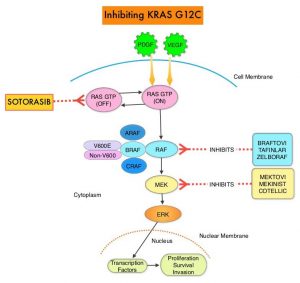
SUMMARY: The KRAS (Kirsten rat sarcoma viral oncogene homologue) proto-oncogene encodes a protein that is a member of the small GTPase super family. The KRAS gene provides instructions for making the KRAS protein, which is a part of a signaling pathway known as the RAS/MAPK pathway. By relaying signals from outside the cell to the cell nucleus, the protein instructs the cell to grow, divide and differentiate. The KRAS protein is a GTPase, and converts GTP into GDP. To transmit signals, the KRAS protein must be turned on, by binding to a molecule of GTP. When GTP is converted to GDP, the KRAS protein is turned off or inactivated, and when the KRAS protein is bound to GDP, it does not relay signals to the cell nucleus. The KRAS gene is in the Ras family of oncogenes, which also includes two other genes, HRAS and NRAS. When mutated, oncogenes have the potential to change normal cells cancerous.
KRAS is the most frequently mutated oncogene in human cancers and are often associated with resistance to targeted therapies and poor outcomes. The KRAS-G12C mutation occurs in approximately 12-15% of Non Small Cell Lung Cancers (NSCLC) and in 3-5% of Colorectal cancers and other solid cancers. G12C is a single point mutation with a Glycine-to-Cysteine substitution at codon 12. This substitution favors the activated state of KRAS, resulting in a predominantly GTP-bound KRAS oncoprotein, amplifying signaling pathways that lead to oncogenesis.Inhibiting-KRAS-G12C

Sotorasib (AMG 510) is a small molecule that specifically and irreversibly inhibits KRAS-G12C and traps KRAS-G12C in the inactive GDP-bound state. Preclinical studies in animal models showed that Sotorasib inhibited nearly all detectable phosphorylation of Extracellular signal-Regulated Kinase (ERK), a key downstream effector of KRAS, leading to durable complete regression of KRAS-G12C tumors.

The authors conducted a multicenter, open label Phase I trial of Sotorasib, in patients with advanced solid tumors harboring the KRAS-G12C mutation. This trial consisted of dose escalation and expansion cohorts and included a total of 129 patients, of whom 59 patients had NSCLC, 42 had Colorectal cancer, and 28 patients had other tumor types (Appendiceal, Endometrial, Pancreatic cancers and Melanoma). Sotorasib was administered orally once daily and each treatment cycle was 21 days. The planned dose levels for the escalation cohorts were 180, 360, 720, and 960 mg. Treatment was continued until disease progression or unacceptable toxicity. The median patient age was 62 years and most of the enrolled patients were heavily pretreated and had received a median of 3 previous lines of anticancer therapy for metastatic disease. Among the NSCLC patient cohort, approximately 90% of patients were current or former smokers and had received anti- Programmed cell Death protein-1 (PD-1) or PD-Ligand 1 (PD-L1) therapies. All patients had received previous platinum-based chemotherapy. The Primary endpoint was safety, including the incidence of dose-limiting toxicities and key Secondary end points were pharmacokinetics and Objective Response Rates. The Sotorasib dose of 960 mg daily was identified as the dose for the expansion cohort. The median follow up was 11.7 months and the median duration of treatment was 3.9 months, with 57% of patients having received treatment for 3 months or more, and 29% of patients, for 6 months or more.

Among those patients with NSCLC, 32.2% of the patients had a confirmed Objective Response (Complete or Partial Response at all dose levels, and 88% had disease control (Objective Response or Stable disease), with a median Progression Free Survival of 6.3 months. Responses were rapid and were seen at week 6, and these responses were durable and ongoing at a median follow up of nearly a year.

Among the colorectal cancer subgroup, at a median follow up of 12.8 months, 7% had a confirmed response, and 74% had disease control, with a median duration of stable disease of 5.4 months and median PFS of 4 months. Responses were also observed in patients with Pancreatic, Endometrial, and Appendiceal cancers and Melanoma. It has been postulated that the inconsistent tumor responses noted between NSCLC and Colorectal cancer suggests either that KRAS-G12C is not the dominant oncogenic driver for colorectal cancer or that other pathways such as Wnt or EGFR pathways may mediate oncogenic signaling beyond KRAS. The authors suggest that a viable option would be to combine Sotorasib with therapies that block additional pathways, as was shown by studies in BRAF V600E-mutant Colorectal cancer. Approximately 57% of patients had treatment-related Adverse Events, of whom, about 12% had Grade 3 or 4 events. These toxicities included abnormal liver function studies, anemia, lymphopenia and diarrhea.

It was concluded Sotorasib showed promising anticancer activity in patients with heavily pretreated advanced solid tumors harboring the KRAS-G12C mutation. Studies evaluating Sotorasib as monotherapy or in combination with various agents in patients with NSCLC or other solid tumors are under way

KRASG12C Inhibition with Sotorasib in Advanced Solid Tumors. Hong DS, Fakih MG, Strickler JH, et al. N Engl J Med 2020; 383:1207-1217.

Five Year Analysis of Adjuvant TAFINLAR® plus MEKINIST® in Stage III Melanoma

SUMMARY: It is estimated that in the US, approximately 100,350 new cases of melanoma will be diagnosed in 2020 and approximately 6,850 patients are expected to die of the disease. The incidence of melanoma has been on the rise for the past three decades. Surgical resection with a curative intent is the standard of care for patients with early stage melanoma, with a 5-year survival rate of 98% for Stage I disease and 90% for Stage II disease. Stage III malignant melanoma is a heterogeneous disease and the risk of recurrence is dependent on the number of positive nodes as well as presence of palpable versus microscopic nodal disease. Further, patients with a metastatic focus of more than 1 mm in greatest dimension in the affected lymph node, have a significantly higher risk of recurrence or death, than those with a metastasis of 1 mm or less. Patients with Stage IIIA disease have a disease-specific survival rate of 78%, whereas those with Stage IIIB and Stage IIIC disease have disease-specific survival rates of 59% and 40% respectively.BRAF-and-MEK-Inhibition-in-MAPK-Pathway

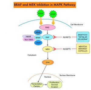
The Mitogen-Activated Protein Kinase pathway (MAPK pathway) is an important signaling pathway which enables the cell to respond to external stimuli. This pathway plays a dual role, regulating cytokine production and participating in cytokine dependent signaling cascade. The MAPK pathway of interest is the RAS-RAF-MEK-ERK pathway. The RAF family of kinases includes ARAF, BRAF and CRAF signaling molecules. BRAF is a very important intermediary of the RAS-RAF-MEK-ERK pathway. BRAF mutations have been demonstrated in 6-8% of all malignancies. The most common BRAF mutation in melanoma is at the V600E/K site and is detected in approximately 50% of melanomas and result in constitutive activation of the MAPK pathway.

TAFINLAR® (Dabrafenib) is a selective oral BRAF inhibitor and MEKINIST® (Trametinib) is a potent and selective inhibitor of MEK gene, which is downstream from RAF in the MAPK pathway. In patients with BRAF V600 mutation-positive unresectable or metastatic melanoma, a combination of TAFINLAR® and MEKINIST® resulted in a median Overall Survival (OS) of more than 2 years, with approximately 20% of the patients remaining progression free at 3 years. These encouraging results led to the study of this combination in patients with Stage III melanoma, with BRAF V600E or V600K mutations, after complete surgical resection.

COMBI-AD, an international, multi-center, randomized, double-blind, placebo-controlled, Phase III trial, in which 870 patients with completely resected, Stage III melanoma and with BRAF V600E or V600K mutations were enrolled. Patients were randomly assigned in a 1:1 to receive TAFINLAR® 150 mg orally twice daily in combination with MEKINIST® 2 mg orally once daily (N=438) or two matched placebos (N=432). Treatment was given for 12 months. Eligible patients had undergone completion lymphadenectomy, with no clinical or radiographic evidence of residual regional node disease. None of the patients had received previous systemic anticancer treatment or radiotherapy for melanoma. BRAF V600 mutation status was confirmed in primary tumor tissue or lymph node tissue by a central reference laboratory. The median age was 50 years. Both treatment groups were well balanced and 18% had Stage IIIA disease, 41% had Stage IIIB disease, and 40% had Stage IIIC disease. Of the enrolled patients, 91% had a BRAF V600E mutation, and 9% had a BRAF V600K mutation. The Primary end point was Relapse Free Survival (RFS) and Secondary end points included Overall Survival (OS), Distant metastasis-free survival, Freedom from relapse, and Safety.

The authors had previously reported that at a median follow up of 2.8 years, the estimated 3-year RFS rate was 58% in the combination therapy group and 39% in the placebo group (HR=0.47; P<0.001), and this represented a 53% lower risk of relapse. At the time of this analysis, median RFS rate had not been reached in the combination therapy group, and was 16.6 months in the placebo group. The improved RFS benefit with the combination therapy was consistent across patient subgroups, regardless of lymph node involvement or primary tumor ulceration. The risk of distant metastases or death was reduced by 49% with the combination therapy versus placebo (HR=0.51; P<0.001).

The authors in this publication reported the results for RFS and Distant metastasis-free survival at 5 years. Overall survival was not analyzed as the data was not mature. The minimum duration of follow up was 59 months. The RFS at 5 years 52% with TAFINLAR® plus MEKINIST® and 36% with placebo (HR for relapse or death=0.51). The Distant metastasis-free survival at 5 years was 65% with TAFINLAR® plus MEKINIST® and 54% with placebo (HR for distant metastasis or death=0.55). As has been reported in previous studies, majority of relapses occurred, within the first 3 years after surgery. There were no clinically meaningful differences noted in the incidence or severity of serious Adverse Events during the follow up period.

It was concluded that in this 5-year analysis of extended follow up from the COMBI-AD trial, 12 months of adjuvant therapy with a combination of TAFINLAR® and MEKINIST® resulted in longer Relapse Free and Distant metastasis-free Survival, compared to placebo, in patients with resected Stage III melanoma with BRAF V600 mutations.

Five-Year Analysis of Adjuvant Dabrafenib plus Trametinib in Stage III Melanoma. Dummer R, Hauschild A, Santinami M, et al. N Engl J Med 2020; 383:1139-1148

DCIS and Risk of Death from Breast Cancer

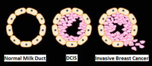
SUMMARY: Breast cancer is the most common cancer among women in the US and about 1 in 8 women (12%) will develop invasive breast cancer during their lifetime. Approximately 279,100 new cases of invasive breast cancer will be diagnosed in 2020 and about 42,690 individuals will die of the disease largely due to metastatic recurrence. Carcinoma in situ of the breast also known as Ductal Carcinoma In Situ (DCIS) is defined as a malignant proliferation of ductal epithelial cells that are confined to the milk ducts without invasion of the basement membrane, and is a precursor lesion to invasive carcinoma. DCIS accounts for approximately 25% of all newly diagnosed breast cancers. Patients with small, screening-detected lesions, are often treated with breast-conserving surgery (to prevent the development of invasive breast cancer), followed by adjuvant radiation and hormonal therapy, although neither of the latter two interventions have been shown to improve survival outcomes. As such, a significant number of patients are over treated. DCIS in itself is not life-threatening but can potentially progress to invasive breast cancer. The two important goals of DCIS treatment therefore are, to prevent invasive ipsilateral cancer recurrence and to prevent death from breast cancer. There remains a large unmet need, to distinguish relatively benign DCIS from DCIS that will develop into invasive breast cancer.DCIS

In a previously published meta-analysis (Cancer Epidemiol Biomarkers Prev. 2019;28:835-845), researchers identified six prognostic factors that were statistically significant and were associated with a 36% to 84% increase in the relative risk of recurrence of invasive disease after a DCIS diagnosis. These six factors included-

1) African American race (43% higher risk)
2) Premenopausal status (59% higher risk)bre
3) Detection by palpation (84% higher risk)
4) Positive margins (63% higher risk)
5) High histologic grade (36% higher risk)
6) High p16 expression (51% higher risk).

This present large cohort study was conducted to determine the risk of death from breast cancer, following diagnosis and treatment of DCIS, compared with the mortality risk among cancer-free women, in the general population. This study included a total of 144,524 women diagnosed with first primary DCIS between 1995 and 2014, from the Surveillance, Epidemiology and End Results (SEER) registries database. Patients with DCIS with microinvasion, Lobular Carcinoma In Situ (LCIS), nonepithelial histological presentations, Paget disease of the nipple, diffuse DCIS, unknown laterality, no surgical intervention on the primary tumor, DCIS diagnosis in women younger than 25 years or aged 80 years or older, were all excluded. Patients with DCIS underwent surgical treatment, and approximately half of these patients also received radiotherapy. These patients were followed from the date of DCIS diagnosis until death from breast cancer, or date of last follow up. These patients were compared with women in the general population without a diagnosis of breast cancer (control group). The mean age at diagnosis was 57.4 years. The Primary outcome was death from breast cancer. Standardized Mortality Ratios (SMR) were estimated by comparing deaths from breast cancer among women diagnosed with DCIS, with expected deaths from breast cancer among women in the general population who did not have cancer.

At a mean follow up period of 9.2 years, the incidence of ipsilateral invasive recurrence events was 3.1%, resulting in a 20-year actuarial risk of 13.9%. There was a 3.8% incidence of contralateral invasive breast cancer events during this follow up period, resulting in a 20-year actuarial risk of 11.3%. The 20-year actuarial risk of breast cancer death among women with DCIS was 3.3%.

The Standardized Mortality Ratio (SMR) for death from breast cancer given a diagnosis of DCIS was 3.36, but varied based on age and race. The SMR for women younger than 40 years was much higher at 11.95, whereas the SMR for women aged 40 to 49 years was 4.15. The SMR for White women was 3.03, for Black women was 7.56, and for East Asian women was 1.89. The SMR for Black women diagnosed with DCIS before age 50 years was 12.10, and the SMR for White women diagnosed with DCIS before age 50 years was 4.21, suggesting that Black women did worse than White woman.

All women with DCIS underwent surgical treatment, and 47.1% also received radiotherapy. Among those patients who were not treated with radiotherapy, the SMR was 4.12, for those treated with unilateral mastectomy and 4.14 for those treated with bilateral mastectomies. Among women who underwent lumpectomy, the SMR was 2.81 for women treated with radiotherapy and 3.42 for those who underwent surgical treatment alone. There were 1540 women who died of breast cancer in the cohort, of whom 45.7% experienced an ipsilateral invasive recurrence or contralateral invasive breast cancer in the interval between DCIS and death from breast cancer. Among the patients who died, 27.8% were known to have undergone a mastectomy.

The annual mortality rate from breast cancer over the entire period of follow up was, 0.12% per year. The mortality rate increased for the first 10 years of the follow-up period and remained constant through years 15 thru 20. The cumulative 20-year risk of breast cancer-specific mortality following DCIS was 3.3% overall, but for Black women diagnosed before age 50 years, the 20-year risk of breast cancer-specific mortality was 8.1%. It has been postulated that the highest risk for recurrence among women who underwent mastectomy may be related to them having more extensive disease with close margins or may have genetic mutations that increase the likelihood of recurrence. Further, patients with DCIS undergoing bilateral mastectomies generally are not treated with endocrine therapy.

It was concluded from this cohort study that women with DCIS had a 3-fold increased risk of death from breast cancer after surgical treatment. The Standardized Mortality Ratio was lower among women who received lumpectomy plus radiation compared with women who received lumpectomy alone. The rate of breast cancer death was nearly 12-fold higher among women diagnosed with DCIS before age 40 years and 7-fold higher in Black women diagnosed with DCIS, compared with the general population.

Association of a Diagnosis of Ductal Carcinoma In Situ With Death From Breast Cancer. Giannakeas V, Sopik V and Narod SA. JAMA Netw Open. 2020;3(9):e2017124. doi:10.1001/jamanetworkopen.2020.17124

Optimal Duration of Immune Checkpoint Inhibitors in Advanced Non Small Cell Lung Cancer

SUMMARY: Lung cancer is the second most common cancer in both men and women and accounts for about 14% of all new cancers and 27% of all cancer deaths. The American Cancer Society estimates that for 2020, about 228, 820 new cases of lung cancer will be diagnosed and 135,720 patients will die of the disease. Lung cancer is the leading cause of cancer-related mortality in the United States. Non-Small Cell Lung Cancer (NSCLC) accounts for approximately 85% of all lung cancers.
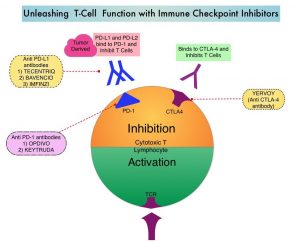
Immunotherapy with Immune Checkpoint Inhibitors (ICIs) has revolutionized cancer care and has become one of the most effective treatment options, by improving Overall Response Rate and prolongation of survival, across multiple tumor types. These agents target Programmed cell Death protein-1 (PD-1), Programmed cell Death Ligand-1 (PD-L1), Cytotoxic T-Lymphocyte-Associated protein-4 (CTLA-4), and many other important regulators of the immune system. Checkpoint inhibitors unleash the T cells resulting in T cell proliferation, activation, and a therapeutic response. Biomarkers predicting responses to ICI’s include Tumor Mutational Burden (TMB), Mismatch Repair (MMR) status, and Programmed cell Death Ligand 1 (PD‐L1) expression. Other biomarkers such as Tumor Infiltrating Lymphocytes (TILs), TIL‐derived Interferon‐γ, Neutrophil‐to‐Lymphocyte ratio, and peripheral cytokines, have also been proposed as predictors of response. The optimal duration of treatment with checkpoint inhibitors across tumor types is currently unknown and finding the balance between efficacy, toxicity and cost of therapy remains an ongoing challenge. There are presently no adequately powered, prospective, checkpoint inhibitor trials, comparing different treatment durations.Unleashing-T-Cell-Function-with-Immune-Checkpoint-Inhibitors

OPDIVO® is a fully human, immunoglobulin G4 monoclonal antibody that binds to the PD-1 receptor and blocks its interaction with PD-L1 and PD-L2, thereby undoing PD-1 pathway-mediated inhibition of the immune response, and unleashing the T cells. The authors in this study explored the impact of duration of treatment with OPDIVO®, on outcomes, in patients with previously treated advanced NSCLC, in a randomized study.

CheckMate 153 is a largely community based, ongoing, Phase IIIb/IV study, reflecting a real-world population, designed to evaluate the efficacy and safety of OPDIVO® monotherapy treatment duration, in previously treated advanced NSCLC. In this study, patients with previously treated advanced or metastatic NSCLC received OPDIVO® 3 mg/kg IV every 2 weeks until disease progression, unacceptable toxicity, or for 1 year. Treatment beyond initial progressive disease was permitted for patients with investigator-assessed clinical benefit, no rapidly progressive disease, and stable ECOG performance status, who were tolerating treatment. Patients who continued to receive treatment at 1 year were randomly assigned, regardless of response status to continue OPDIVO®, or to stop treatment (1-year fixed duration group), with the option of receiving OPDIVO® retreatment on study after disease progression. The Primary end point of safety was previously reported. Exploratory post-random assignment end points were added. Safety and tolerability, Progression Free survival (PFS), Overall Survival (OS), and Objective Response Rate (ORR) were assessed from the time of random assignment of those patients who continued to receive treatment at 1 year. The comparison was between a fixed 1-year treatment regimen and continuous therapy.

Of the 1,428 patients who received OPDIVO® in this study, 252 patients were randomly assigned to continuous treatment (N=127) or 1-year fixed-duration treatment (N=125). With minimum post-random assignment follow up of 13.5 months, median PFS was longer with continuous treatment versus 1-year fixed duration treatment (24.7 months versus 9.4 months; HR=0.56). Median Overall Survival from random assignment was also longer with continuous treatment versus 1-year fixed duration treatment in the Progression-Free Survival population (Not Reached versus 32.5 months; HR, 0.61), as well as in the Intent To Treat population (Not reached versus 28.8 months; HR, 0.62). New onset treatment-related Adverse Events occurred in a few patients and no new safety signals were identified.

The authors concluded that the above findings from an exploratory analysis represent the first randomized data on continuous versus fixed-duration immunotherapy, in previously treated patients with advanced NSCLC, and suggest that continuing OPDIVO® beyond 1 year improves outcomes.

Continuous Versus 1-Year Fixed-Duration Nivolumab in Previously Treated Advanced Non–Small-Cell Lung Cancer: CheckMate 153. Waterhouse DM, Garon EB, Chandler J, et al. DOI: 10.1200/JCO.20.00131 Journal of Clinical Oncology. Published online September 10, 2020.

Late Breaking Abstract – ESMO 2020. VERZENIO® Plus Endocrine Therapy Improves Disease Free Survival in Early Breast Cancer

SUMMARY: Breast cancer is the most common cancer among women in the US and about 1 in 8 women 12%) will develop invasive breast cancer during their lifetime. Approximately 279,100 new cases of invasive breast cancer will be diagnosed in 2020 and about 42,690 individuals will die of the disease largely due to metastatic recurrence. About 70% of breast tumors express Estrogen Receptors and/or Progesterone Receptors, and Hormone Receptor (HR)-positive/HER2-negative breast cancer is the most frequently diagnosed molecular subtype. Majority of these patients are diagnosed with early stage disease and are often cured with a combination of surgery, radiotherapy, chemotherapy, and hormone therapy. However approximately 20% of patients will experience local recurrence or distant relapse during the first 10 years of treatment. This may be more relevant for those with high risk disease, among whom the risk of recurrence is even greater during the first 2 years while on adjuvant endocrine therapy, due to primary endocrine resistance. More than 75% of the early recurrences are seen at distant sites.

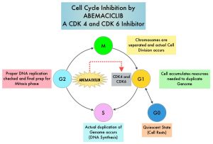
Cyclin Dependent Kinases (CDK) play a very important role to facilitate orderly and controlled progression of the cell cycle. Genetic alterations in these kinases and their regulatory proteins have been implicated in various malignancies. CDK 4 and 6 phosphorylate RetinoBlastoma protein (RB), and initiate transition from the G1 phase to the S phase of the cell cycle. RetinoBlastoma protein has antiproliferative and tumor-suppressor activity and phosphorylation of RB protein nullifies its beneficial activities. CDK4 and CDK6 are activated in hormone receptor positive breast cancer, promoting breast cancer cell proliferation. Further, there is evidence to suggest that endocrine resistant breast cancer cell lines depend on CDK4 for cell proliferation. The understanding of the role of Cyclin Dependent Kinases in the cell cycle, has paved the way for the development of CDK inhibitors.MOA-of_ABEMACICLIB

VERZENIO® (Abemaciclib) is an oral, selective inhibitor of CDK4 and CDK6 kinase activity, and prevents the phosphorylation and subsequent inactivation of the Rb tumor suppressor protein, thereby inducing G1 cell cycle arrest and inhibition of cell proliferation. VERZENIO® is structurally distinct from other CDK 4 and 6 inhibitors (such as Ribociclib and Palbociclib) and is 14 times more potent against cyclin D1/CDK 4 and cyclin D3/CDK 6, in enzymatic assays, but potentially less toxic than earlier pan-CDK inhibitors. At higher doses, only VERZENIO® causes significant cancer cell death, compared with other CDK4/6 inhibitors, suggesting that this drug may be affecting proteins, other than CDK4/6. Additionally, preclinical studies have demonstrated that VERZENIO® may have additional therapeutic benefits for a subset of tumors that are unresponsive to treatment or have grown resistant to other CDK4/6 inhibitors. It has also been shown to cross the blood-brain barrier.

VERZENIO® is presently approved by the FDA as monotherapy as well as in combination with endocrine therapy for patients with HR-positive, HER2- negative advanced breast cancer. The addition of VERZENIO® to FASLODEX® resulted in a statistically significant improvement in Overall Survival among patients with HR-positive, HER2-negative advanced breast cancer, who had progressed on prior endocrine therapy. The goal of monarchE was to evaluate the additional benefit of adding a CDK4/6 inhibitor to endocrine therapy in the adjuvant setting, for patients with HR-positive, HER2-negative early breast cancer.

The international monarchE trial, is an open-label, randomized, Phase III study, which included 5637 patients, who were pre- and postmenopausal, with HR-positive, HER2-negative early breast cancer, and with clinical and/or pathologic risk factors that rendered them at high risk for relapse. The researchers defined high risk as the presence of four or more positive axillary lymph nodes, or 1-3 three positive axillary lymph nodes, with either a tumor size of 5 cm or more, histologic Grade 3, or centrally tested high proliferation rate (Ki-67 of 20% or more). Following completion of primary therapy which included both adjuvant and neoadjuvant chemotherapy and radiotherapy, patients were randomly assigned (1:1) to VERZENIO® 150 mg orally twice daily for 2 years plus 5 to 10 years of physicians choice of endocrine therapy as clinically indicated (N=2808), or endocrine therapy alone (N=2829). The median patient age was 51 years, about 43% of the patients were premenopausal, and 95% of patients had prior chemotherapy. Approximately 60% of patients had 4 or more positive lymph nodes. The Primary endpoint was Invasive Disease Free Survival (IDFS), and Secondary end points included distant Relapse Free Survival, Overall Survival, and safety. The authors in this publication reported the first results, following a preplanned interim analysis.

The addition of VERZENIO® to endocrine therapy resulted in an IDFS of 92.2% at 2 years compared with 88.7% with endocrine therapy alone, and this was statistically significant (HR=0.75; P=0.01). This suggested a 25% reduction in the risk of developing an IDFS event, relative to endocrine therapy alone, and a 3.5% absolute improvement in 2-year IDFS rates. VERZENIO® plus endocrine therapy combination also reduced the risk of metastatic recurrence especially in bone and liver (distant recurrences or Distant Relapse Free Survival) by a clinically meaningful 28% compared to endocrine therapy alone. This clinical benefit was observed in all prespecified subgroups, and among the 43% of patients who were premenopausal at diagnosis, there was a significant 37% reduction in the risk of recurrence compared to endocrine therapy alone. The safety was consistent with the known profile of VERZENIO® and included diarrhea, neutropenia, and fatigue. Diarrhea was well managed with antidiarrheal medications and dose adjustments.

It was concluded that VERZENIO® when combined with endocrine therapy demonstrated a significant improvement in Invasive Disease Free Survival, compared to endocrine therapy alone, in patients with high risk HR-positive, HER2-negative early breast cancer. The researchers also plan to look at genomic signatures in the tissue and plasma samples of enrolled patients and response to VERZENIO®.

Abemaciclib Combined With Endocrine Therapy for the Adjuvant Treatment of HR+, HER2−, Node-Positive, High-Risk, Early Breast Cancer (monarchE). Johnston SRD, Harbeck N, Hegg R, et al. DOI: 10.1200/JCO.20.02514 Journal of Clinical Oncology – published online before print September 20, 2020

OPDIVO® plus CABOMETYX® Combination Doubles Progression Free Survival in Newly Diagnosed Advanced Kidney Cancer

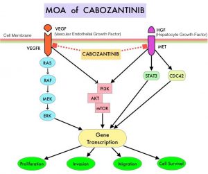
SUMMARY: The American Cancer Society estimates that 73,750 new cases of kidney and renal pelvis cancers will be diagnosed in the United States in 2020 and about 14,830 people will die from the disease. Renal Cell Carcinoma (RCC) is by far the most common type of kidney cancer and is about twice as common in men as in women. Modifiable risk factors include smoking, obesity, workplace exposure to certain substances and high blood pressure. The five year survival of patients with advanced RCC is less than 10% and there is significant unmet need for improved therapies for this disease. SUTENT® (Sunitinib) is a MultiKinase Inhibitor (MKI) which simultaneously targets the tumor cell wall, vascular endothelial cell wall as well as the pericyte/fibroblast/vascular/smooth vessel cell wall and is capable of specifically binding to tyrosine kinases, inhibiting the earlier signaling events and thereby inhibits phosphorylation of VEGF receptor, PDGF receptor, FLT-3 and c-KIT. SUTENT® is the standard first-line intervention for treatment naïve patients with advanced RCC. In a large, multi-center, randomized, Phase III study, the median Progression Free Survival (PFS) with SUTENT® was 9.5 months, the Objective Response Rate (ORR) was 25%, and the median Overall Survival was 29.3 months, when compared with Interferon Alfa, in patients with treatment-naïve Renal Cell Carcinoma. This was however associated with a high rate of hematological toxicities.MOA-of-CABOZANTINIB

The FDA in 2018, approved combination immunotherapy, OPDIVO® (Nivolumab) plus YERVOY® (Ipilimumab), for the treatment of intermediate or poor-risk, previously untreated advanced Renal Cell Carcinoma (RCC), based on significantly higher Overall Survival (OS) and Objective Response Rates (ORR), compared with SUTENT® (CheckMate 214). Subsequently, two studies, a combination of BAVENCIO® (Avelumab) and INLYTA® (Axitinib) – JAVELIN Renal 101, and KEYTRUDA® (Pembrolizumab) and INLYTA® (KEYNOTE-426), demonstrated superior OS, compared to SUTENT®, and for the first time set the stage for the use of a combination of checkpoint inhibitor and targeted therapy in this patient population.

OPDIVO®, an anti-PD-1 checkpoint inhibitor and CABOMETYX® (Cabozantinib), a small-molecule Tyrosine Kinase Inhibitor, are both approved as single agents, for the second-line treatment of Renal Cell Carcinoma. The rationale for combining these two agents is that OPDIVO® unleashes the immune system and restores antitumor immune response, whereas CABOMETYX® has both antiangiogenic and immunomodulatory properties and may counteract tumor-induced immunosuppression.

CheckMate 9ER study is a multinational, randomized, Phase III trial, in which a combination of OPDIVO® plus CABOMETYX® was compared with single agent SUTENT®, in treatment naïve patients with advanced clear cell Renal Cell Carcinoma. This study included 651 treatment naïve patients with advanced Renal Cell Carcinoma with a clear cell component, who were randomly assigned in a 1:1 ratio to receive OPDIVO® 240 mg IV every 2 weeks along with CABOMETYX® 40 mg orally daily (N=323) or SUTENT® 50 mg orally daily in 4-week-on, 2-week-off cycles (N=328). Treatment was continued until disease progression or unacceptable toxicity. Patients with any IMDC (International Metastatic RCC Database Consortium) risk score were included. Patients with sarcomatoid tumor features were allowed. Patients were stratified by IMDC risk score and tumor PD-L1 expression. The median patient age was 62 years, 58% of patients were in the IMDC intermediate risk category and 75% of patients had tumor PD-L1 expression of less than 1%. The Primary endpoint was Progression Free Survival (PFS) and Secondary endpoints included Overall Survival (OS), Objective Response Rate (ORR) and safety.

At a median follow up of 18.1 months, the median PFS was 16.6 months with OPDIVO® plus CABOMETYX® combination versus 8.3 months with single agent SUTENT® (HR=0.51; P<0.0001), suggesting a doubling of PFS, with a 49% reduction in the risk of disease progression or death. The median Overall Survival, a secondary endpoint, was not reached in either treatment group, but at this first analysis, patients randomized to the OPDIVO® plus CABOMETYX® combination had significantly longer OS, than those receiving SUTENT® (median Not Reached; HR=0.60; P=0.001), suggesting a 40% reduction in the risk of death. These benefits were seen consistently across pre-specified subgroups defined according to IMDC risk categories and PD-L1 expression. The Objective Response Rate (ORR) was also significantly higher and doubled among patients receiving the OPDIVO® plus CABOMETYX® combination, compared to those receiving SUTENT® (55.7% versus 27.1%, P<0.0001). Complete response rates were also higher among those receiving the OPDIVO® plus CABOMETYX® combination (8.0% versus 4.6%), with a shorter median time to response, and longer duration of response. Grade 3 or more Adverse Events were higher among those receiving OPDIVO® plus CABOMETYX® combination, compared to those receiving SUTENT® (60.6% versus 50.9%).

It was concluded that a combination of OPDIVO® plus CABOMETYX® demonstrated superior Progression Free Survival, Overall Survival and Overall Response Rate, compared to SUTENT®, in treatment naïve patients with advanced Renal Cell Carcinoma, and provides a new treatment option for this patient group.

Nivolumab + cabozantinib vs sunitinib in first-line treatment for advanced renal cell carcinoma: first results from the randomized phase 3 CheckMate 9ER trial. Choueiri TK, Powles T, Burotto M, et al. Ann Oncol. 2020;31(4). Abstract 696O.

FDA Approves GAVRETO® for Metastatic RET Fusion-Positive Non Small Cell Lung Cancer

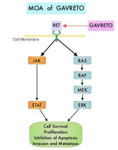
SUMMARY: The FDA on September 4, 2020, granted accelerated approval to GAVRETO® (Pralsetinib) for adult patients with metastatic RET fusion-positive Non Small Cell Lung Cancer (NSCLC), as detected by an FDA approved test. The FDA also approved the Oncomine Dx Target (ODxT) Test as a companion diagnostic for GAVRETO®. Lung cancer is the second most common cancer in both men and women MOA-of-GAVRETOand accounts for about 14% of all new cancers and 27% of all cancer deaths. The American Cancer Society estimates that for 2020, about 228, 820 new cases of lung cancer will be diagnosed and 135,720 patients will die of the disease. Lung cancer is the leading cause of cancer-related mortality in the United States. Non-Small Cell Lung Cancer (NSCLC) accounts for approximately 85% of all lung cancers.

In addition to the well characterized gene fusions involving ALK and ROS1 in NSCLC, genetic alterations involving other kinases including EGFR, BRAF, RET, NTRK, are all additional established targetable drivers. These genetic alterations are generally mutually exclusive, with no more than one predominant driver in any given cancer. The hallmark of all of these genetic alterations is oncogene addiction, in which cancers are driven primarily, or even exclusively, by aberrant oncogene signaling, and are highly susceptible to small molecule inhibitors.

RET kinase is a transmembrane Receptor Tyrosine Kinase and plays an important role during the development and maintenance of a variety of tissues, including neural and genitourinary tissues. RET signaling activates downstream pathways such as JAK/STAT3 and RAS/RAF/MEK/ERK and leads to cellular proliferation, survival, invasion, and metastasis. Oncogenic alterations to the RET proto-oncogene results in uncontrolled cell growth and enhanced tumor invasiveness. RET alterations include RET rearrangements, leading to RET fusions, and activating point mutations occurring across multiple tumor types. RET fusions have been identified in approximately 2% of NSCLCs, 10-20% of non-medullary thyroid cancers. Activating RET point mutations account for approximately 60% of sporadic Medullary Thyroid Cancers (MTC) and more than 90% of inherited MTCs. Other cancers with documented RET alterations include colorectal, breast, and several hematologic malignancies.

GAVRETO® is an oral, highly potent, selective RET kinase inhibitor targeting oncogenic RET alterations, including fusions and mutations, regardless of the tissue of origin. The efficacy of GAVRETO® was investigated in a multicenter, open-label, multi-cohort, Phase I/II basket clinical trial (ARROW), in patients with tumors showing RET alterations. Identification of RET gene alterations was prospectively determined in local laboratories using either Next Generation Sequencing (NGS), Fluorescence In Situ Hybridization (FISH), or other tests. (In a basket trial, tumors with different histologies and single biomarker are placed in different baskets and receive a single treatment). The main efficacy outcome measures were Overall Response Rate (ORR) and response duration, as determined by a blinded Independent Review Committee, using RECIST criteria.

The efficacy for RET fusion-positive NSCLC was evaluated in 87 patients previously treated with platinum-based chemotherapy. Patients received GAVRETO® 400 mg orally once daily. The ORR was 57%, with a Complete Response (CR) rate of 5.7% and 80% of responding patients had responses lasting 6 months or longer. The median Duration of Response was not reached. Efficacy was also evaluated in 27 patients who never received systemic treatment and the ORR in this patient group was 70% with 11% CR rate and 58% of responding patients had responses lasting 6 months or longer. The most common adverse reactions (25% or more) were fatigue, constipation, musculoskeletal pain and hypertension.

It was concluded that patients treated with GAVRETO® had a rapid, potent, and durable clinical response, in patients with advanced RET fusion positive NSCLC, regardless of RET fusion partner, presence of brain metastases, or prior therapies.

Gainor JF, Curigliano G, Kim D-W, et al. DOI: 10.1200/JCO.2020.38.15_suppl.9515 Journal of Clinical Oncology 38, no. 15_suppl (May 20, 2020) 9515-9515.

FDA Approves DARZALEX® plus KYPROLIS® and Dexamethasone for Multiple Myeloma

SUMMARY: The FDA on August 20, 2020, approved KYPROLIS® (Carfilzomib) and DARZALEX® (Daratumumab), in combination with Dexamethasone, for adult patients with Relapsed or Refractory multiple myeloma, who have received one to three lines of therapy. Multiple Myeloma is a clonal disorder of plasma cells in the bone marrow and the American Cancer Society estimates that in the United States, 32, 270 new cases will be diagnosed in 2020 and 12,830 patients are expected to die of the disease. Multiple Myeloma (MM) in 2020 remains an incurable disease. The therapeutic goal therefore is to improve Progression Free Survival (PFS) and Overall Survival (OS). Multiple Myeloma is a disease of the elderly, with a median age at diagnosis of 69 years and characterized by intrinsic clonal heterogeneity. Almost all patients eventually will relapse, and patients with a high-risk cytogenetic profile or refractory disease have the worst outcomes. The median survival for patients with Myeloma is over 10 years.

REVLIMID® (Lenalidomide) in combination with VELCADE® (Bortezomib) and Dexamethasone is the preferred regimen according to the NCCN guidelines, for both transplant and non-transplant candidates with newly diagnosed Multiple Myeloma, and when given continuously or with maintenance therapy, has improved survival outcomes. Nonetheless, a significant number of patients progress while on these agents or discontinue therapy due to toxicities. There is therefore a need for effective and tolerable regimens for patients who are exposed or refractory to REVLIMID® or VELCADE®.Mechanism-of-Action-of-Daratumumab

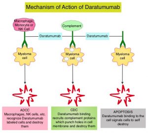
KYPROLIS® (Carfilzomib) is a second generation selective, epoxyketone Proteasome Inhibitor and unlike VELCADE®, proteasome inhibition with KYPROLIS® is irreversible. DARZALEX® (Daratumumab) is a human IgG1 antibody that targets CD38, a transmembrane glycoprotein abundantly expressed on malignant plasma cells and with low levels of expression on normal lymphoid and myeloid cells. DARZALEX® exerts its cytotoxic effect on myeloma cells by multiple mechanisms, including Antibody Dependent Cellular Cytotoxicity (ADCC), Complement Dependent Cytotoxicity (CDC) and direct Apoptosis. Additionally, DARZALEX® may play a role in immunomodulation, by depleting CD38-positive regulator immune suppressor cells, and thereby expanding T cells, in patients responding to therapy. Both KYPROLIS® and DARZALEX® are approved as single agents, as well as in combination with other drugs, for the treatment of patients with Relapsed/Refractory Multiple Myeloma. In a Phase I study, KYPROLIS® in combination with Dexamethasone and DARZALEX® demonstrated safety and efficacy in patients Relapsed/Refractory Multiple Myeloma.

The efficacy of KYPROLIS® and DARZALEX® along with Dexamethasone was evaluated in two clinical trials, CANDOR and EQUULEUS. CANDOR is a multicenter, open-label, Phase III trial, which included Relapsed/Refractory Multiple Myeloma patients with measurable disease who had received 1-3 prior lines of therapy, with Partial Response or better to one or more lines of therapy. A total of 466 patients were randomly assigned 2:1 to receive triplet of KYPROLIS®, Dexamethasone, and DARZALEX® (KdD)- N=312 or KYPROLIS® and Dexamethasone (Kd) alone- N=154. All patients received KYPROLIS® as a 30 minute IV infusion on days 1, 2, 8, 9, 15, and 16 of each 28-day cycle (20 mg/m2 on days 1 and 2 during cycle 1 and 56 mg/m2 thereafter). DARZALEX® 8 mg/kg was administered IV on days 1 and 2 of cycle 1 and at 16 mg/kg once weekly for the remaining doses of the first 2 cycles, then every 2 weeks for 4 cycles (cycles 3-6), and every 4 weeks thereafter. All patients received Dexamethasone 40 mg oral or IV weekly (20 mg for patients over 75 years of age). The median age was 64 years, 42% and 90% received prior REVLIMID® and VELCADE® (Bortezomib) containing regimens respectively, and a third of patients were refractory to REVLIMID®. The Primary endpoint was Progression Free Survival (PFS) and Secondary endpoints included Overall Response Rate (ORR), Minimal Residual Disease (MRD)-negative status, Complete Response (CR) rate at 12 months, Overall Survival (OS), Duration of Response, and Safety.

After a median follow up of 17 months, the study met its Primary endpoint and the median PFS was not reached for the KdD arm and was 15.8 months for the Kd arm (HR=0.63; P=0.0027). This represented a 37% reduction in the risk of progression or death in the KdD group. The PFS benefit of KdD was maintained across prespecified subgroups, particularly among REVLIMID®-exposed and REVLIMID®-refractory patients. The ORR was 84.3% in the KdD group versus 74.7% in the Kd group (P=0.004), with a CR rate or better of 28.5% versus 10.4% respectively. The median time to first response was one month in both treatment groups. Patients treated with KdD achieved deeper responses which was nearly 10 times higher, with a MRD-negative Complete Response rate at 12 months of 12.5% for KdD versus 1.3% for Kd (P<0.0001). The median treatment duration was longer in the KdD group compared to the Kd group (70.1 versus 40.3 wks). The median OS was not reached in either groups, at a median follow up time of 17 months. Toxicities were generally manageable and the incidence of Adverse Events leading to treatment discontinuation was similar in both treatment groups.

EQUULEUS is an open label, multicohort trial which evaluated the combination of KYPROLIS® administered weekly on days 1, 8, and 15 of each 28-day cycle (20 mg/m2 IV on cycle 1, day 1, and if tolerated, increased to 70 mg/m2 on Cycle 1 Day 8 and thereafter) along with DARZALEX® IV and Dexamethasone (KdD). Efficacy was based on Overall Response Rate (ORR). Of the 85 patients with Relapsed or Refractory multiple myeloma who had received 1 to 3 prior lines of therapy enrolled in the KdD cohort, the ORR was 81%, with response duration of 27.5 months.

It was concluded that a combination of KYPROLIS® along with Dexamethasone and DARZALEX® resulted in a significant PFS benefit over KYPROLIS® and Dexamethasone alone, with deeper responses, and the PFS benefit of KdD was maintained across prespecified, clinically important subgroups, particularly REVLIMID®-exposed and REVLIMID®-refractory patients. The authors added that KdD regimen should be considered as a novel, efficacious, and tolerable immunomodulatory-free treatment option for Relapsed/Refractory Multiple Myeloma patients.

Carfilzomib, dexamethasone, and daratumumab versus carfilzomib and dexamethasone for patients with relapsed or refractory multiple myeloma (CANDOR): results from a randomised, multicentre, open-label, phase 3 study. Dimopoulos M, Quach H, Mateos M-V, et al. The Lancet 2020;396:186-197.

Precision Medicine in Pancreatic Cancer May Improve Overall Survival

SUMMARY: The American Cancer Society estimates that for 2020, about 57,600 people will be diagnosed with pancreatic cancer and about 47,050 people will die of the disease. Pancreatic cancer is the fourth most common cause of cancer-related deaths in the United States and Western Europe. Unfortunately, unlike other malignancies, very little progress has been made and outcome for patients with advanced pancreatic cancer has been dismal, with a 5-year survival rate for metastatic pancreatic cancer of approximately 9%.

Patients with metastatic Pancreatic Ductal AdenoCarcinoma (PDAC) are often treated with chemotherapy and treatment regimens include FOLFIRINOX and Gemcitabine with nab-Paclitaxel (ABRAXANE®). However, resistance to current treatment modalities is common, and the median Overall Survival (OS) remains less than 1 year, suggesting that treatment with chemotherapy alone probably may not increase response rates and Overall Survival.

In an attempt to improve outcomes in patients with metastatic PDAC, molecular profiling using Next Generation Sequencing (NGS) and protein IHC panel-based examination of patients’ tumors, has enabled grouping patients into molecular subgroups with therapeutically actionable molecular alterations. It is estimated that approximately 25% of pancreatic cancers harbor actionable molecular alterations, defined as molecular alterations for which there is clinical or strong preclinical evidence of a predictive benefit from a specific therapy. However, less than 5% of patients with pancreatic cancer having actionable molecular alterations receive targeted therapies. This may be because of the aggressive nature of the disease or economic and logistical barriers. The commonly altered pathways include DNA repair (15%), cell cycle (11%), and AKT/mTOR (19%). Molecular targets have included Homologous Recombination Repair genes (14-17%), HER2 amplification genes (2%) and MisMatch Repair gene deficiency (MicroSatellite Instability 2-3%). Mutations in DNA repair genes are the most common “highly actionable” alterations (15%). The most frequently mutated DNA repair genes are ATM (4.5%) and BRCA2 (2.9%). Some examples of available targeted agents for patients with metastatic PDAC include PARP inhibitors for BRCA1 and BRCA2 mutations, TRK inhibitors for NTRK1, NTRK2, or NTRK3 fusions, and Immune Checkpoint Inhibitors for MMR-deficient or MSI-H tumors. Patients with these genetic alterations constitute about 8% of patients, with pancreatic cancer.

Know Your Tumor (KYT) is a precision medicine program, which is a collaboration between Perthera Inc. and the Pancreatic Cancer Action Network (PanCAN), and utilizes Perthera’s precision medicine system for multiomic molecular profiling of a nonselected patient population. Multiomics is data analysis at multiple levels such as genome, epigenome, transcriptome, proteome, and metabolome, to comprehensively understand human health and diseases, by interpreting molecular intricacy and variations. The intent of the KYT program is to match patients with appropriate clinical trials and therapies, based on actionable molecular alterations, treatment history, and geographical locations. The purpose of this study was to determine whether patients with pancreatic cancer whose tumors harbored actionable molecular alterations and who received molecularly matched therapy, had a longer median Overall Survival, than similar patients who did not receive molecularly matched therapy.

In this program, of the 1082 patients who received reports on their tumor genomic profile, outcomes were available for 677 patients, of whom 189 patients had actionable molecular alterations. At a median follow up of 383 days, patients with actionable molecular alterations who received a matched therapy (N=46) had a significantly longer median Overall Survival, compared to those patients who only received unmatched therapies (N=143), and this was statistically significant (2.58 years versus 1.51 years; HR=0.42: P=0.0004). The 46 patients who received a matched therapy also had significantly longer Overall Survival than the 488 patients who did not have an actionable molecular alteration (2.58 years versus 1.32 years; HR=0.34; P<0.0001). The median Overall Survival was not different between the patients who received unmatched therapy and those without an actionable molecular alteration (HR=0.82; P=0.10).

It was concluded from these Real-World outcomes that a matched therapy for patients with actionable molecular alterations can have a substantial effect on survival, in patients with pancreatic cancer. The authors acknowledged that only 2% of patients who were referred to undergo molecular profiling ultimately received a matched therapy and 143 patients with actionable molecular alterations received only unmatched therapies due to a variety of reasons including logistical issues and economic barriers.

Overall survival in patients with pancreatic cancer receiving matched therapies following molecular profiling: a retrospective analysis of the Know Your Tumor registry trial. Pishvaian MJ, Blais EM, Brody JR, et al. Lancet Oncol. 2020 Apr;21(4):508-518.doi: 10.1016/S1470-2045(20)30074-7. Epub 2020 Mar 2.