Maintenance RUBRACA® in Patients with Advanced Pancreatic Cancer with a Pathogenic Germline or Somatic Variant in BRCA1, BRCA2, or PALB2

SUMMARY: The American Cancer Society estimates that for 2021, about 60,430 people will be diagnosed with pancreatic cancer and about 48,220 people will die of the disease. Pancreatic cancer is the fourth most common cause of cancer-related deaths in the United States and Western Europe. Unfortunately, unlike other malignancies, very little progress has been made and outcome for patients with advanced pancreatic cancer has been dismal, with a 5-year survival rate for metastatic pancreatic cancer of approximately 2%. Pancreatic cancer has surpassed breast cancer as the third leading cause of cancer death in the United States and is on track to surpass colorectal cancer, to move to the second leading cause of cancer related deaths in the United States around 2021.

DNA damage is a common occurrence in daily life by UV light, ionizing radiation, replication errors, chemical agents, etc. This can result in single and double strand breaks in the DNA structure which must be repaired for cell survival. The two vital pathways for DNA repair in a normal cell are BRCA1/BRCA2 and PARP. BRCA1 and BRCA2 genes recognize and repair double strand DNA breaks via Homologous Recombination Repair (HRR) pathway. Homologous Recombination is a type of genetic recombination, and is a DNA repair pathway utilized by cells to accurately repair DNA double-stranded breaks during the S and G2 phases of the cell cycle, and thereby maintain genomic integrity. Homologous Recombination Deficiency (HRD) is noted following mutation of genes involved in HR repair pathway. At least 15 genes are involved in the Homologous Recombination Repair (HRR) pathway including BRCA1, BRCA2, PALB2, CHEK2 and ATM genes. BRCA1 and BRCA2 are tumor suppressor genes located on chromosome 17 and chromosome 13 respectively and functional BRCA proteins repair damaged DNA, and play an important role in maintaining cellular genetic integrity. They regulate cell growth and prevent abnormal cell division and development of malignancy. Mutations in these genes predispose an individual to develop malignant tumors. It is well established that the presence of BRCA1 and BRCA2 mutations can significantly increase the lifetime risk for developing breast and ovarian cancer, as high as 85% and 40% respectively. BRCA1/2 mutations have been detected in 4-7% of patients with pancreatic cancer, with a 2-6 fold increase in risk, associated with these mutations. These patients tend to be younger. Among pancreatic cancer patients with Ashkenazi Jewish ancestry, the prevalence of BRCA1/2 mutations is 6-19%, with mutations more common for BRCA2. NCCN guideline recommends that germline testing should be considered for all patients with pancreatic cancer and is especially recommended for those with a personal history of cancer, family history or clinical suspicion of a family history of pancreatic cancer. Approximately 10% of pancreatic cancer cases have a familial component. When hereditary cancer syndrome is suspected in patients with pancreatic cancer, genetic counseling should be considered.

BRCA mutations can either be inherited (Germline) and present in all individual cells or can be acquired and occur exclusively in the tumor cells (Somatic). Somatic mutations account for a significant portion of overall BRCA1 and BRCA2 aberrations. Loss of BRCA function due to frequent somatic aberrations likely deregulates HR pathway, and other pathways then come in to play, which are less precise and error prone, resulting in the accumulation of additional mutations and chromosomal instability in the cell, with subsequent malignant transformation. Homologous Recombination Deficiency therefore indicates an important loss of DNA repair function.

The PARP (Poly ADP Ribose Polymerase), family of enzymes include, PARP1 and PARP2, and is a related enzymatic pathway that repairs single strand breaks in DNA. In a BRCA mutant, the cancer cell relies solely on PARP pathway for DNA repair to survive. PARP inhibitors trap PARP onto DNA at sites of single-strand breaks, preventing their repair and generating double-strand breaks that cannot be repaired accurately in tumors harboring defects in Homologous Recombination Repair pathway genes, such as BRCA1 or BRCA2 mutations, and this leads to cumulative DNA damage and tumor cell death.

LYNPARZA® (Olaparib) is a PARP inhibitor and is presently approved as maintenance therapy for patients with advanced pancreatic cancer demonstrating a germline BRCA1 or BRCA2 pathogenic variant. However, previously published studies have demonstrated the benefit of PARP inhibitors in breast, prostate and ovarian cancer patients, beyond germline BRCA pathogenic variants. Further, there is an unmet need to expand the group of patients with pancreatic cancer who may benefit from therapy with a PARP inhibitor, beyond those with germline BRCA pathogenic variants.

This investigator-initiated, single-arm Phase II study was conducted to assess the role of oral, small molecule PARP inhibitor RUBRACA® (Rucaparib), as maintenance therapy in advanced pancreatic cancer with germline or somatic pathogenic variants in BRCA1, BRCA2, or PALB2 genes. This study enrolled 46 patients with advanced pancreatic cancer with germline or somatic pathogenic variants in BRCA1, BRCA2, or PALB2, and had received at least 16 weeks of platinum-based chemotherapy without evidence of platinum resistance, which was defined as growing tumors, new lesions, or a steadily rising tumor marker during or within 8 weeks of platinum therapy. The median age was 62 years, approximately 17% had germline BRCA1, 64% had germline BRCA2, 14% had germline PALB2 and 5% had somatic BRCA2 pathogenic variants. Majority of patients (95%) had metastatic disease and 5% had locally advanced disease. Ashkenazi Jewish founder mutation was found in 24% of patients. The Primary end point was Progression Free Survival (PFS) at 6 months (PFS). Secondary end points included Safety, Objective Response Rate (ORR), Disease Control Rate, Duration of Response, and Overall Survival.

The PFS at 6 months was 59.5% and the PFS at 12 months was 54.8%. The median PFS was 13.1 months and median Overall Survival was 23.5 months. The ORR in those with measurable disease was 42%, and the Disease Control Rate was 67%. The median Duration of Response was 17.3 months. These responses were noted across all germline and somatic pathogenic variants in BRCA1, BRCA2, and PALB2 genes, and no new safety signals were noted.

It was concluded from this study that maintenance RUBRACA® is a safe and effective therapy for platinum-sensitive, advanced pancreatic cancer patients, with a pathogenic variant in BRCA1, BRCA2, or PALB2. The authors added that the finding of efficacy in patients with germline PALB2 and somatic BRCA2 pathogenic variants, expands the population of patients likely to benefit from PARP inhibitors, beyond those with germline BRCA1 and BRCA2 pathogenic variants.

Phase II Study of Maintenance Rucaparib in Patients With Platinum-Sensitive Advanced Pancreatic Cancer and a Pathogenic Germline or Somatic Variant in BRCA1, BRCA2, or PALB2. Reiss KA, Mick R, O’Hara MH, et al. J Clin Oncol. 2021;39:2497-2505.

FDA Approves TRODELVY® for Advanced Urothelial Cancer

SUMMARY: The FDA on April 13, 2021, granted accelerated approval to TRODELVY® (Sacituzumab Govitecan) for patients with locally advanced or metastatic Urothelial Cancer who previously received a Platinum-containing chemotherapy, and either a Programmed Death receptor-1 (PD-1) or a Programmed Death-Ligand 1 (PD-L1) inhibitor. The American Cancer Society estimates that in the United States for 2021, about 83,730 new cases of bladder cancer will be diagnosed and approximately 17,200 patients will die of the disease. Bladder cancer is the fourth most common cancer in men, but it is less common in women. A third of the patients initially present with locally invasive or metastatic disease. Patients with Urothelial Carcinoma are currently treated in the first line setting with a Platinum based chemotherapy regimen, and a checkpoint Inhibitor (PD-1 or PD-L1 inhibitor) in the second line setting. Treatment options for patients who progress after first and second line therapies are limited, with poor outcomes. The response rates with standard chemotherapy in this patient population, is about 10%, with a median Overall Survival (OS) of 7-8 months.

Two new agents approved by the FDA include BALVERSA® (Erdafitinib), a pan-Fibroblast Growth Factor Receptor (FGFR) inhibitor, for patients with locally advanced or metastatic Urothelial Carcinoma with susceptible FGFR3 or FGFR2 genetic alterations, that has progressed during or following Platinum-containing chemotherapy, as well as PADCEV® (Enfortumab Vedotin), an Antibody-Drug Conjugate (ADC) that targets Nectin-4, a cell adhesion molecule, highly expressed in Urothelial Cancers and other solid tumors. These two agents have Objective Response Rates (ORRs) of approximately 40%, and most patients will progress on these therapies. Further, FGFR alterations occur in only 20% of patients with metastatic Urothelial Carcinoma, limiting the use of BALVERSA®. Hence, there is an unmet need for novel therapies.

Trop-2 is a transmembrane glycoprotein and calcium signal transducer. It stimulates cancer-cell growth, and this cell surface receptor is overexpressed in several epithelial cancers including cancers of the Breast, Colon, Lung and Urothelial Cancer, and has limited expression in normal human tissues. TRODELVY® is an Antibody-Drug Conjugate (ADC) in which SN-38, an active metabolite of Irinotecan, a Topoisomerase I inhibitor, is coupled to the humanized Anti-Trophoblast cell-surface antigen 2 (Trop-2) monoclonal antibody (hRS7 IgG1κ), through the cleavable CL2A linker. SN-38 cannot be given directly to patients because of its toxicity and poor solubility. Upon binding to Trop-2, the anti-TROP-2 monoclonal antibody is internalized and delivers SN-38 directly into the tumor cell, making it a suitable transporter for the delivery of cytotoxic drugs. Further, the cleavable linker enables SN-38 to be released both intracellularly into the tumor cells, as well as the tumor microenvironment, thereby allowing for the delivery of therapeutic concentrations of the active drug in bystander cells to which the conjugate has not bound. Thus, TRODELVY®-bound tumor cells are killed by intracellular uptake of SN-38, whereas the adjacent tumor cells are killed by the extracellular release of SN-38.

TRODELVY® in a Phase I/II trial involving patients with advanced epithelial cancers, showed encouraging clinical activity across various solid tumors and was associated with a Objective Response Rate (ORR) of 31% in patients with relapsed or refractory metastatic Urothelial Carcinoma, including a 27% ORR among patients who had received prior checkpoint inhibitor and Platinum based therapy. The TROPHY-U-01 Phase II trial was designed to assess the activity of TRODELVY® and confirm these findings in patients with locally advanced unresectable or metastatic Urothelial Carcinoma. This trial includes 5 patient cohorts, evaluating the role of TRODELVY® in various groups of patients and in combination with various agents including checkpoint inhibitors. The authors in this publication reported the primary results from the full Cohort 1 of the TROPHY-U-01 study in patients with metastatic Urothelial Cancer who progressed after prior Platinum based and checkpoint inhibitor based therapies.

Cohort 1 included 113 patients who had received a median of three prior therapies. Patients received TRODELVY® 10 mg/kg IV, on days 1 and 8 of a 21-day treatment cycle, until disease progression or unacceptable toxicities. The median patient age was 66 years and 66% of patients had visceral metastases. The main efficacy endpoints were Objective Response Rate (ORR) and Duration of Response (DOR), evaluated by Independent Review, using RECIST 1.1 criteria.

At a median follow up of 9.1 months, the ORR was 27.7%, and 77% of patients had decrease in measurable disease. The Complete Response rate was 5.4% and 22.3% had Partial Responses. The median DOR was 7.2 months. The median Progression Free Survival was 5.4 months and Overall Survival was 10.9 months. Important Grade 3 or more treatment related adverse events included, neutropenia, anemia, diarrhea, and febrile neutropenia, with 6% of patients discontinuing treatment due to adverse events because of treatment-related adverse events.

It was concluded that TRODELVY® is an active agent and has notable efficacy, compared with historical controls, in pretreated metastatic Urothelial Cancer, that has progressed on both prior Platinum regimens and checkpoint inhibitors, and has manageable safety profile.

TROPHY-U-01: A Phase II Open-Label Study of Sacituzumab Govitecan in Patients With Metastatic Urothelial Carcinoma Progressing After Platinum-Based Chemotherapy and Checkpoint Inhibitors. Tagawa, ST, Balar AV, Petrylak DP, et al. J Clin Oncol. 2021;39:2474-2485.

Noninvasive Evaluation of Myelodysplastic Syndrome in Patients with Unexplained Anemia

SUMMARY: It is estimated that in the US approximately 13,000 people are diagnosed with MyeloDysplastic Syndromes (MDS) each year. The prevalence has been estimated to be from 60,000 to 170,000 in the US. MyeloDysplastic Syndromes are a heterogenous group of stem cell disorders characterized by marrow failure resulting in cytopenias, mainly symptomatic anemia, with associated cytogenetic abnormalities, and abnormal cellular maturation with morphologic changes in clonal cells. Majority of the individuals diagnosed with MDS are 65 years or older and die as a result of infection and/or bleeding, consequent to bone marrow failure. About a third of patients with MDS develop Acute Myeloid Leukemia (AML).

Elderly patients with mildly symptomatic anemia (macrocytic anemia) or pancytopenia are initially evaluated for B12, folate and iron deficiency, as well as hypothyroidism and hemolysis. The next recommended test for unexplained anemia is bone marrow examination, which is the current gold standard for diagnosis of MDS. However, this procedure is invasive and can be painful, and is occasionally associated with infectious and bleeding complications. For these reasons, many patients and their physicians may avoid this procedure, potentially delaying access to effective treatments.

The researchers in this study developed a noninvasive algorithm, to help diagnose or exclude MDS, without bone marrow evaluation. To develop this web-based app, 502 patients diagnosed with MDS based on bone marrow evaluation were randomly selected from the European MDS registry and this sample was combined with 502 controls with unexplained anemia, aged 50 years and older, who had normal findings on bone marrow evaluation. Patients with bone marrow involvement as a part of a hematological or other disease or with any degree of bone marrow dysplasia could not serve as controls. The authors using a logistic regression model were able to classify patients into 1 of 3 categories: probable MDS, probably not MDS, and indeterminate. The initial model that was developed by the researchers was further improved using the new Gradient-Boosted Models (GBMs), adding more variables based on their known association with MDS. They included 10 routinely measured and readily available demographic, clinical, and laboratory variables such as Age, Sex, Hemoglobin, White Blood Cell count, Platelet count, Mean Corpuscular Volume, Neutrophils, Monocytes, Glucose, and Creatinine, as well as including more patients. A web app was developed that would help clinicians diagnose, and more importantly rule out MDS noninvasively, without bone marrow examination.

The researchers then calculated Positive Predictive Values and Negative Predictive Values assuming a 20% prevalence of MDS within the population of patients to which the model would be applied in practice (patients with unexplained anemia, in whom other causes of anemia have been excluded, who would likely undergo bone marrow examination in clinical practice). Approximately 90% of the MDS patients had anemia, about 35-40% of them had neutropenia, thrombocytopenia or bicytopenia, and about 15% had pancytopenia, all according to WHO criteria. Using the more severe cytopenia criteria as would be used for the International Prognostic Scoring System (IPSS) score, about 50% of MDS patients were severely anemic, about 20-25% of patients were neutropenic, thrombocytopenic, or bicytopenic, and 5% were pancytopenic.

It was noted that this tool was reliably able to separate patients with and without MDS. This model had a sensitivity of 88% and specificity of 95%. In this patient population with unexplained anemia, probable MDS and probably not MDS could be determined in 86% of patients, leaving it to the patient and the physician to discuss whether the bone marrow evaluation should be performed in the indeterminate group, to make the definitive diagnosis. The researchers also determined the robustness of this model in patients with neutropenia, thrombocytopenia, as well as in those with bicytopenia and pancytopenia. It was noted that the predictive model continued to be reliable, especially in its ability to rule out MDS in almost all of these categories, with Negative Predictive Values all above 90% and relatively narrow 95% Confidence Intervals (CIs). Moreover, the lower boundaries of the 95% CI were all above 90%. However, when it came to making a diagnosis of MDS, the accuracy was somewhat diminished. The researchers attributed this to smaller number of patients in these groups, and further added that patients with multiple cytopenias should have bone marrow evaluation, irrespective of the model prediction. Based on this algorithm, the researchers developed a web-based predictor calculator which would serve as a practical tool for clinicians. The limitations of this algorithm are that morphology, blast percentage, genetics, and cytogenetics have not yet been integrated into the model.

It was concluded that based on this MDS model, a web-based computer app has been developed, to help the physician community to primarily exclude MDS in a cytopenic individual and also predict the possibility of MDS, without performing an invasive bone marrow evaluation. The authors plan to not only improve the predictive power of the model by increasing the number of measured variables, but also validate this model with independent prospective patient data, and develop a predictive prognostic tool, in addition to diagnosis.

A predictive algorithm using clinical and laboratory parameters may assist in ruling out and in diagnosing MDS. Oster HS, Crouch S, Smith A, et al. Blood Adv. 2021;5:3066-3075.

FDA Approves TIBSOVO® for IDH1 Mutated Advanced Cholangiocarcinoma

SUMMARY: The FDA on August 25, 2021, approved TIBSOVO® (Ivosidenib) for adult patients with previously treated, locally advanced or metastatic Cholangiocarcinoma, with an Isocitrate DeHydrogenase-1 (IDH1) mutation, as detected by an FDA-approved test. The FDA also approved the Oncomine Dx Target Test (Life Technologies Corporation) as a companion diagnostic device, to aid in selecting patients with Cholangiocarcinoma for treatment with TIBSOVO®.

Bile Duct cancer (Cholangiocarcinoma), comprise about 30% of all primary liver tumors and includes both intrahepatic and extrahepatic bile duct cancers. Klatskin tumor is a type of Cholangiocarcinoma that begins in the hilum, at the junction of the left and right bile ducts. It is the most common type of Cholangiocarcinoma, accounting for more than half of all cases. About 8,000 people in the US are diagnosed with Cholangiocarcinoma each year and approximately 20% of the cases are suitable for surgical resection. The 5-year survival is less than 10%, with limited progress made over the past two decades. There is therefore an unmet need for new effective therapies.

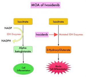
Isocitrate DeHydrogenase (IDH) is a metabolic enzyme that helps generate energy from glucose and other metabolites, by catalyzing the conversion of Isocitrate to Alpha-Ketoglutarate. Alpha-ketoglutarate is required to properly regulate DNA and histone methylation, which in turn is important for gene expression and cellular differentiation. IDH mutations lead to aberrant DNA methylation and altered gene expression thereby preventing cellular differentiation, with resulting immature undifferentiated cells. IDH mutations can thus promote leukemogenesis in Acute Myeloid Leukemia (AML) and tumorigenesis in solid tumors and can result in inferior outcomes. There are three isoforms of IDH. IDH1 is mainly found in the cytoplasm, as well as in peroxisomes, whereas IDH2 and IDH3 are found in the mitochondria, and are a part of the Krebs cycle. Approximately 20% of patients with AML, 70% of patients with Low-grade Glioma and secondary Glioblastoma, 50% of patients with Chondrosarcoma, 20% of patients with Intrahepatic Cholangiocarcinoma, 30% of patients with Angioimmunoblastic T-cell lymphoma and 8% of patients with Myelodysplastic syndromes/Myeloproliferative neoplasms, are associated with IDH mutations.MOA-of-Ivosidenib

TIBSOVO® (Ivosidenib) is an oral, targeted, small-molecule inhibitor of mutant IDH1. A previously published Phase I study demonstrated the safety and activity of TIBSOVO® in patients with IDH1 mutated advanced Cholangiocarcinoma. ClarIDHy is an international, randomized, double-blind, Phase III study, in which 187 previously treated patients with advanced Cholangiocarcinoma with an IDH1 mutation were randomly assigned 2:1 to receive TIBSOVO® 500 mg orally once daily (N=126) or matched placebo (N=61). All patients had advanced unresectable Cholangiocarcinoma. The median age was 62 years, 91% had intrahepatic Cholangiocarcinoma, 93% of patients had metastatic disease and 47% had received two prior therapies. The patient’s disease must have progressed following at least one, but not more than two prior regimens, including at least one Gemcitabine or 5-FU containing regimen. The Primary endpoint was Progression Free Survival (PFS) and Secondary endpoints included Safety, Objective Response Rate (ORR) and Overall Survival (OS). Crossover from placebo to TIBSOVO® was permitted upon radiographic disease progression.

This study met its Primary endpoint and the median PFS was 2.7 months for patients treated with TIBSOVO® compared to 1.4 months with placebo (HR=0.37; P<0.0001). More importantly, the median PFS at 6 and 12 months were 32% and 22% in the TIBSOVO® group, whereas no patients randomized to the placebo group were progression-free for 6 or more months, at the time of data cutoff.
The authors also reported the results of final analysis which showed an improvement in the secondary endpoint of OS, favoring patients randomized to TIBSOVO® compared to those randomized to placebo. However, statistical significance was not reached. The median OS for patients in the TIBSOVO® arm was 10.3 months compared to 7.5 months for patients in the placebo arm (HR=0.79; 1-sided P=0.093). A high proportion of patients in the placebo arm (70.5%) crossed over to TIBSOVO®. After adjusting for crossover from placebo to TIBSOVO®, the median OS for patients in the placebo arm was 5.1 months (HR=0.49; 1-sided P<0.0001).

The 6-month survival rate for patients in the TIBSOVO® arm was 69% compared to 57% of patients in the placebo arm, not adjusted for crossover. The 12-month survival rate for patients in the TIBSOVO® arm was 43% compared to 36% for patients in the placebo arm, not adjusted for crossover. Treatment with TIBSOVO® preserved patients’ physical functioning from baseline, as assessed by the EORTC QLQ-C30 questionnaire, whereas patients in the placebo arm experienced decline from baseline starting cycle 2. The most common Adverse Events of any grade for TIBSOVO® were nausea (38%), diarrhea (33.1%) and fatigue (28.9%). Adverse Events leading to discontinuation were more common with placebo compared with total TIBSOVO® (8.5% versus 6.6%).

It was concluded that treatment with TIBSOVO® in patients with advanced Cholangiocarcinoma with an IDH1 mutation, resulted in significant improvement in Progression Free Survival as well as favorable Overall Survival trend, when compared to Placebo, despite a high rate of crossover. This is the first pivotal study demonstrating the clinical benefit of targeting IDH1 mutation in this patient group. This new oral, non-cytotoxic, targeted treatment option, with a tolerable safety profile, will be a welcome addition to treat this aggressive disease, for which there is an unmet need for new therapies.

Final results from ClarIDHy, a global, phase III, randomized, double-blind study of ivosidenib (IVO) versus placebo (PBO) in patients (pts) with previously treated cholangiocarcinoma (CCA) and an isocitrate dehydrogenase 1 (IDH1) mutation. Zhu A, Macarulla T, Javle MM, et al. J Clin Oncol 39, 2021 (suppl 3; abstr 266)

Myelodysplastic Syndromes: Managing Anemia due to Ineffective Erythropoiesis in Patients with MDS Requiring RBC Transfusions

Dr-M-Yair-Levy

Written by: Dr. M. Yair Levy, Texas Oncology
Promotional Content Sponsored by: Bristol Myers Squibb
Dr. Levy is a paid consultant for BMS and was compensated for his contribution in drafting this article.


Myelodysplastic syndromes (MDS) are a heterogeneous group of myeloid malignancies characterized by multilineage cytopenias, including anemia.1 In MDS, stem cells lack the ability for differentiation and maturation, resulting in bone marrow dysfunction and poor blood cell production, in particular red blood cells (RBCs).2 Anemia is present in the majority of patients with MDS and, at diagnosis, anemia is the most common cytopenia present in patients with MDS.1 Anemia in MDS is linked to bone marrow dysfunction characterized by ineffective erythropoiesis.2

Ineffective erythropoiesis in MDS may lead to anemia requiring RBC transfusions and is characterized by increased proliferation of erythroid progenitors, increased death of erythroid precursors, and impaired erythroid maturation.3,4 In fact, 94% (515/546) of patients with MDS received RBC transfusions in the SEER-Sound registry from 2001 to 2007, 13% of whom had ring sideroblasts.5 Ring sideroblasts are erythroblasts with iron-loaded mitochondria associated with anemia that can be identified by iron staining and the results can be found on pathology reports.6

The presence of anemia despite increased proliferation of progenitor cells is indicative of ineffective erythropoiesis in MDS.3,4 There is a need to help address anemia due to ineffective erythropoiesis in patients with MDS requiring RBC transfusions after erythropoiesis stimulating agent (ESA) failure. REBLOZYL® (luspatercept-aamt), the first and only erythroid maturation agent, is approved for the treatment of anemia failing an ESA and requiring 2 or more RBC units over 8 weeks in adult patients with very low- to intermediate-risk myelodysplastic syndromes with ring sideroblasts (MDS-RS) or with myelodysplastic/myeloproliferative neoplasm with ring sideroblasts and thrombocytosis (MDS/MPN-RS-T).7 REBLOZYL is not indicated for use as a substitute for RBC transfusions in patients who require immediate correction of anemia.7 The approval of REBLOZYL by the FDA marked the first new treatment indicated for patients with MDS in 14 years.8 In my clinical experience, the results that I’ve seen in patients with lower-risk MDS-RS are consistent with those seen in the MEDALIST clinical trial, as discussed below.

As the first and only erythroid maturation agent, REBLOZYL enhances erythroid maturation through differentiation of late-stage erythroid precursors. REBLOZYL works by binding several TGF-β superfamily ligands, thereby diminishing Smad2/3 signaling and increasing the number and quality of mature RBCs in preclinical models.7

REBLOZYL was FDA approved for MDS-associated anemia based on the efficacy and safety outcomes of the pivotal phase 3 MEDALIST trial.7,9 The MEDALIST trial was a multicenter, randomized, double-blind, placebo-controlled, phase 3 trial of 229 adult patients with IPSS-R very low-, low-, or intermediate-risk MDS-RS (<5% bone marrow blasts, presence of ring sideroblasts of ≥15% or ≥5% with an SF3B1 mutation) who required RBC transfusions (≥2 RBC units/8 weeks) were randomized 2:1 to REBLOZYL (n = 153) or placebo (n = 76).7,9 Patients were also required to have had an inadequate response to prior treatment with an ESA (defined as response that is no longer maintained after at least 8 doses of recombinant human erythropoietin or 4 doses of darbepoetin alfa), be intolerant of ESAs, or be ineligible for ESAs (serum EPO >200 U/L).7,9 The MEDALIST trial excluded patients who had del 5q MDS, a white blood cell count >13 Gi/L, neutrophils <0.5 Gi/L, platelets <50 Gi/L, or who had prior use of a disease-modifying agent for treatment of MDS.7

REBLOZYL was administered 1 mg/kg subcutaneously every 3 weeks for at least 24 weeks or until unacceptable toxicity, loss of efficacy, or disease progression. Patients could have their dose increased to 1.33 mg/kg and then to 1.75 mg/kg. Patients received dose increases if they did not achieve transfusion independence after two doses or 6 weeks at 1 mg/kg and 1.33 mg/kg. All patients received best supportive care, which included RBC transfusions as needed.7

In MEDALIST, 36% (83/229) of all patients in the trial were 75 years of age or older, including patients up to 95 years.7,9 95.2% (218/229) of all patients in the trial were ESA-exposed, while only 4.8% (11/229) were ESA-naive, with serum EPO >200 U/L.7,9 All patients in the trial had ring sideroblasts (≥15% ring sideroblasts or ≥5% ring sideroblasts with an SF3B1 mutation), and the majority (206/229) had an SF3B1 mutation.7,9 All patients except 1 were classified as having very low- to intermediate-risk MDS by the IPSS-R criteria.7 57% (130/229) of patients had a baseline RBC transfusion burden <6 RBC units/8 weeks.7

The primary endpoint in MEDALIST was RBC transfusion independence (RBC-TI), defined as the absence of any RBC transfusion during any consecutive 8-week period occurring entirely within the first 24 weeks of treatment.7 Approximately 3 times greater percentage of patients receiving REBLOZYL achieved the primary endpoint of RBC transfusion independence than placebo: 37.9% (58/153) vs 13.2% (10/76; common risk difference [95% CI]: 24.6 [14.5, 34.6]; P < 0.0001), respectively.7 These data support that in patients requiring ≥2 RBC units/8 weeks, REBLOZYL should be started after at least 2 to 3 months of an inadequate response to ESAs.7,9

Key secondary endpoints in MEDALIST were based on RBC transfusion independence (absence of any RBC transfusions) during any consecutive 12-week period occurring entirely within weeks 1 to 24 and 1 to 48. 28.1% (43/153) of patients receiving REBLOZYL achieved transfusion independence ≥12 weeks occurring entirely within weeks 1 to 24 vs 7.9% (6/76) of patients receiving placebo (common risk difference [95% CI]: 20.0 [10.9, 29.1]; P = 0.0002). For weeks 1 to 48,* 33.3% (51/153) of patients receiving REBLOZYL achieved transfusion independence ≥12 weeks vs 11.8% (9/76) of patients receiving placebo (common risk difference [95% CI]: 21.4 [11.2, 31.5]; P = 0.0003).7
*The median (range) duration of treatment was 49 weeks (6–114 weeks) on the REBLOZYL arm and 24 weeks (7-89 weeks) on the placebo arm.

REBLOZYL provided RBC transfusion independence vs placebo in patients with MDS-RS and MDS/MPN-RS-T, based on the WHO 2016 classification. Of patients who were diagnosed with MDS-RS, 34.1% (46/135; 95% CI 26.1, 42.7) of patients receiving REBLOZYL achieved transfusion independence vs 12.3% (8/65; 95% CI 5.5, 22.8) receiving placebo. Of patients who were diagnosed with MDS/MPN-RS-T, 64.3% (9/14; 95% CI 35.1, 87.2) of patients receiving REBLOZYL achieved transfusion independence vs 22.2% (2/9; 95% CI 2.8, 60.0) receiving placebo. Of patients who were diagnosed with other types of MDS (MDS-EB-1, MDS-EB-2, and MDS-U), 75% (3/4; 95% CI 19.4, 99.4) of patients receiving REBLOZYL achieved transfusion independence vs 0% (0/2; 95% CI 0.0, 84.2) receiving placebo.7

RBC transfusion independence was also examined by baseline RBC transfusion burden. Of patients requiring 2 to 3 RBC units/8 weeks at baseline,† 80.4% (37/46; 95% CI 66.1, 90.6) of patients receiving REBLOZYL achieved transfusion independence vs 40% (8/20; 95% CI 19.1, 63.9) receiving placebo. Of patients requiring 4 to 5 RBC units/8 weeks at baseline,‡ 36.6% (15/41; 95% CI 22.1, 53.1) of patients receiving REBLOZYL achieved transfusion independence vs 4.3% (1/23; 95% CI 0.1, 21.9) receiving placebo. Of patients requiring ≥6 RBC units/8 weeks, 9.1% (6/66; 95% CI 3.4, 18.7) of patients receiving REBLOZYL achieved transfusion independence vs 3% (1/33; 95% CI 0.1, 15.8) receiving placebo.7

†Includes patients who received 3.5 units.
‡Includes patients who received 5.5 units.

The safety of REBLOZYL at the recommended dose and schedule was evaluated in 242 patients with MDS-RS (n = 192) or other myeloid neoplasms (n = 50). The median time on treatment with REBLOZYL was 50.4 weeks (range, 3-221 weeks), with 67% of patients exposed for 6 months or longer and 49% exposed for >1 year.7

Among the 242 patients treated with REBLOZYL, 5 (2.1%) had a fatal adverse reaction. 4.5% (11/242) of patients discontinued REBLOZYL due to an adverse reaction and 2.9% (7/242) of patients had their REBLOZYL dose reduced due to adverse reactions. The most common (≥10%) all-grade adverse reactions included fatigue, musculoskeletal pain, dizziness, diarrhea, nausea, hypersensitivity reactions, hypertension, headache, upper respiratory tract infection, bronchitis, and urinary tract infection. The majority of adverse reactions with REBLOZYL were Grade 1 or 2 (mild to moderate). The most common (≥2%) Grade ≥3 adverse reactions included fatigue, hypertension, syncope, and musculoskeletal pain.7

IMPORTANT SAFETY INFORMATION
WARNINGS AND PRECAUTIONS
Thrombosis/Thromboembolism
In adult patients with beta thalassemia, thromboembolic events (TEE) were reported in 8/223 (3.6%) REBLOZYL-treated patients. TEEs included deep vein thrombosis, pulmonary embolus, portal vein thrombosis, and ischemic stroke. Patients with known risk factors for thromboembolism (splenectomy or concomitant use of hormone replacement therapy) may be at further increased risk of thromboembolic conditions. Consider thromboprophylaxis in patients at increased risk of TEE. Monitor patients for signs and symptoms of thromboembolic events and institute treatment promptly.

Hypertension
Hypertension was reported in 10.7% (61/571) of REBLOZYL-treated patients. Across clinical studies, the incidence of Grade 3 to 4 hypertension ranged from 1.8% to 8.6%. In adult patients with MDS with normal baseline blood pressure, 26 (29.9%) patients developed SBP ≥130 mm Hg and 23 (16.4%) patients developed DBP ≥80 mm Hg. Monitor blood pressure prior to each administration. Manage new or exacerbations of preexisting hypertension using anti-hypertensive agents.

Embryo-Fetal Toxicity
REBLOZYL may cause fetal harm when administered to a pregnant woman. REBLOZYL caused increased post-implantation loss, decreased litter size, and an increased incidence of skeletal variations in pregnant rat and rabbit studies. Advise pregnant women of the potential risk to a fetus. Advise females of reproductive potential to use effective contraception during treatment and for at least 3 months after the final dose.

ADVERSE REACTIONS
Grade ≥3 (≥2%) adverse reactions included fatigue, hypertension, syncope and musculoskeletal pain. A fatal adverse reaction occurred in 5 (2.1%) patients.

The most common (≥10%) adverse reactions included fatigue, musculoskeletal pain, dizziness, diarrhea, nausea, hypersensitivity reactions, hypertension, headache, upper respiratory tract infection, bronchitis, and urinary tract infection

LACTATION
It is not known whether REBLOZYL is excreted into human milk or absorbed systemically after ingestion by a nursing infant. REBLOZYL was detected in milk of lactating rats. When a drug is present in animal milk, it is likely that the drug will be present in human milk. Because many drugs are excreted in human milk, and because of the unknown effects of REBLOZYL in infants, a decision should be made whether to discontinue nursing or to discontinue treatment. Because of the potential for serious adverse reactions in the breastfed child, breastfeeding is not recommended during treatment and for 3 months after the last dose.

Please see full Prescribing Information for REBLOZYL

References:
1. Greenberg PL, Tuechler H, Schanz J, et al. Revised international prognostic scoring system for myelodysplastic syndromes. Blood. 2012;120(12):2454-2465.
2. Cazzola M, Malcovati L. Myelodysplastic syndromes—coping with ineffective hematopoiesis. N Engl J Med. 2005;352(6):536-538.
3. Santini V. Anemia as the main manifestation of myelodysplastic syndromes. Semin Hematol. 2015;52(4):348-356.
4. Fontenay-Roupie M, Bouscary D, Guesnu M, et al. Ineffective erythropoiesis in myelodysplastic syndromes: correlation with Fas expression but not with lack of erythropoietin receptor signal transduction. Br J Haematol. 1999;106(2):464-473.
5. Ramsey SD, McCune JS, Blough DK, et al. Patterns of blood product use among patients with myelodysplastic syndrome. Vox Sang. 2012;102(4):331-337.
6. Malcovati L, Cazzola M. Recent advances in the understanding of myelodysplastic syndromes with ring sideroblasts. Br J Haematol. 2016;174(6):847-858.
7. REBLOZYL [Prescribing Information]. Summit, NJ: Celgene Corporation; 2020.
8. Steensma, D.P. Myelodysplastic syndromes current treatment algorithm 2018. Blood Cancer J. 2018;8(5):47.
9. Data on file, Celgene Corporation. Summit, New Jersey.

© 2021 Celgene Corporation.
REBLOZYL is a trademark of Celgene Corporation, a Bristol Myers Squibb company.
REBLOZYL is licensed from Acceleron Pharma Inc.
08/21 2007-US-2100270