Oral Relugolix Superior to Leuprolide in Advanced Prostate Cancer

SUMMARY: Prostate cancer is the most common cancer in American men with the exclusion of skin cancer, and 1 in 9 men will be diagnosed with prostate cancer during their lifetime. It is estimated that in the United States, about 191,930 new cases of prostate cancer will be diagnosed in 2020 and 33,330 men will die of the disease. The development and progression of prostate cancer is driven by androgens. Androgen Deprivation Therapy (ADT) or testosterone suppression has therefore been the cornerstone of treatment of advanced prostate cancer, and is the first treatment intervention. Androgen Deprivation Therapies have included bilateral orchiectomy or Gonadotropin Releasing Hormone (GnRH) analogues, with or without first generation Androgen Receptor (AR) inhibitors such as CASODEX® (Bicalutamide), NILANDRON® (Nilutamide) and EULEXIN® (Flutamide) or with second-generation, anti-androgen agents, which include ZYTIGA® (Abiraterone), XTANDI® (Enzalutamide), ERLEADA® (Apalutamide) and NUBEQA® (Darolutamide).

Androgen Deprivation Therapies such as GnRH analogs/Luteinizing Hormone Releasing Hormone (LHRH) agonists are standard treatment for patients with advanced prostate cancer. These agents when first administered cause an initial surge in Luteinizing Hormone, Follicle Stimulating Hormone (FSH), and testosterone levels. With continuous administration, LHRH agonists desensitize the pituitary receptor and suppress the production of Luteinizing Hormone and testosterone, thus blocking the pulsatile secretion of GnRH by the hypothalamus. LHRH agonists however do not fully suppress FSH which is a potential mitogenic growth factor for prostate cancer cells. The initial testosterone surge may result in flaring up of symptoms such as bone pain, obstructive urinary symptoms, and rarely spinal cord compression. For this reason, anti-androgen agents are recommended for the first few weeks after initiation of an LHRH agonist. LHRH agonists have been shown to increase the near-term risk of major adverse cardiovascular events, by promoting plaque destabilization and rupture.

Degarelix (FIRMAGON®) is a GnRH antagonist, and the depot injection was approved by the FDA in December 2018. Degarelix suppresses both Luteinizing Hormone and FSH, resulting in rapid testosterone suppression, without an initial testosterone surge. This agent however has to be administered monthly and approximately 40% of patients experience reactions at the injection site.MOA-of-GnRH-Agonists-and-Antagonists

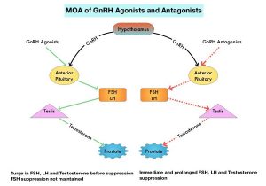
Relugolix is a highly selective, GnRH antagonist that can be given orally once daily, and has a half-life of 25 hours. In multiple Phase I and Phase II studies, Relugolix has been shown to lower testosterone levels by rapidly inhibiting the pituitary release of Luteinizing Hormone and FSH. The HERO trial is a multinational, randomized, open-label, Phase III study, which evaluated the efficacy and safety of Relugolix, an oral GnRH antagonist, as compared with those of Leuprolide (LUPRON®) (GnRH agonist), in men with advanced prostate cancer. In this study, a total of 930 patients were randomly assigned in a 2:1 ratio to receive either Relugolix 120 mg orally once daily, after a single oral loading dose of 360 mg (N=622) or Leuprolide acetate 22.5 mg IM every 3 months (N=308), for 48 weeks.
Eligible patients had one of three clinical disease presentations: 1) Evidence of biochemical (PSA) or clinical relapse after local primary intervention with curative intent 2) Newly diagnosed hormone-sensitive metastatic disease, or 3) Advanced localized disease unlikely to be cured by local primary intervention with curative intent. Patients with major adverse cardiovascular events within 6 months before trial initiation were excluded. Patients were stratified according to the presence or absence of metastatic disease, and age (75 yrs or less and over 75 years). Approximately 32% of patients had metastatic disease and 50% had biochemical recurrence after definitive treatment.
The Primary endpoint was sustained testosterone suppression to castrate levels (less than 50 ng/dL) through 48 weeks. Secondary end points included noninferiority of Relugolix to Leuprolide with respect to sustained castration rate, castrate levels of testosterone on day 4, and profound castrate levels (less than 20 ng/dL) on day 15. Testosterone recovery after discontinuation of the trial drug was to be evaluated in a subgroup of patients. The median follow up time in both groups, including the 30-day safety follow-up period for adverse events, was 52 weeks.

Relugolix was associated with a significantly higher rate of maintained castrate levels of testosterone, when compared to Leuprolide. Castrate levels of testosterone were maintained through 48 weeks in 96.7% of patients in the Relugolix group compared to 88.8% of patients in the Leuprolide group. The difference of 7.9 percentage points showed noninferiority as well as superiority of Relugolix (P<0.001 for superiority) over Leuprolide. All other key Secondary end points showed superiority of Relugolix over Leuprolide (P<0.001). These endpoints included the percentage of patients with castrate levels of testosterone on day 4 (56% versus 0%) and on day 15 (98.7% versus 12%), testosterone suppression to less than 20ng/dL on day 15 (78.4% versus 1%) and confirmed PSA response of more than 50% decrease at day 15 (79.4% versus 19.8%; P<0.001). In the subgroup of 184 patients followed for testosterone recovery, the mean testosterone levels 90 days after treatment discontinuation were 288.4 ng/dL in the Relugolix group and 58.6 ng/dL in the Leuprolide group. The incidence of major adverse cardiovascular events among all the patients was 2.9% in the Relugolix group and 6.2% in the Leuprolide group (HR=0.46).

The authors concluded that in this trial involving men with advanced prostate cancer, Relugolix achieved rapid and sustained suppression of testosterone levels, that was superior to that with Leuprolide, with a 54% lower risk of major adverse cardiovascular events.

Oral Relugolix for Androgen-Deprivation Therapy in Advanced Prostate Cancer. Shore ND, Saad F, Cookson MS, et al. for the HERO Study Investigators. N Engl J Med 2020; 382:2187-2196.

FDA Approves RUBRACA® for BRCA-Mutated Metastatic Castrate Resistant Prostate Cancer

SUMMARY: The FDA on May 15, 2020, granted accelerated approval to RUBRACA® (Rucaparib) for patients with deleterious BRCA mutation (germline and/or somatic)-associated metastatic Castration-Resistant Prostate Cancer (mCRPC), who have been treated with Androgen Receptor-directed therapy and a taxane-based chemotherapy. Prostate cancer is the most common cancer in American men with the exclusion of skin cancer, and 1 in 9 men will be diagnosed with prostate cancer during their lifetime. It is estimated that in the United States, about 191,930 new cases of prostate cancer will be diagnosed in 2020 and 33,330 men will die of the disease.

The development and progression of Prostate cancer is driven by androgens. Androgen Deprivation Therapy (ADT) or testosterone suppression has therefore been the cornerstone of treatment of advanced Prostate cancer and is the first treatment intervention. Androgen Deprivation Therapies have included bilateral orchiectomy or Gonadotropin Releasing Hormone (GnRH) analogues, with or without first generation Androgen Receptor (AR) inhibitors such as CASODEX® (Bicalutamide), NILANDRON® (Nilutamide) and EULEXIN® (Flutamide) or with second-generation, anti-androgen agents, which include, ZYTIGA® (Abiraterone), XTANDI® (Enzalutamide) and ERLEADA® (Apalutamide). ZYTIGA® inhibits CYP17A1 enzyme and depletes adrenal and intratumoral androgens, thereby impairing AR signaling. XTANDI® and ERLEADA® compete with Testosterone and Dihydrotestosterone and avidly bind to the Androgen Receptor, thereby inhibiting AR signaling, and in addition inhibit translocation of the AR into the nucleus and thus inhibits the transcriptional activities of the AR. Approximately 10-20% of patients with advanced Prostate cancer will progress to Castration Resistant Prostate Cancer (CRPC) within five years during ADT, and over 80% of these patients will have metastatic disease at the time of CRPC diagnosis. The estimated mean survival of patients with CRPC is 9-36 months, and there is therefore an unmet need for new effective therapies.

DNA damage is a common occurrence in daily life by UV light, ionizing radiation, replication errors, chemical agents, etc. This can result in single and double strand breaks in the DNA structure which must be repaired for cell survival. The two vital pathways for DNA repair in a normal cell are BRCA1/BRCA2 and PARP. The PARP (Poly ADP Ribose Polymerase), family of enzymes include, PARP1, PARP2 and PARP3. BRCA1 and BRCA2 genes recognize and repair double strand DNA breaks via Homologous Recombination (HR) pathway. Homologous Recombination is a type of genetic recombination, and is a DNA repair pathway utilized by cells to accurately repair DNA double-stranded breaks during the S and G2 phases of the cell cycle, and thereby maintain genomic integrity. Homologous Recombination Deficiency (HRD) is noted following mutation of genes involved in HR repair pathway. At least 15 genes are involved in the Homologous Recombination Repair (HRR) pathway including BRCA1, BRCA2 and ATM genes. The BRCA1 gene is located on the long (q) arm of chromosome 17 whereas BRCA2 is located on the long arm of chromosome 13. BRCA1 and BRCA2 are tumor suppressor genes and functional BRCA proteins therefore repair damaged DNA, and play an important role in maintaining cellular genetic integrity. They regulate cell growth and prevent abnormal cell division and development of malignancy.

Recently published data has shown that deleterious germline and/or somatic mutations in BRCA1, BRCA2, ATM, or other Homologous Recombination DNA-repair genes, are present in about 25% of patients with advanced prostate cancer, including mCRPC. Mutations in BRCA1 and BRCA2 also account for about 20-25% of hereditary breast cancers, about 5-10% of all breast cancers, and 15% of ovarian cancers. BRCA mutations can either be inherited (Germline) and present in all individual cells or can be acquired and occur exclusively in the tumor cells (Somatic). Somatic mutations account for a significant portion of overall BRCA1 and BRCA2 aberrations. Loss of BRCA function due to frequent somatic aberrations likely deregulates HR pathway, and other pathways then come in to play, which are less precise and error prone, resulting in the accumulation of additional mutations and chromosomal instability in the cell, with subsequent malignant transformation. HRD therefore indicates an important loss of DNA repair function. PARP is a related enzymatic pathway that repairs single strand breaks in DNA. In a BRCA mutant, the cancer cell relies solely on PARP pathway for DNA repair. In the presence of a PARP inhibitor, there is synthetic lethality because of the loss of both repair pathway genes, leading to cell death. Thus PARP inhibitors are only harmful to cancer cells.MOA-of-RUBRACA

RUBRACA® is an oral, small molecule inhibitor of Poly-Adenosine diphosphate [ADP] Ribose Polymerase (PARP), presently approved by the FDA for ovarian, fallopian tube or primary peritoneal cancers. This recent FDA approval for prostate cancer patients was based on TRITON2, which is an ongoing international, multicenter, open-label, single arm, Phase II trial, in patients with BRCA-mutated mCRPC, who had been treated with Androgen Receptor-directed therapy and taxane-based chemotherapy. In this study, 115 mCRPC patients with either germline or somatic BRCA mutations were enrolled, of whom 62 patients had measurable disease at baseline. Patients received RUBRACA® 600 mg orally twice daily and concomitant GnRH analog or had prior bilateral orchiectomy. Treatment was continued until disease progression or unacceptable toxicity. The median age was 73 years, majority of patients had an ECOG performance status of 0 or 1, 18% of patients had lung metastases, 21% had liver metastases, 24% had metastases to lymph nodes alone and 40% had 10 or more bone lesions at baseline. The major efficacy outcomes of the study were Objective Response Rate (ORR) and Duration of Response (DOR) in the 62 patients with measurable disease. The median duration of follow up was 13.1 months

The confirmed ORR was 44% and the median DOR was not evaluable. Fifty six percent (56%) of patients with confirmed Objective Responses had a DOR of 6 months or more.

It was concluded that RUBRACA® demonstrates promising efficacy in patients with mCRPC with deleterious BRCA mutations. TRITON3 study is evaluating RUBRACA® versus physician’s choice of second-line AR-directed therapy or Docetaxel, in chemotherapy-naïve patients with mCRPC and alterations in BRCA1/2, who progressed on one prior AR-directed therapy.

ESMO 2019: Preliminary Results from the TRITON2 Study of Rucaparib in Patients with DNA Damage Repair-deficient mCRPC: Updated Analyses. Abida W, Campbell D, Patnaik A, et al. 2019 ESMO Annual Meeting, #ESMO19, 27 Sept – 1 Oct 2019 in Barcelona, Spain.

LYNPARZA® (Olaparib)

The FDA on May 19, 2020 approved LYNPARZA® for adult patients with deleterious or suspected deleterious germline or somatic Homologous Recombination Repair (HRR) gene-mutated metastatic Castration-Resistant Prostate Cancer (mCRPC), who have progressed following prior treatment with Enzalutamide or Abiraterone. LYNPARZA® is a product of AstraZeneca Pharmaceuticals, LP.

RUBRACA® (Rucaparib)

The FDA on May 15, 2020 granted accelerated approval to RUBRACA® for patients with deleterious BRCA mutation (germline and/or somatic)-associated metastatic Castration-Resistant Prostate Cancer (mCRPC), who have been treated with androgen receptor-directed therapy and a taxane-based chemotherapy. RUBRACA® is a product of Clovis Oncology, Inc.

Bone-Targeting Radioisotope, XOFIGO® Improves Overall Survival in CRPC Patients with Bone Metastases 

SUMMARY: Prostate cancer is the most common cancer in American men with the exclusion of skin cancer, and 1 in 9 men will be diagnosed with prostate cancer during their lifetime. It is estimated that in the United States, about 191,930 new cases of prostate cancer will be diagnosed in 2020 and 33,330 men will die of the disease. The skeletal system is the most common site for distant metastases among patients with prostate cancer and over 90% of patients with advanced prostate cancer develop bone metastases, which are osteoblastic (or sclerotic), characterized by deposition of new bone. Bone scan is the most common and cost effective modality for the diagnosis of bone metastases and Technetium (Tc) 99m-labeled methylene diphosphonate is the most widely used bone scanning agent. Agents such as ZOMETA® (Zoledronic acid) and XGEVA® (Denosumab) can prevent or delay Skeletal Related Events (SRE’s) and External Beam Radiation Therapy (EBRT) is often utilized to treat symptomatic SRE’s. EBRT can however damage the bone marrow in the radiated field, resulting in cytopenias, and consequently can potentially preclude patients from receiving cytotoxic chemotherapy.
Radium Ra 223 dichloride (XOFIGO®) is a bone seeking alpha particle emitter and by virtue of its chemical similarity to calcium is preferentially taken up by the bone and forms complexes with bone mineral, hydroxyapatite, in areas where there is increased bone turnover, such as bone metastases. XOFIGO® induces double stranded DNA breaks resulting in antitumor effects and has a very short range in tissues (around 2 and 10 cells), quickly losing energy, compared to beta or gamma radiation. The end result is less damage to the adjacent healthy tissues. Further, unlike Ra-226 which was first isolated by Madame Curie, XOFIGO® has a short half life of 11.4 days and rapidly decays, preventing significant radiation exposure.
Strontium-89 (METASTRON®) is a pure beta emitter and imitates the bio-distribution of calcium in vivo, and is avidly taken up into bony metastases where it has a biological half-life of about 50 days. The biological half-life in the normal bone is approximately 14 days. METASTRON® in two Phase III studies significantly reduced the appearance of new painful metastases, analgesic requirements, and serum PSA levels, compared with radiotherapy alone, among patients with metastatic prostate cancer, suggesting that METASTRON® is an effective systemic radiopharmaceutical for the palliation of bony metastases.Types-of-Radiation-and-Their-Penetrating-Power
In this publication, the authors assessed the Overall Survival (OS) benefit of both alpha emitting and beta emitting bone-targeted RadioIsotopes (RIs) in men with bone metastases from CRPC (Castrate Resistant Prostate Cancer), treated with bone-targeted RIs, and further compared the effects of alpha emitting RIs with beta emitting RIs. This meta-analysis included Individual Patient Data of 2081 patients with CRPC and bone metastases from 6 randomized clinical trials, conducted between January 1993 and June 2013, and data was collected via PubMed, Cochrane Library, ClinicalTrials.gov, and meeting proceedings. Patients included in this study had histologically proven diagnosis of prostate cancer and disease progression after both, surgical or chemical castration, and have evidence of bone metastasis. The median age of patients was 70 years and the proportion of patients with more than 6 bone metastases ranged from 67% to 86%. The median follow up was 26.7 months. The Primary end point of the study was Overall Survival (OS), and Secondary end points included Symptomatic Skeletal Event (SSE)-Free Survival and adverse events.
This analysis showed that an alpha emitting bone- targeted RI (XOFIGO®) was associated with a significantly higher Overall Survival and higher symptomatic Skeletal Event-Free Survival, whereas a beta emitting RI (METASTRON®) was not associated with these significant outcomes. Treatment with the alpha emitter (XOFIGO®) was associated with a significant 30% decreased risk of death when compared with no radioisotope use, whereas use of the beta emitter (METASTRON®) did not confer a significant survival benefit. Further, treatment with XOFIGO® was associated with a significant 35% decreased risk of symptomatic Skeletal Event-Free Survival, whereas treatment with METASTRON® was not associated with such benefit. In the subgroup analyses, men with the lowest serum PSA values appeared to benefit significantly more with the use of bone-targeted RI therapy compared with those with the highest serum PSA values. Hematological toxicities were more frequently observed in patients treated by RI compared with those treated without RI, and the type of radiation did not result in significant differences.
It was concluded from this meta-analysis that among patients with metastatic CRPC, a significant improvement of Overall Survival and symptomatic Skeletal Event-Free Survival was noted with bone-targeted alpha emitting bone- targeted RI (XOFIGO®), but not beta emitting RIs (METASTRON®). Overall Survival in Men With Bone Metastases From Castration-Resistant Prostate Cancer Treated With Bone-Targeting Radioisotopes: A Meta-analysis of Individual Patient Data From Randomized Clinical Trials. Terrisse S, Karamouza E, Parker CC, et al. for the MORPHEP Collaborative Group. JAMA Oncol. 2020;6:206-216

Combination of MRI-Targeted and Systematic Biopsy Increases Detection of Clinically Significant Prostate Cancer 

SUMMARY: Prostate cancer is the most common cancer in American men with the exclusion of skin cancer, and 1 in 9 men will be diagnosed with prostate cancer during their lifetime. It is estimated that in the United States, about 191,930 new cases of prostate cancer will be diagnosed in 2020 and 33,330 men will die of the disease. TransRectal UltraSound (TRUS) guided biopsy has been the standard of care for diagnosing prostate cancer in men with a clinical suspicion of prostate cancer, based on an abnormal Digital Rectal Examination and/or an elevated Prostate Specific Antigen (PSA) level. TransRectal UltraSound (TRUS) guided biopsy is a blind biopsy of the lateral and posterior peripheral zone of the prostate using a template, and 10 to 12 cores of prostate tissue is obtained (Systematic Biopsy). Even though this may result in a higher rate of prostate cancer detection, many detected cancers are low grade tumors that do not benefit from treatment, and these patients are on active surveillance for their low risk disease. The major limitation of this biopsy procedure is the risk of under-sampling a more significant tumor that is located in a region of the prostate not usually targeted with a template. Further, in patients with a rising PSA with a prior negative biopsy, patients are often subjected to a repeat blind biopsy with the same limitations as the original biopsy. Since biopsy access is through the rectum and only specific zones of the prostate are sampled, large areas of the prostate, especially the anterior and central prostate, are not routinely sampled and clinically significant higher-grade cancers are sometimes missed.
Multiparametric MRI (mp-MRI) combines anatomic imaging in the form of T2-weighted imaging, with functional imaging and is being used to detect or rule out cancer in men who have persistent concern for prostate cancer. Previously published studies have shown that MRI-targeted biopsies alone have shown similar or higher rates of detection of clinically SIGNIFICANT cancer in the prostate gland (high grade cancers) and lower rates of detection of clinically INSIGNIFICANT cancer, when compared to systematic biopsy (standard TRUS guided biopsy). This interesting advantage appears to allow the use of mp-MRI as a triage test to avoid a biopsy if the results were negative, and if positive could be used for targeting abnormal areas in the prostate during biopsy. Despite this advantage, debate persists whether MRI-targeted biopsy should be used in place of systematic biopsy or in conjunction with it.
The Trio Study is a substudy of a larger clinical trial called, Use of Tracking Devices to Locate Abnormalities During Invasive Procedures. In this substudy, the authors assessed the use of MRI-targeted, systematic, or combined MRI-targeted and systematic prostate biopsy, in an attempt to define the most effective method for prostate cancer diagnosis.
In this study a total of 2732 men with abnormal PSA or Digital Rectal Exam underwent prostate MRI. Among these patients, 2103 men had MRI-visible lesions and subsequently underwent both MRI-targeted and systematic biopsies. Grade group 1 refers to clinically INSIGNIFICANT disease (Gleason score, 3+3=6), Grade group 2 or higher refers to cancer with favorable intermediate risk or worse (Gleason score, 3+4=7), and Grade group 3 or higher refers to clinically SIGNIFICANT cancer with unfavorable intermediate risk or worse (Gleason score, 4+3=7). The Primary outcome of this study was cancer detection rates according to Grade group (Clustering of Gleason grades) for each biopsy method, and in combination.
Among all 2103 patients who underwent the two biopsy methods, prostate cancer was diagnosed in 52.5% with systematic biopsy alone and in 51.5% with MRI-targeted biopsy alone. The addition of MRI-targeted biopsy to systematic biopsy led to 208 more prostate cancer diagnoses for a total prostate cancer diagnosis of 62.4%. MRI-targeted biopsy detected more clinically SIGNIFICANT Grade group 3 or higher cancers than systematic biopsy (P=0.004) and detected fewer cancers in Grade group 1 (P<0.001). Of the 404 patients who subsequently underwent radical prostatectomy, disease upgrading on histopathology occurred in 41.6% of patients when compared to findings on systematic biopsy alone, 30.9% when compared with findings on MRI-targeted biopsy alone and 14.4% when compared with findings on combined systematic and MRI-targeted biopsy.
The authors concluded that among patients with MRI-visible lesions, a combination of systematic and MRI-targeted biopsy increases the detection of clinically significant prostate cancers, compared to either strategy alone. MRI-Targeted, Systematic, and Combined Biopsy for Prostate Cancer Diagnosis. Ahdoot M, Wilbur AR, Reese SE, et al. N Engl J Med 2020; 382:917-928

Association Between Pseudoprogression and Outcomes in Men with Metastatic Castration-Resistant Prostate Cancer Treated with XTANDI®

SUMMARY: Prostate cancer is the most common cancer in American men with the exclusion of skin cancer, and 1 in 9 men will be diagnosed with prostate cancer during their lifetime. It is estimated that in the United States, about 174,650 new cases of prostate cancer were diagnosed in 2019 and 31,620 men died of the disease. The development and progression of prostate cancer is driven by androgens. Androgen Deprivation Therapy (ADT) or testosterone suppression has therefore been the cornerstone of treatment of advanced prostate cancer and is the first treatment intervention. Androgen Deprivation Therapies have included bilateral orchiectomy or Gonadotropin Releasing Hormone (GnRH) analogues, with or without first generation Androgen Receptor (AR) inhibitors such as CASODEX® (Bicalutamide), NILANDRON® (Nilutamide) and EULEXIN® (Flutamide) or with second-generation, anti-androgen agents, which include, ZYTIGA® (Abiraterone), XTANDI® (Enzalutamide), ERLEADA® (Apalutamide) and NUBEQA® (Darolutamide). Approximately 10-20% of patients with advanced prostate cancer will progress to Castration Resistant Prostate Cancer (CRPC) within five years during ADT, and over 80% of these patients will have metastatic disease at the time of CRPC diagnosis (mCRPC). Among those patients without metastases at CRPC diagnosis, 33% are likely to develop metastases within two years. The estimated mean survival of patients with CRPC is 9-36 months.
The skeletal system is the most common site for distant metastases among patients with prostate cancer and over 80% of patients with advanced prostate cancer develop bone metastases, which are osteoblastic (or sclerotic), characterized by deposition of new bone. Bone scan is the most common and cost effective modality for the diagnosis of bone metastases and Technetium (Tc) 99m-labeled methylene diphosphonate is the most widely used bone scanning agent. Bone scans are commonly used to both diagnose and monitor disease progression in the bone, among patients with advanced prostate cancer, with a sensitivity ranging from 60-90% but with lower specificity. Bone scan however is more sensitive and specific than plain films and CT scans, whereas MRI is superior in evaluating vertebral metastases. Bone scan provides information on osteoblastic activity and skeletal vascularity, with preferential uptake at sites of active bone formation, reflecting the metabolic reaction of bone to the disease activity, regardless of whether it is neoplastic, traumatic or inflammatory. It is for these reasons it has been well known that bone scans can be misleading in determining whether a patient with bone metastases is benefiting from a treatment, particularly endocrine therapy. The Prostate Cancer Working Group (PCWG) recommended that the assessment of disease progression in bone in the absence of other signs of progression, requires that new lesions detected on the first post-treatment scan be confirmed with the documentation of additional new lesions on the next follow-up scan, in the absence of other signs of disease progression. This is because the new lesions detected on the first post-treatment scan may either reflect true progression or can be the result of bone healing known as pseudoprogression (also known as bone scan flare) that can be misinterpreted as treatment failure, and lead to the premature discontinuation of an effective therapy. Even though the occurrence of pseudoprogression is well documented, its association with clinical outcomes in large prospective studies has not been evaluated.
The authors therefore conducted a post hoc retrospective analysis of the PREVAIL (A Safety and Efficacy Study of Oral MDV3100 in Chemotherapy-Naive Patients With Progressive Metastatic Prostate Cancer) and AFFIRM (Safety and Efficacy Study of MDV3100 in Patients With Castration-Resistant Prostate Cancer Who Have Been Previously Treated With Docetaxel-based Chemotherapy) studies to determine the association between new unconfirmed lesions detected on a follow up bone scan, and clinical outcomes in XTANDI® (Enzalutamide)-treated men with mCRPC. The PREVAIL and AFFIRM trials were both designed in accordance with the PCWG guidelines. This analysis included 643 patients from the PREVAIL study who had not received Docetaxel and 404 men from the AFFIRM study who had previously received Docetaxel. Eligible patients had stable disease or response to therapy based on non-bone disease criteria, including assessment of PSA and soft-tissue disease response. Pseudoprogression was defined as detection of one or more lesions on a first post-treatment bone scan (at week 9 in PREVAIL or 13 in AFFIRM) or a second bone scan (at week 17 in PREVAIL or 25 in AFFIRM), without subsequent new lesions detected at later assessments. The authors evaluated the association of the new lesions detected on the first and second bone scans, with radiographic Progression Free Survival (rPFS), Overall Survival (OS), PSA decline, Objective Response in soft tissue, and Quality of Life.
In the PREVAIL study, new unconfirmed bone lesions were detected on bone scans in 27.5% of Docetaxel-naive patients. The rPFS, OS and time to PSA progression among these patients was similar to those without new lesions, suggesting pseudoprogression. In the AFFIRM study, new, unconfirmed lesions were detected in 18.1% of Docetaxel-treated patients and the rPFS, and time to PSA progression among these patients was similar to those without new lesions on bone scans. However, the OS was significantly worse among these patients, compared with those without new lesions on bone scan, suggesting true disease progression. Most lesions were detected on the first follow up bone scan and investigators were unable to identify any pretreatment factor associated with the development of new, unconfirmed lesions in patients responding to XTANDI®, in either clinical setting.
It was concluded that new unconfirmed lesions detected on follow up bone scans within the first 4 months of treatment initiation may represent pseudoprogression in men with mCRPC and are indicative of a favorable treatment response to XTANDI®. However, new unconfirmed bone lesions in men with mCRPC who were previously treated with Docetaxel may reflect disease heterogeneity and true progression with associated worse Overall Survival. Treatment discontinuation can be considered in this patient group, taking into consideration other disease manifestations such as changes in PSA level, finding on soft tissue imaging, symptoms, and patient preferences. These findings reinforce the importance of functional imaging for diagnosing bone metastases. Association Between New Unconfirmed Bone Lesions and Outcomes in Men With Metastatic Castration-Resistant Prostate Cancer Treated With Enzalutamide: Secondary Analysis of the PREVAIL and AFFIRM Randomized Clinical Trials. Armstrong AJ, Al-Adhami M, Lin P, et al. JAMA Oncol. 2019 Dec 12. doi: 10.1001/jamaoncol.2019.4636. [Epub ahead of print]

Higher Levels of IGF-1 and Free Testosterone Increase the Risk of Prostate Cancer

SUMMARY: Prostate cancer is the most common cancer in American men with the exclusion of skin cancer, and 1 in 9 men will be diagnosed with Prostate cancer during their lifetime. It is estimated that in the United States, about 174,650 new cases of Prostate cancer will be diagnosed in 2019 and 31,620 men will die of the disease. The etiology of Prostate cancer remains unclear, and North American Blacks are at the highest risk whereas risk is lowest in Asians, with the risk among Caucasians somewhere in between.

The Insulin-like Growth Factor (IGF) system has been reported to regulate normal and malignant cell growth, proliferation and differentiation, tissue homeostasis and cellular metabolism, and its relevance in carcinogenesis has been well established. The role of IGF system in Prostate cancer was initially recognized from epidemiological studies which showed that higher serum IGF-1 concentrations correlated with an increased risk of Prostate cancer. Testosterone in the circulation is bound primarily to Sex Hormone Binding Globulin (SHBG) while the unbound, or free testosterone, is the most bioavailable and active form.

The authors in this large prospective study investigated the association of circulating levels of IGF-I, free (biologically active testosterone) and total testosterone, and Sex Hormone Binding Globulin (SHBG) with Prostate cancer incidence and mortality, in a large cohort of patients. This study included 200,452 male participants who were free of cancer when they were enrolled in the study, and were not taking any hormone therapy. Baseline blood samples were obtained from these men and tested for levels of total testosterone and IGF-1. Free testosterone level was calculated from measured total testosterone and binding protein concentrations.

After a mean follow up of 6.9 years, 5412 men were diagnosed with Prostate cancer and 296 had died from the disease. It was noted that higher levels of circulating IGF-I was associated with an elevated risk of Prostate cancer diagnosis, as well as Prostate cancer mortality. Higher free testosterone level was associated with an elevated risk of incident Prostate cancer, whereas higher SHBG was associated with a lower risk, but neither was associated with Prostate cancer mortality. Total testosterone levels were not associated with Prostate cancer incidence or mortality. For every 5 nanomoles increase in the concentration of IGF-1 per liter of blood (5 nmol/L), men were 9% more likely to develop Prostate cancer. For every 50 picomoles increase of free testosterone per liter of blood (50 pmol/L), there was a 10% increase in Prostate cancer risk. The researchers added that their findings correspond to a 25% greater risk of Prostate cancer in men who have the highest levels of IGF-1, compared to those with the lowest, and men with the highest free testosterone levels have a 18% greater risk of Prostate cancer, compared to those with the lowest levels.

It was concluded from this study that circulating levels of IGF-I and free testosterone play a role in the development of Prostate cancer and these two hormones could be one of the several mechanisms that links diet, lifestyle, and body size with the risk of Prostate cancer, taking us one step closer to Prostate cancer prevention. Serum hormones and prostate cancer incidence and mortality in UK Biobank. Travis R, Watts E, Fensom G, et al. Presented at the 2019 NCRI Cancer Conference. Abstract#2904

Late breaking Abstract – ESMO 2019 Targeted Therapy with LYNPARZA® Delays Progression of Advanced Prostate Cancer

SUMMARY: Prostate cancer is the most common cancer in American men with the exclusion of skin cancer, and 1 in 9 men will be diagnosed with Prostate cancer during their lifetime. It is estimated that in the United States, about 174,650 new cases of Prostate cancer will be diagnosed in 2019 and 31,620 men will die of the disease. The development and progression of Prostate cancer is driven by androgens. Androgen Deprivation Therapy (ADT) or testosterone suppression has therefore been the cornerstone of treatment of advanced Prostate cancer and is the first treatment intervention. Androgen Deprivation Therapies have included bilateral orchiectomy or Gonadotropin Releasing Hormone (GnRH) analogues, with or without first generation Androgen Receptor (AR) inhibitors such as CASODEX® (Bicalutamide), NILANDRON® (Nilutamide) and EULEXIN® (Flutamide) or with second-generation, anti-androgen agents, which include, ZYTIGA® (Abiraterone), XTANDI® (Enzalutamide) and ERLEADA® (Apalutamide). ZYTIGA® inhibits CYP17A1 enzyme and depletes adrenal and intratumoral androgens, thereby impairing AR signaling. XTANDI® and ERLEADA® compete with Testosterone and Dihydrotestosterone and avidly binds to the Androgen Receptor, thereby inhibiting AR signaling, and in addition inhibits translocation of the AR into the nucleus and thus inhibits the transcriptional activities of the AR.

Approximately 10-20% of patients with advanced Prostate cancer will progress to Castration Resistant Prostate Cancer (CRPC) within five years during ADT, and over 80% of these patients will have metastatic disease at the time of CRPC diagnosis. Among those patients without metastases at CRPC diagnosis, 33% are likely to develop metastases within two years. The estimated mean survival of patients with CRPC is 9-36 months and there is therefore an unmet need for new effective therapies.

The malignant transformation of prostatic epithelial cell as well as the development of CRPC has been attributed to deleterious alterations in a variety of genes including loss-of-function alterations in Homologous Recombination repair genes such as BRCA1, BRCA2 and ATM. Homologous Recombination (HR) is a type of genetic recombination, and this DNA repair pathway is used by cells to accurately repair DNA double-stranded breaks (DSBs) and thereby maintain genomic integrity. Homologous Recombination Deficiency (HRD) is noted following mutation of genes involved in HR repair pathway. At least 15 genes are involved in the Homologous Recombination Repair (HRR) pathway including BRCA1 and BRCA2 genes. BRCA1 and BRCA2 are tumor suppressor genes and functional BRCA proteins repair damaged DNA, and play an important role in maintaining cellular genetic integrity. They regulate cell growth and prevent abnormal cell division and development of malignancy. Mutations in BRCA1 and BRCA2 account for about 20-25% of hereditary breast cancers and about 5-10% of all breast cancers. They also account for 15% of ovarian cancers, in addition to other cancers such as Colon and Prostate. BRCA mutations can either be inherited (Germline) and present in all individual cells or can be acquired and occur exclusively in the tumor cells (Somatic). Somatic mutations account for a significant portion of overall BRCA1 and BRCA2 aberrations. Loss of BRCA function due to frequent somatic aberrations likely deregulates Homologous Recombination (HR) pathway and increases sensitivity to platinum drugs. Majority of the individuals with Germline BRCA mutations (gBRCA) are positive for HR deficiency.MOA-of_LYNPARZA

The PARP (Poly ADP Ribose Polymerase) family of enzymes which include PARP1 and PARP2 repair damaged DNA. PARP inhibitors inhibit the PARP protein in cancer cells and kill tumors defective in the BRCA1 or BRCA2 genes through the concept of synthetic lethality. LYNPARZA® (Olaparib) is a first-in-class PARP inhibitor and blocks DNA damage response in tumors harboring a deficiency in Homologous Recombination Repair, as is noted in those with mutations such as BRCA1 and/or BRCA2. LYNPARZA® showed promising results in a Phase II trial (TOPARP), when given as monotherapy, in patients with BRCA1/2 or ATM gene-mutated mCRPC, who had received a prior Taxane-based chemotherapy, and at least one newer hormonal agent (ZYTIGA® or XTANDI®).

PROfound is a prospective, multicentre, randomized, open-label, Phase III trial in which the efficacy and safety of LYNPARZA® was compared with physician’s choice of either XTANDI® or ZYTIGA® in two groups of patients with mCRPC, who had progressed on prior treatment with new hormonal anticancer treatments, and had a qualifying tumor mutation in one of 15 genes involved in the Homologous Recombination Repair (HRR) pathway. Those in Cohort A (N=245) had alterations in BRCA1, BRCA2 or ATM genes while those in Cohort B (N=142) had alterations in any one of 12 other genes known to be involved in DNA repair which included BRIP1, BARD1, CDK12, CHEK1, CHEK2, FANCL, PALB2, PPP2R2A, RAD51B, RAD51C, RAD51D or RAD54L. Patients were randomized 2:1 within each cohort to receive LYNPARZA® 300 mg orally BID or physician’s choice of XTANDI® 160 mg orally QD or ZYTIGA® 1000 mg orally QD along with Prednisone 5 mg orally BID. Patient characteristics were well-balanced between arms in each treatment group, median patient age was 68 years, approximately 25% of patients had de novo metastatic disease, about 65% of patients received prior Taxane therapy and more than 20% had received two lines of chemotherapy. Patients were allowed to cross over to LYNPARZA® upon progression. The Primary endpoint was radiographic Progression-Free Survival (rPFS) in Cohort A, assessed by Blinded Independent Central Review (BICR). Secondary endpoints included Objective Response Rate (ORR), Time to pain progression and Overall Survival (OS) in Cohort A.

It was noted that in Cohort A, the median PFS was 7.4 months with LYNPARZA®, compared to 3.5 months with hormonal treatment (HR=0.34, P<0.0001). This represented a 66% greater delay in disease progression compared to hormonal therapy. In the overall population (Cohort A+B), median PFS was 5.8 months versus 3.5 months respectively (HR=0.49, P<0.0001). The interim Overall Survival analysis in Cohort A showed that median OS was 18.5 months with LYNPARZA® compared to 15 months with hormonal treatment (HR=0.64, P=0.0173). Median OS in the overall population (Cohort A+B) was 17.5 months versus 14.2 months with LYNPARZA® versus hormonal treatment, respectively (HR 0.67, P=0.0063). In Cohort A, the Objective Response Rate (ORR) was 33.3% with LYNPARZA® compared with 2.3% with the hormonal therapies (P<0.0001). The median Time to pain progression was not yet reached with LYNPARZA® compared with 9.9 months for the hormonal agents. This suggested a 56% reduction in the risk of pain progression (HR=0.44; P=0.019). The most common Adverse Events associated with LYNPARZA® were nausea, decreased appetite, anemia and fatigue.

It was concluded that in this landmark, molecularly targeted, Phase III trial, LYNPARZA® improved Progression Free Survival and Objective Response Rates, with a trend for Overall Survival, among patients with heavily pretreated metastatic CRPC, and alterations in the Homologous Recombination Repair genes. PROFOUND: Phase III study of olaparib versus enzalutamide or abiraterone for metastatic castration-resistant prostate cancer (mCRPC) with homologous recombination repair (HRR) gene alterations. Hussain M, Mateo J, Fizazi K, et al. Presented at ESMO Congress 2019; September 27-October 1, 2019; Barcelona, Spain. Abstract LBA12_PR.